Power Point Presentation Holes Human Anatomy and Physiology


- Slides: 51
Power. Point Presentation Hole’s Human Anatomy and Physiology, 9/e Shier, Butler, and Lewis
Unit 02 Joints of the Skeletal System
Joints • Joints or articulations are the functional junctions between bones. • They bind parts of the skeletal system, make bone growth possible, permit the skeleton to change shape during childbirth.
Fibrous Joints • Fibrous joints: bound with dense connective tissue, allow little movement. • Syndesmosis: joined with an interosseous ligament, allow some movement, amphiarthrotic. Ex: distal ends of the tibia and fibula, tibiofibular articulation.
Fibrous Joints • Suture: flat skull bones joined with a sutural ligament (synarthrotic or immoveable joint).
Fibrous Joints • Gomphosis: teeth anchored to the jaw with a periodontal ligament in a synarthrotic joint.
Cartilaginous Joints • Hyaline or fibrocartilage connects bones. • Synchondrosis: hyaline cartilage joins the epiphyses to the diaphysis at the epiphyseal disc for bone growth (synarthrotic joint). • Symphysis: thin layer of hyaline cartilage with a pad of fibrocartilage in an amphiarthrotic joint. Ex: Intervertebral disk and pubic symphysis.
Synovial Joints • Most joints are synovial joints. • They are diarthrotic and allow free movement. • They consists of articular cartilage, joint capsule, and a synovial membrane which secretes synovial fluid.
General Synovial Joint Structure • Articular cartilage: thin layer of hyaline cartilage lining the ends of the epiphyses. • Joint capsule: two layer capsule, outer layer is dense connective tissue. • Synovial membrane: Inner layer of the joint capsule, vascular loose connective tissue. • Ligaments: collagenous fibers that reinforce the joint capsule.
General Synovial Joint Structure • Synovial cavity: closed sac surrounded by the synovial membrane. • Synovial fluid: clear, viscous fluid that moistens and lubricates articular surfaces. • Menisci: fibrocartilage located between articular surfaces. • Bursae: fluid-filled sacs between the skin and underlying bony prominences.
Types of Synovial Joints • Ball-andsocket joint: permits movement in all planes, ex: hip and shoulder.
Types of Synovial Joints • Condyloid joint: movement in several planes, does not allow rotation, ex: joint between metacarpals and phalanges.
Types of Synovial Joints • Gliding joints: sliding and twisting movements, ex: wrist and ankle.
Types of Synovial Joints • Hinge joint: movement in one plane like a door hinge, ex: elbow, between phalanges
Types of Synovial Joints • Pivot joint: rotation around a central axis, ex: joint between the radius and the ulna
Types of Synovial Joints • Saddle joint: movements in two planes, ex: carpal and metacarpal of the thumb
Joint Movements • Flexion: bending at a joint decreasing the angle, ex: bending the lower leg at the knee
Joint Movements • Extension: straightening a joint increasing the angle, ex: straightening the leg at the knee
Joint Movements • Hyperextension: excess extension beyond anatomical position, ex: bending the head back
Joint Movements • Dorsiflexion: bending the foot upward at the ankle • Plantar flexion: bending the foot downward at the ankle
Joint Movements • Abduction: moving a part away from midline, ex: lifting the arm at the shoulder
• Adduction: moving a part toward midline, ex: lowering the arm at the shoulder Joint Movements
• Rotation: moving a part around an axis, ex: twisting the head from side to side Joint Movements
Joint Movements • Circumduction: moving a part so the end moves in a circular path, ex: moving the finger in a circle without moving the hand
• Supination: turning the palm upward • Pronation: turning the palm downward Joint Movements
Joint Movements • Eversion: turning the foot so the sole faces laterally • Inversion: turning the foot so the sole faces medially • Protraction: moving a part forward
Joint Movements • Retraction: moving a part backward • Elevation: raising a part, shrug the shoulder • Depression: lowering a part, droop the shoulder
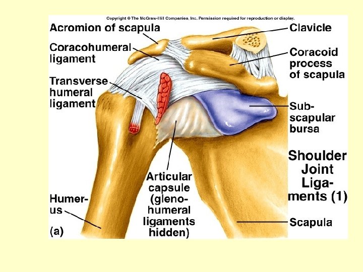
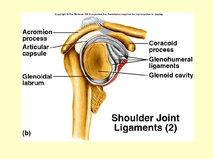
Shoulder Joint • Ball and socket joint made up of the rounded head of the humerus and the glenoid cavity of the scapula.
Shoulder Joint • The joint capsule is loose. Muscles and tendons reinforce the joint. • Shoulder joint is capable of a wide range of movements including flexion, extension, abduction, adduction, rotation, and circumduction.
Shoulder Joint • Ligaments: coracohumeral ligament, glenohumeral ligaments, transverse humeral ligament, and glenoid labrum • Bursae: subscapular, subdeltoid, subacromial, subcorocoid bursae
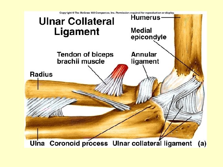
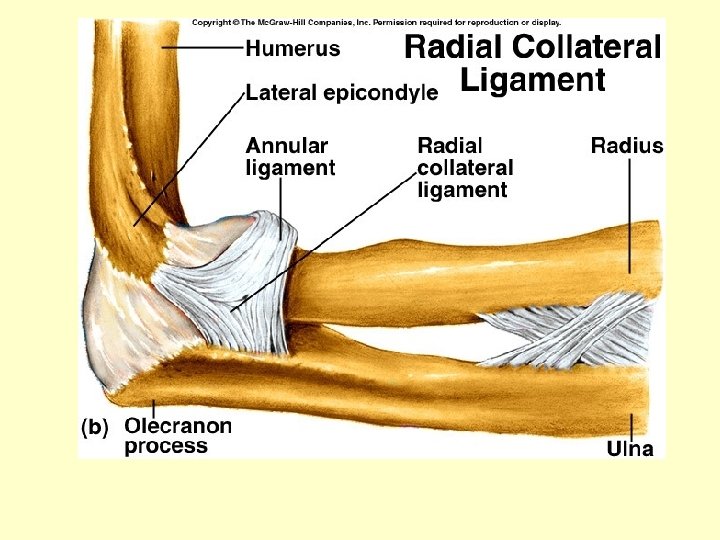
Elbow Joint • The elbow joint includes two articulations. • Hinge joint between the troclea of the humerus and the trochlear notch of the ulna.
Elbow Joint • Gliding joint between the capitulum of the humerus and a fovea on the radius head. • Movements include flexion and extension between the humerus and ulna. The radius allows rotation and supination of the hand. • Ligaments include the ulnar collateral and the radial collateral ligament.
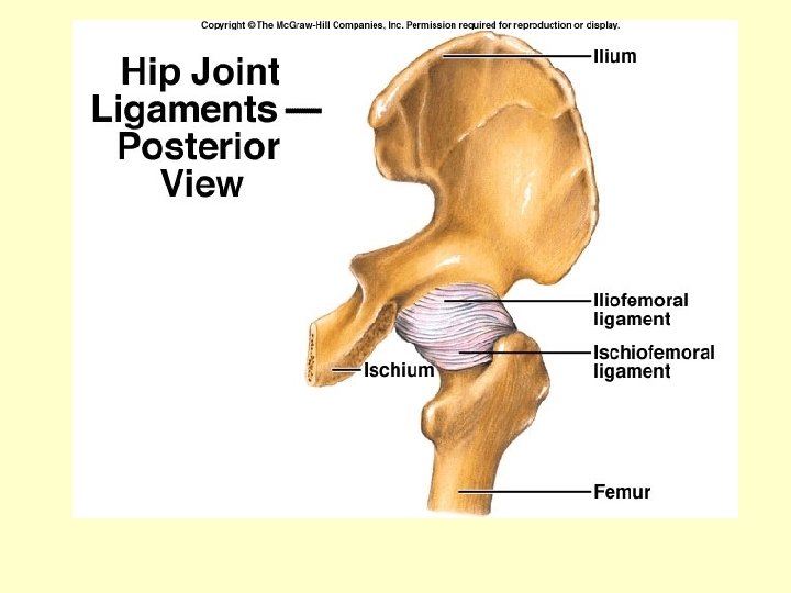
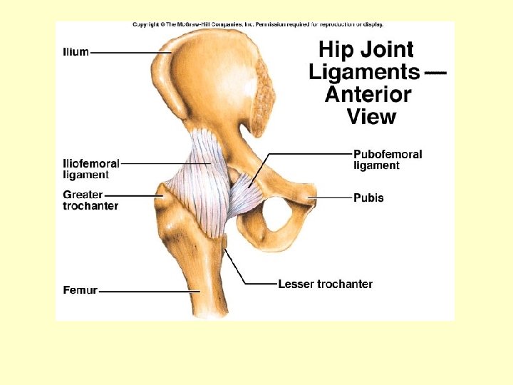
Hip Joint • Ball and socket joint consisting of the head of the femur and the acetabulum of the coxal bones • Muscles surround the joint capsule • Movements: flexion, extension, abduction, adduction, rotation, and circumduction • Ligaments: iliofemoral, pubofemoral, ischiofemoral ligaments
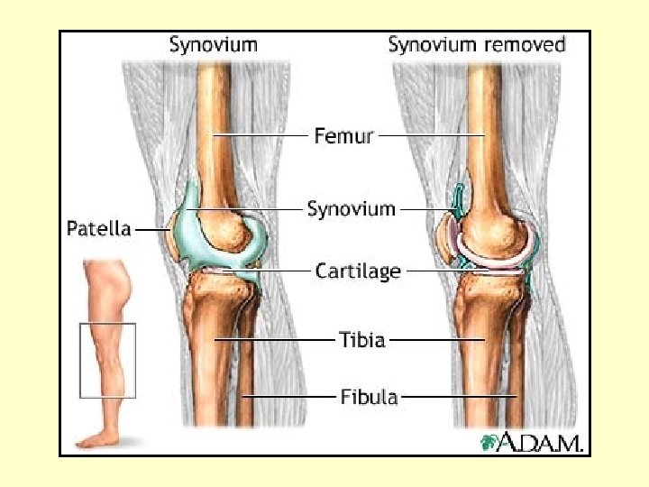
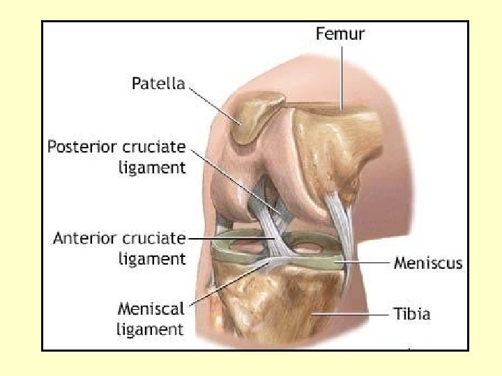
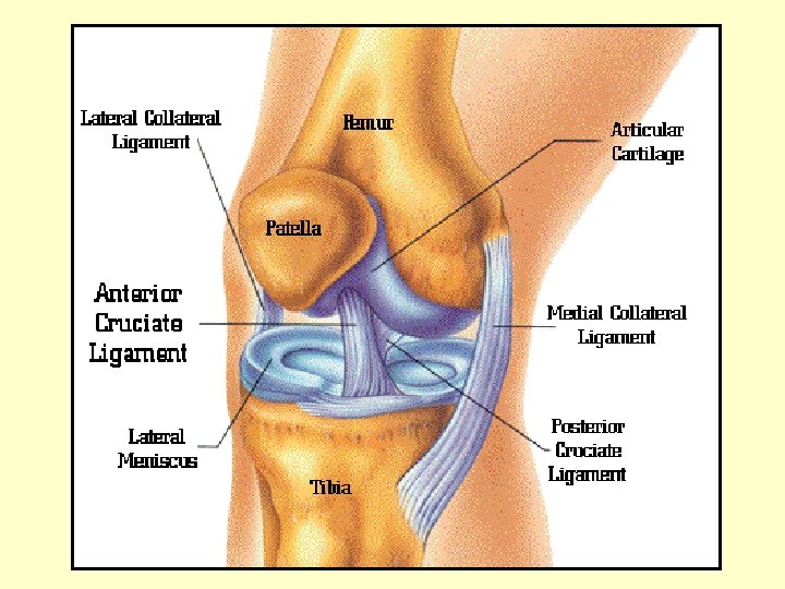
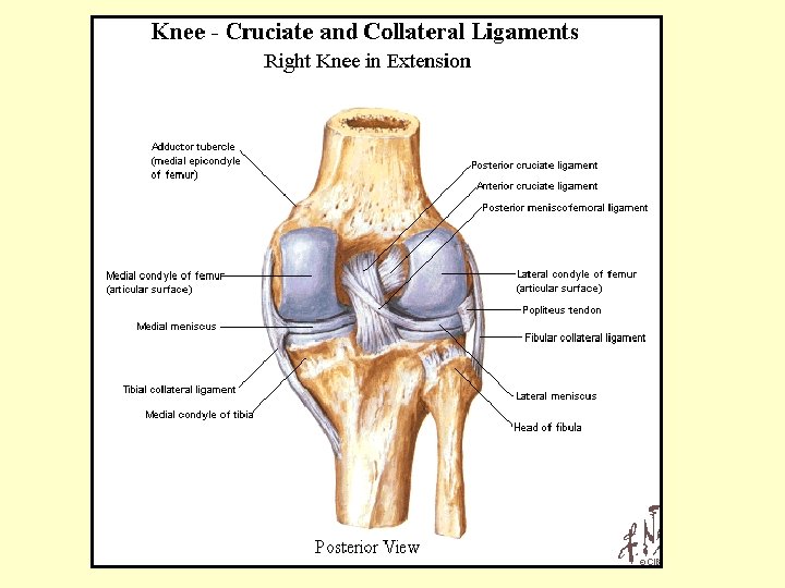
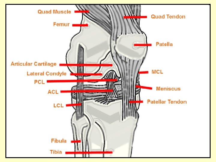
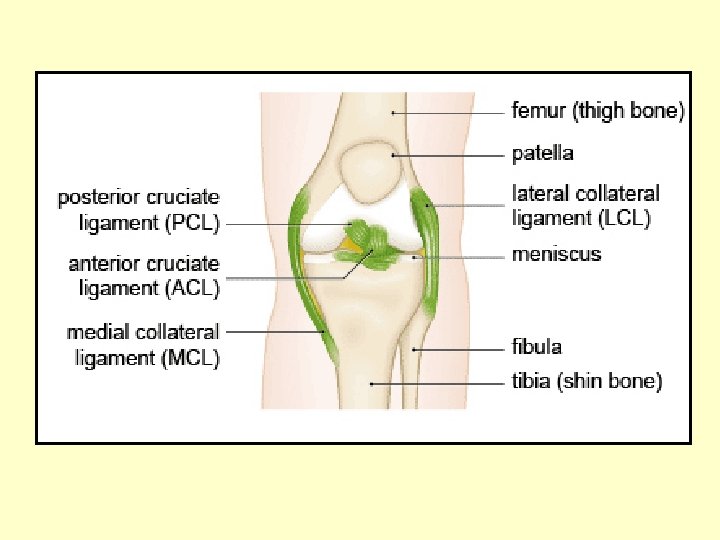
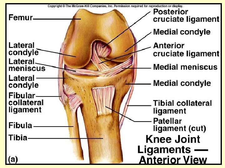
Knee Joint • The knee is the largest and most complex synovial joint. • It consists of the medial and lateral condyles at the proximal end of the tibia. The femur articulates with the patella. • The joint capsule is thin and strengthened by muscles and tendons.
Knee Joint • Ligaments of the knee joint: patella, oblique popliteal, arcuate popliteal, tibial collateral, fibular collateral ligament strengthen the joint capsule. • Cruciate ligaments prevent displacement of articulating surfaces. • Two fibrocartilaginous menisci separate the articulating surfaces.
Life-Span Changes • Joint stiffness occurs due to a change in collagen structure. • Fibrous joints strengthen over a lifetime.
Life-Span Changes • Synchondrosis disappear over time as part of skeletal growth and development. • Symphysis joints may lose water and flexibility may decrease.