
Power Point Lecture Slides prepared by Vince Austin
Power. Point® Lecture Slides prepared by Vince Austin, Bluegrass Technical and Community College CHAPTER Elaine N. Marieb Katja Hoehn 21 PART B Human Anatomy & Physiology SEVENTH EDITION Copyright © 2006 Pearson Education, Inc. , publishing as Benjamin Cummings The Immune System: Innate and Adaptive Body Defenses
Antibodies § Also called immunoglobulins § § Constitute the gamma globulin portion of blood proteins Are soluble proteins secreted by activated B cells and plasma cells in response to an antigen Are capable of binding specifically with that antigen There are five classes of antibodies: Ig. D, Ig. M, Ig. G, Ig. A, and Ig. E Copyright © 2006 Pearson Education, Inc. , publishing as Benjamin Cummings
Classes of Antibodies § § § Ig. D – monomer attached to the surface of B cells, important in B cell activation Ig. M – pentamer released by plasma cells during the primary immune response Ig. G – monomer that is the most abundant and diverse antibody in primary and secondary response; crosses the placenta and confers passive immunity Copyright © 2006 Pearson Education, Inc. , publishing as Benjamin Cummings
Classes of Antibodies § § Ig. A – dimer that helps prevent attachment of pathogens to epithelial cell surfaces Ig. E – monomer that binds to mast cells and basophils, causing histamine release when activated Copyright © 2006 Pearson Education, Inc. , publishing as Benjamin Cummings
Basic Antibody Structure NOTE: Study handout § Consists of four looping polypeptide chains linked together with disulfide bonds § § Two identical heavy (H) chains and two identical light (L) chains The four chains bound together form an antibody monomer Each chain has a variable (V) region at one end a constant (C) region at the other Variable regions of the heavy and light chains combine to form the antigen-binding site Copyright © 2006 Pearson Education, Inc. , publishing as Benjamin Cummings
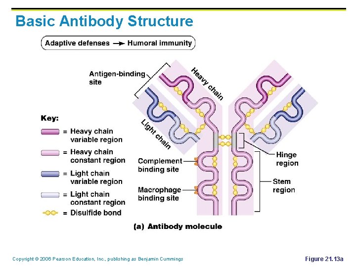
Basic Antibody Structure Copyright © 2006 Pearson Education, Inc. , publishing as Benjamin Cummings Figure 21. 13 a
Antibody Structure § Antibodies responding to different antigens have different V regions but the C region is the same for all antibodies in a given class Copyright © 2006 Pearson Education, Inc. , publishing as Benjamin Cummings
Antibody Structure § C regions form the stem of the Y-shaped antibody and: § Determine the class of the antibody § Serve common functions in all antibodies § § Dictate the cells and chemicals that the antibody can bind to Determine how the antibody class will function in elimination of antigens Copyright © 2006 Pearson Education, Inc. , publishing as Benjamin Cummings
Mechanisms of Antibody Diversity § § § Plasma cells make over a billion types of antibodies However, each cell only contains 100, 000 genes that code for these polypeptides To code for this many antibodies, somatic recombination takes place: § § Gene segments are shuffled and combined in different ways by each B cell as it becomes immunocompetent Information of the newly assembled genes is expressed as B cell receptors and as antibodies Copyright © 2006 Pearson Education, Inc. , publishing as Benjamin Cummings
Antibody Diversity § § § Random mixing of gene segments makes unique antibody genes that: § Code for H and L chains § Account for part of the variability in antibodies V gene segments, called hypervariable regions, mutate and increase antibody variation Plasma cells can switch H chains, making two or more classes with the same V region Copyright © 2006 Pearson Education, Inc. , publishing as Benjamin Cummings
Antibody Targets § § § Antibodies themselves do not destroy antigen; they inactivate and tag it for destruction All antibodies form an antigen-antibody (immune) complex This triggers complement fixation and cell lysis Copyright © 2006 Pearson Education, Inc. , publishing as Benjamin Cummings
Other Mechanisms of Antibody Action § Agglutination – antibodies bind the same determinant on more than one antigen § § § Makes antigen-antibody complexes that are crosslinked into large lattices Cell-bound antigens are cross-linked, causing clumping (agglutination) Example: Blood transfusions – when Type B blood is given to Type A recipient, etc. Copyright © 2006 Pearson Education, Inc. , publishing as Benjamin Cummings
Monoclonal Antibodies § § Commercially prepared antibodies are used: § To provide passive immunity § In research, clinical testing, and cancer treatment Monoclonal antibodies are pure antibody preparations § Specific for a single antigenic determinant § Produced from descendents of a single cell Copyright © 2006 Pearson Education, Inc. , publishing as Benjamin Cummings
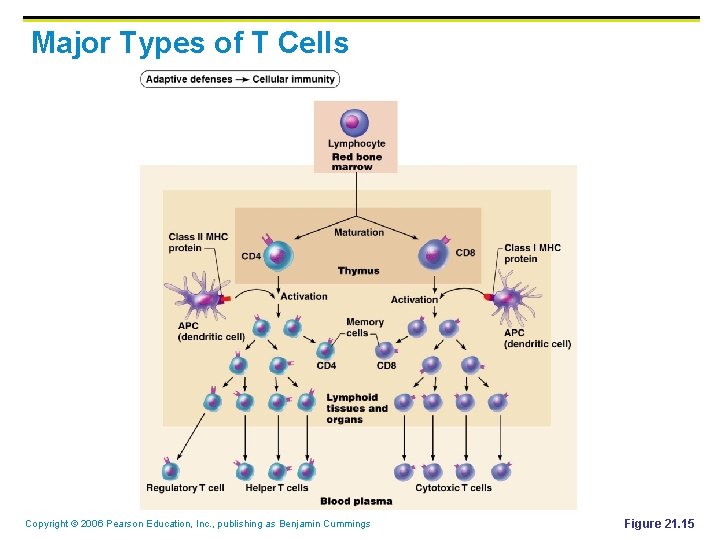
Major Types of T Cells Copyright © 2006 Pearson Education, Inc. , publishing as Benjamin Cummings Figure 21. 15
Importance of Cellular Response § § T cells recognize and respond only to processed fragments of antigen displayed on the surface of body cells T cells are best suited for cell-to-cell interactions, and target: § Cells infected with viruses, bacteria, or intracellular parasites § Abnormal or cancerous cells § Cells of infused or transplanted foreign tissue Copyright © 2006 Pearson Education, Inc. , publishing as Benjamin Cummings
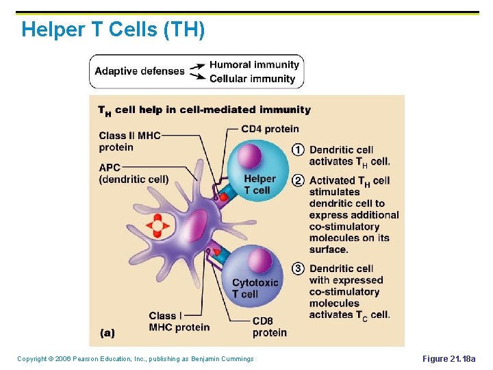
T Cell Activation: Step One – Antigen Binding § § MHC restriction – TH and TC bind to different classes of MHC proteins TH cells bind to antigen linked to class II MHC proteins Mobile APCs (Langerhans’ cells) quickly alert the body to the presence of antigen by migrating to the lymph nodes and presenting antigen NOTE: Study handout Copyright © 2006 Pearson Education, Inc. , publishing as Benjamin Cummings
T Cell Activation: Step One – Antigen Binding § § TC cells are activated by antigen fragments complexed with class I MHC proteins APCs produce co-stimulatory molecules that are required for TC activation TCR that acts to recognize the self-antiself complex is linked to multiple intracellular signaling pathways Other T cell surface proteins are involved in antigen binding (e. g. , CD 4 and CD 8 help maintain coupling during antigen recognition) Copyright © 2006 Pearson Education, Inc. , publishing as Benjamin Cummings
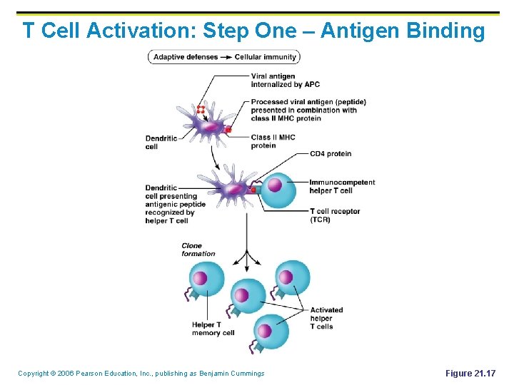
T Cell Activation: Step One – Antigen Binding Copyright © 2006 Pearson Education, Inc. , publishing as Benjamin Cummings Figure 21. 17
T Cell Activation: Step Two – Co-stimulation § T cells that are activated: § § Enlarge, proliferate, and form clones Differentiate and perform functions according to their T cell class Copyright © 2006 Pearson Education, Inc. , publishing as Benjamin Cummings
T Cell Activation: Step Two – Co-stimulation § § § Primary T cell response peaks within a week after signal exposure T cells then undergo apoptosis between days 7 and 30 Effector activity wanes as the amount of antigen declines The disposal of activated effector cells is a protective mechanism for the body Memory T cells remain and mediate secondary responses to the same antigen Copyright © 2006 Pearson Education, Inc. , publishing as Benjamin Cummings
Cytokines § § § Mediators involved in cellular immunity, including hormonelike glycoproteins released by activated T cells and macrophages Some are co-stimulators of T cells and T cell proliferation Interleukin 1 (IL-1) released by macrophages costimulates bound T cells to: § Release interleukin 2 (IL-2) § Synthesize more IL-2 receptors Copyright © 2006 Pearson Education, Inc. , publishing as Benjamin Cummings
Cytokines § IL-2 is a key growth factor, which sets up a positive feedback cycle that encourages activated T cells to divide § § It is used therapeutically to enhance the body’s defenses against cancer Other cytokines amplify and regulate immune and nonspecific responses Copyright © 2006 Pearson Education, Inc. , publishing as Benjamin Cummings
Cytokines § Examples include: § § § Perforin and lymphotoxin – cell toxins Gamma interferon – enhances the killing power of macrophages Inflammatory factors Copyright © 2006 Pearson Education, Inc. , publishing as Benjamin Cummings
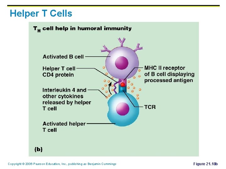
Helper T Cells (TH) § § Regulatory cells that play a central role in the immune response Once primed by APC presentation of antigen, they: § § § Chemically or directly stimulate proliferation of other T cells Stimulate B cells that have already become bound to antigen Without TH, there is no immune response Copyright © 2006 Pearson Education, Inc. , publishing as Benjamin Cummings
Helper T Cells (TH) Copyright © 2006 Pearson Education, Inc. , publishing as Benjamin Cummings Figure 21. 18 a
Helper T Cell § § § TH cells interact directly with B cells that have antigen fragments on their surfaces bound to MHC II receptors TH cells stimulate B cells to divide more rapidly and begin antibody formation B cells may be activated without TH cells by binding to T cell–independent antigens Most antigens, however, require TH co-stimulation to activate B cells Cytokines released by TH amplify nonspecific defenses Copyright © 2006 Pearson Education, Inc. , publishing as Benjamin Cummings
Helper T Cells Copyright © 2006 Pearson Education, Inc. , publishing as Benjamin Cummings Figure 21. 18 b
Cytotoxic T Cell (Tc) § § § TC cells, or killer T cells, are the only T cells that can directly attack and kill other cells They circulate throughout the body in search of body cells that display the antigen to which they have been sensitized Their targets include: § Virus-infected cells § Cells with intracellular bacteria or parasites § Cancer cells § Foreign cells from blood transfusions or transplants Copyright © 2006 Pearson Education, Inc. , publishing as Benjamin Cummings
Cytotoxic T Cells § § Bind to self-antiself complexes on all body cells Infected or abnormal cells can be destroyed as long as appropriate antigen and co-stimulatory stimuli (e. g. , IL-2) are present Natural killer cells activate their killing machinery when they bind to MICA receptor – MHC-related cell surface protein in cancer cells, virus-infected cells, and cells of transplanted organs Copyright © 2006 Pearson Education, Inc. , publishing as Benjamin Cummings
Mechanisms of Tc Action § In some cases, TC cells: § Bind to the target cell and release perforin into its membrane § § In the presence of Ca 2+ perforin causes cell lysis by creating transmembrane pores Other TC cells induce cell death by: § § Secreting lymphotoxin, which fragments the target cell’s DNA Secreting gamma interferon, which stimulates phagocytosis by macrophages Copyright © 2006 Pearson Education, Inc. , publishing as Benjamin Cummings
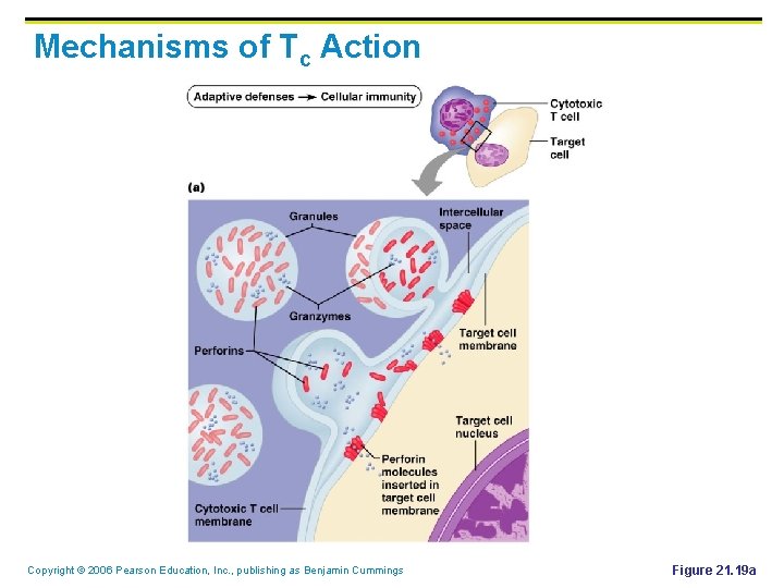
Mechanisms of Tc Action Copyright © 2006 Pearson Education, Inc. , publishing as Benjamin Cummings Figure 21. 19 a
Other T Cells § § Suppressor T cells (TS) – regulatory cells that release cytokines, which suppress the activity of both T cells and B cells Gamma delta T cells (Tgd) – 10% of all T cells found in the intestines that are triggered by binding to MICA receptors Copyright © 2006 Pearson Education, Inc. , publishing as Benjamin Cummings
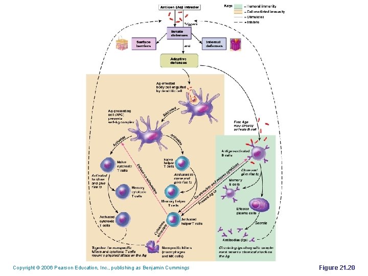
Copyright © 2006 Pearson Education, Inc. , publishing as Benjamin Cummings Figure 21. 20
Organ Transplants § The four major types of grafts are: § § Autografts – graft transplanted from one site on the body to another in the same person Isografts – grafts between identical twins Allografts – transplants between individuals that are not identical twins, but belong to same species Xenografts – grafts taken from another animal species Copyright © 2006 Pearson Education, Inc. , publishing as Benjamin Cummings
Prevention of Rejection § § Prevention of tissue rejection is accomplished by using immunosuppressive drugs However, these drugs depress patient’s immune system so it cannot fight off foreign agents Copyright © 2006 Pearson Education, Inc. , publishing as Benjamin Cummings
Immunodeficiencies § Congenital and acquired conditions in which the function or production of immune cells, phagocytes, or complement is abnormal § SCID – severe combined immunodeficiency (SCID) syndromes; genetic defects that produce: § A marked deficit in B and T cells § Abnormalities in interleukin receptors § Defective adenosine deaminase (ADA) enzyme § § Metabolites lethal to T cells accumulate SCID is fatal if untreated; treatment is with bone marrow transplants Copyright © 2006 Pearson Education, Inc. , publishing as Benjamin Cummings
Acquired Immunodeficiencies § § Hodgkin’s disease – cancer of the lymph nodes leads to immunodeficiency by depressing lymph node cells Acquired immune deficiency syndrome (AIDS) – cripples the immune system by interfering with the activity of helper T (CD 4) cells § § Characterized by severe weight loss, night sweats, and swollen lymph nodes Opportunistic infections occur, including pneumocystis pneumonia and Kaposi’s sarcoma Copyright © 2006 Pearson Education, Inc. , publishing as Benjamin Cummings
AIDS § Caused by human immunodeficiency virus (HIV) transmitted via body fluids – blood, semen, and vaginal secretions § § HIV enters the body via: § Blood transfusions § Contaminated needles § Intimate sexual contact, including oral sex HIV: § Destroys TH cells § Depresses cell-mediated immunity Copyright © 2006 Pearson Education, Inc. , publishing as Benjamin Cummings
AIDS § HIV multiplies in lymph nodes throughout the asymptomatic period § Symptoms appear in a few months to 10 years § Attachment § § HIV’s coat protein (gp 120) attaches to the CD 4 receptor of T helper cell A nearby protein (gp 41) fuses the virus to the target cell Copyright © 2006 Pearson Education, Inc. , publishing as Benjamin Cummings
AIDS § § HIV enters the cell and uses reverse transcriptase to produce DNA from viral RNA This DNA (provirus) directs the host cell to make viral RNA (and proteins), enabling the virus to reproduce and infect other cells Copyright © 2006 Pearson Education, Inc. , publishing as Benjamin Cummings
AIDS § HIV reverse transcriptase is not accurate and produces frequent transcription errors § § This high mutation rate causes resistance to drugs Treatments include: § Reverse transcriptase inhibitors (AZT) § Protease inhibitors (saquinavir and ritonavir) § New drugs currently being developed that block HIV’s entry to helper T cells Copyright © 2006 Pearson Education, Inc. , publishing as Benjamin Cummings
Autoimmune Diseases § § § Loss of the immune system’s ability to distinguish self from nonself The body produces autoantibodies and sensitized TC cells that destroy its own tissues Examples include multiple sclerosis, Graves’ disease, Type I (juvenile) diabetes mellitus, lupus, Crohn’s disease, and rheumatoid arthritis (RA), celiac disease Copyright © 2006 Pearson Education, Inc. , publishing as Benjamin Cummings
Mechanisms of Autoimmune Diseases § § Ineffective lymphocyte programming – selfreactive T and B cells that should have been eliminated in the thymus and bone marrow escape into the circulation New self-antigens appear, generated by: § § Gene mutations that cause new proteins to appear Changes in self-antigens by hapten attachment or as a result of infectious damage Copyright © 2006 Pearson Education, Inc. , publishing as Benjamin Cummings
Mechanisms of Autoimmune Diseases § If the determinants on foreign antigens resemble self-antigens: § Antibodies made against foreign antigens crossreact with self-antigens Copyright © 2006 Pearson Education, Inc. , publishing as Benjamin Cummings
Hypersensitivity § § Immune responses that cause tissue damage Different types of hypersensitivity reactions are distinguished by: § § Their time course Whether antibodies or T cells are the principle immune elements involved Antibody-mediated allergies are immediate and subacute hypersensitivities The most important cell-mediated allergic condition is delayed hypersensitivity Copyright © 2006 Pearson Education, Inc. , publishing as Benjamin Cummings
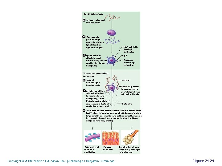
Immediate Hypersensitivity § § Acute (type I) hypersensitivities begin in seconds after contact with allergen Anaphylaxis – initial allergen contact is asymptomatic but sensitizes the person § Subsequent exposures to allergen cause: § § Release of histamine and inflammatory chemicals Systemic or local responses Copyright © 2006 Pearson Education, Inc. , publishing as Benjamin Cummings
Immediate Hypersensitivity § The mechanism involves IL-4 secreted by T cells § IL-4 stimulates B cells to produce Ig. E § Ig. E binds to mast cells and basophils causing them to degranulate, resulting in a flood of histamine release and inducing the inflammatory response Copyright © 2006 Pearson Education, Inc. , publishing as Benjamin Cummings
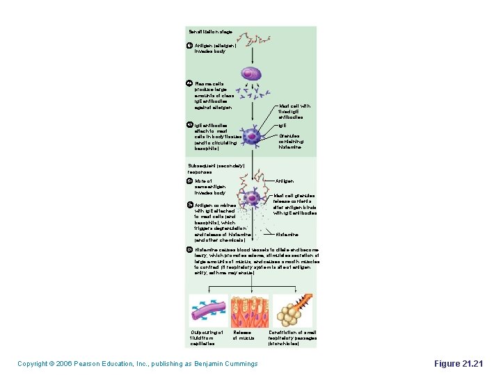
Sensitization stage 1 Antigen (allergen) invades body. 2 Plasma cells produce large amounts of class Ig. E antibodies against allergen. 3 Ig. E antibodies attach to mast cells in body tissues (and to circulating basophils). Mast cell with fixed Ig. E antibodies Ig. E Granules containing histamine Subsequent (secondary) responses 4 More of same antigen invades body. Antigen 5 Antigen combines with Ig. E attached to mast cells (and basophils), which triggers degranulation and release of histamine (and other chemicals). Mast cell granules release contents after antigen binds with Ig. E antibodies Histamine 6 Histamine causes blood vessels to dilate and become leaky, which promotes edema; stimulates secretion of large amounts of mucus; and causes smooth muscles to contract (if respiratory system is site of antigen entry, asthma may ensue). Outpouring of fluid from capillaries Release of mucus Copyright © 2006 Pearson Education, Inc. , publishing as Benjamin Cummings Constriction of small respiratory passages (bronchioles) Figure 21. 21
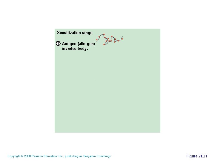
Sensitization stage 1 Antigen (allergen) invades body. Copyright © 2006 Pearson Education, Inc. , publishing as Benjamin Cummings Figure 21. 21
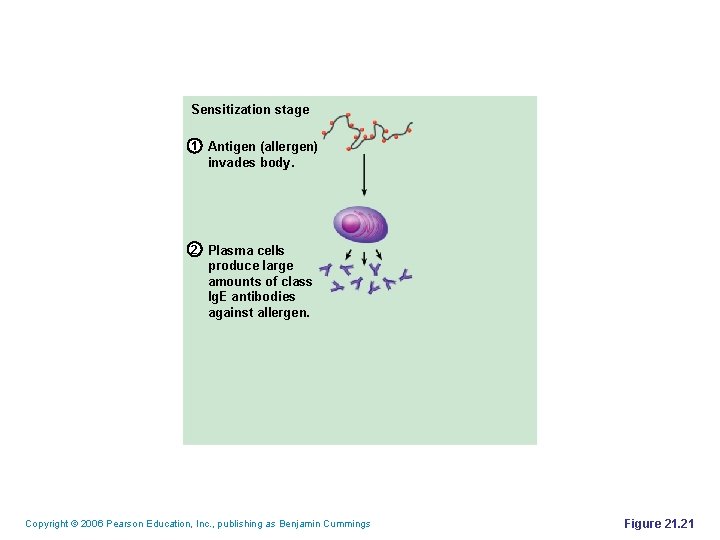
Sensitization stage 1 Antigen (allergen) invades body. 2 Plasma cells produce large amounts of class Ig. E antibodies against allergen. Copyright © 2006 Pearson Education, Inc. , publishing as Benjamin Cummings Figure 21. 21
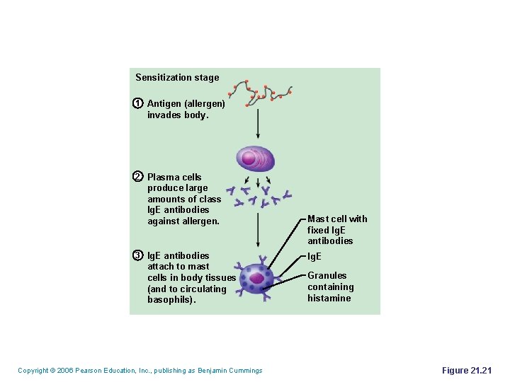
Sensitization stage 1 Antigen (allergen) invades body. 2 Plasma cells produce large amounts of class Ig. E antibodies against allergen. 3 Ig. E antibodies attach to mast cells in body tissues (and to circulating basophils). Copyright © 2006 Pearson Education, Inc. , publishing as Benjamin Cummings Mast cell with fixed Ig. E antibodies Ig. E Granules containing histamine Figure 21. 21
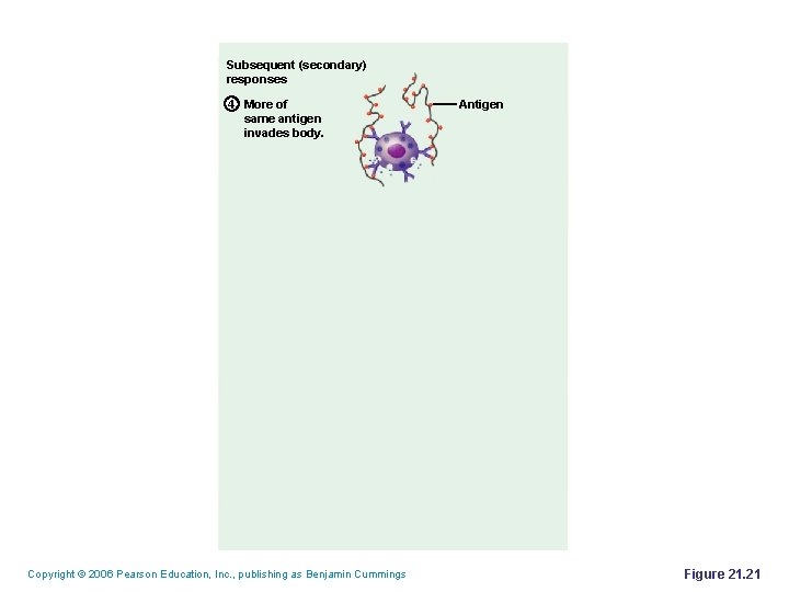
Subsequent (secondary) responses 4 More of same antigen invades body. Copyright © 2006 Pearson Education, Inc. , publishing as Benjamin Cummings Antigen Figure 21. 21
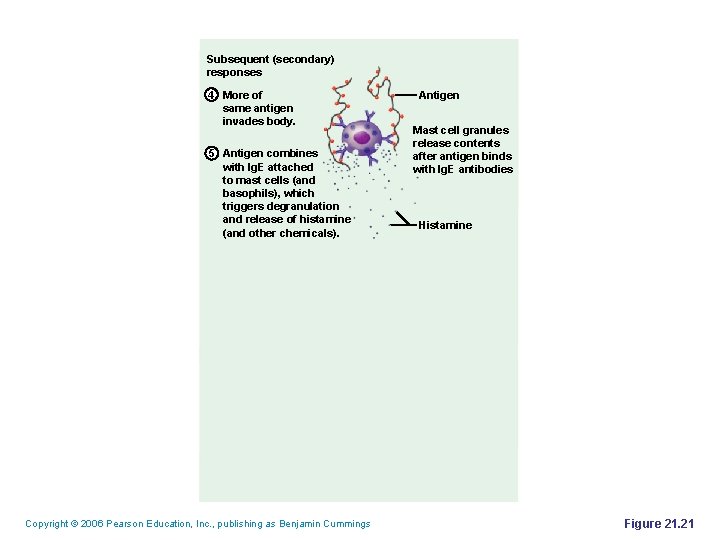
Subsequent (secondary) responses 4 More of same antigen invades body. 5 Antigen combines with Ig. E attached to mast cells (and basophils), which triggers degranulation and release of histamine (and other chemicals). Copyright © 2006 Pearson Education, Inc. , publishing as Benjamin Cummings Antigen Mast cell granules release contents after antigen binds with Ig. E antibodies Histamine Figure 21. 21
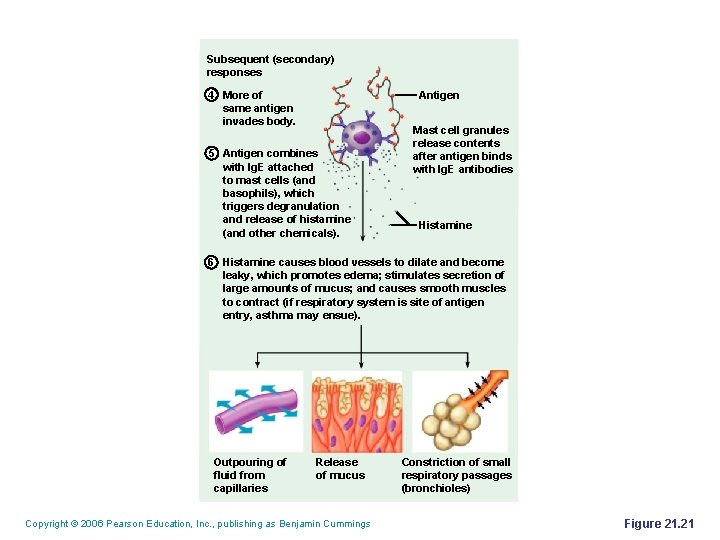
Subsequent (secondary) responses 4 More of same antigen invades body. Antigen 5 Antigen combines with Ig. E attached to mast cells (and basophils), which triggers degranulation and release of histamine (and other chemicals). Mast cell granules release contents after antigen binds with Ig. E antibodies Histamine 6 Histamine causes blood vessels to dilate and become leaky, which promotes edema; stimulates secretion of large amounts of mucus; and causes smooth muscles to contract (if respiratory system is site of antigen entry, asthma may ensue). Outpouring of fluid from capillaries Release of mucus Copyright © 2006 Pearson Education, Inc. , publishing as Benjamin Cummings Constriction of small respiratory passages (bronchioles) Figure 21. 21
Sensitization stage 1 Antigen (allergen) invades body. 2 Plasma cells produce large amounts of class Ig. E antibodies against allergen. 3 Ig. E antibodies attach to mast cells in body tissues (and to circulating basophils). Mast cell with fixed Ig. E antibodies Ig. E Granules containing histamine Subsequent (secondary) responses 4 More of same antigen invades body. Antigen 5 Antigen combines with Ig. E attached to mast cells (and basophils), which triggers degranulation and release of histamine (and other chemicals). Mast cell granules release contents after antigen binds with Ig. E antibodies Histamine 6 Histamine causes blood vessels to dilate and become leaky, which promotes edema; stimulates secretion of large amounts of mucus; and causes smooth muscles to contract (if respiratory system is site of antigen entry, asthma may ensue). Outpouring of fluid from capillaries Release of mucus Copyright © 2006 Pearson Education, Inc. , publishing as Benjamin Cummings Constriction of small respiratory passages (bronchioles) Figure 21. 21
Anaphylaxis § § Reactions include runny nose, itching reddened skin, and watery eyes If allergen is inhaled, asthmatic symptoms appear– constriction of bronchioles and restricted airflow If allergen is ingested, cramping, vomiting, or diarrhea occur Antihistamines counteract these effects Copyright © 2006 Pearson Education, Inc. , publishing as Benjamin Cummings
Anaphylactic Shock § § § Response to allergen that directly enters the blood (e. g. , insect bite, injection) Basophils and mast cells are enlisted throughout the body Systemic histamine releases may result in: § § Constriction of bronchioles Sudden vasodilation and fluid loss from the bloodstream Hypotensive shock and death Treatment – epinephrine is the drug of choice Copyright © 2006 Pearson Education, Inc. , publishing as Benjamin Cummings
Subacute Hypersensitivities § § Caused by Ig. M and Ig. G, and transferred via blood plasma or serum § Onset is slow (1– 3 hours) after antigen exposure § Duration is long lasting (10– 15 hours) Cytotoxic (type II) reactions § § Antibodies bind to antigens on specific body cells, stimulating phagocytosis and complementmediated lysis of the cellular antigens Example: mismatched blood transfusion reaction Copyright © 2006 Pearson Education, Inc. , publishing as Benjamin Cummings
Subacute Hypersensitivities § Immune complex (type III) hypersensitivity § § § Antigens are widely distributed through the body or blood Insoluble antigen-antibody complexes form Complexes cannot be cleared from a particular area of the body Intense inflammation, local cell lysis, and death may result Example: systemic lupus erythematosus (SLE) Copyright © 2006 Pearson Education, Inc. , publishing as Benjamin Cummings
Delayed Hypersensitivities (Type IV) § § Onset is slow (1– 3 days) Mediated by mechanisms involving delayed hypersensitivity T cells and cytotoxic T cells Cytokines from activated TC are the mediators of the inflammatory response Antihistamines are ineffective and corticosteroid drugs are used to provide relief Copyright © 2006 Pearson Education, Inc. , publishing as Benjamin Cummings
Delayed Hypersensitivities (Type IV) § § Example: allergic contact dermatitis (e. g. , poison ivy) Involved in protective reactions against viruses, bacteria, fungi, protozoa, cancer, and rejection of foreign grafts or transplants Copyright © 2006 Pearson Education, Inc. , publishing as Benjamin Cummings
Developmental Aspects § § § Immune system cells develop in the liver and spleen by the ninth week Later, bone marrow becomes the primary source of stem cells Lymphocyte development continues in the bone marrow and thymus system begins to wane Copyright © 2006 Pearson Education, Inc. , publishing as Benjamin Cummings
Developmental Aspects § § § TH 2 lymphocytes predominate in the newborn, and the TH 1 system is educated as the person encounters antigens The immune system is impaired by stress and depression With age, the immune system begins to wane Copyright © 2006 Pearson Education, Inc. , publishing as Benjamin Cummings
- Slides: 63