
Power Point Lecture Slides prepared by Barbara Heard
Power. Point® Lecture Slides prepared by Barbara Heard, Atlantic Cape Community College 29 CHAPTER Heredity © Annie Leibovitz/Contact Press Images © 2013 Pearson Education, Inc.
Genetics • Study of mechanism of heredity • Basic principles proposed by Mendel in mid-1800 s © 2013 Pearson Education, Inc.
The Vocabulary of Genetics • Diploid number (46) of chromosomes in all cells except gametes – 23 pairs of homologous chromosomes – 1 pair-sex chromosomes • Determine genetic sex (XX = female, XY = male) – 22 pairs–autosomes • Guide expression of most other traits © 2013 Pearson Education, Inc.
The Vocabulary of Genetics • Karyotype diploid chromosomal complement displayed in homologous pairs • Genom -genetic (DNA) makeup; two sets of genetic instructions (maternal and paternal) © 2013 Pearson Education, Inc.
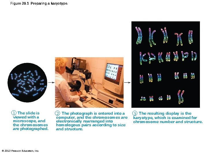
Figure 29. 1 Preparing a karyotype. 1 The slide is viewed with a microscope, and the chromosomes are photographed. © 2013 Pearson Education, Inc. 2 The photograph is entered into a computer, and the chromosomes are electronically rearranged into homologous pairs according to size and structure. 3 The resulting display is the karyotype, which is examined for chromosome number and structure.
Gene Pairs • Alleles – Matched genes at same locus (location) on homologous chromosomes – Homozygous–alleles same for single trait – Heterozygous–alleles different for single trait © 2013 Pearson Education, Inc.
Gene Pairs – Dominant-one allele masks (suppresses) its recessive partner • Dominant denoted by capital letter; recessive by same letter in lower case • Dominant expressed if one or both alleles dominant • Recessive expressed only if both alleles recessive © 2013 Pearson Education, Inc.
Genotype and Phenotype • Genotype–genetic makeup • Phenotype–expression of genotype © 2013 Pearson Education, Inc.
Sexual Sources of Genetic Variation • Each person unique due to three events – Independent assortment of chromosomes – Crossover of homologues – Random fertilization of eggs by sperm © 2013 Pearson Education, Inc.
Chromosome Segregation and Independent Assortment • During gametogenesis, maternal and paternal chromosomes randomly distributed to daughter cells • Alleles for each trait segregated during meiosis I Gamete receives only one allele of the four alleles (2 maternal / 2 paternal) for each trait • Alleles on different pairs of homologous chromosomes distributed independently © 2013 Pearson Education, Inc.
Segregation and Independent Assortment • Independent assortment incredible variety – Number of gamete types = 2 n, where n = number of homologous pairs – In testes, 2 n = 2223 = 8. 5 million © 2013 Pearson Education, Inc.
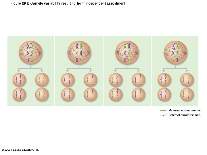
Figure 29. 2 Gamete variability resulting from independent assortment. Maternal chromosomes Paternal chromosomes © 2013 Pearson Education, Inc.
Crossover and Genetic Recombination • Genes on same chromosome linked – Normally pass to daughter cells as unit • Homologous chromosomes can break, even between linked genes precisely exchange gene segments (crossover or chiasma) Recombinant chromosomes • Chromosomes have mixed contributions from each parent Tremendous variability © 2013 Pearson Education, Inc.
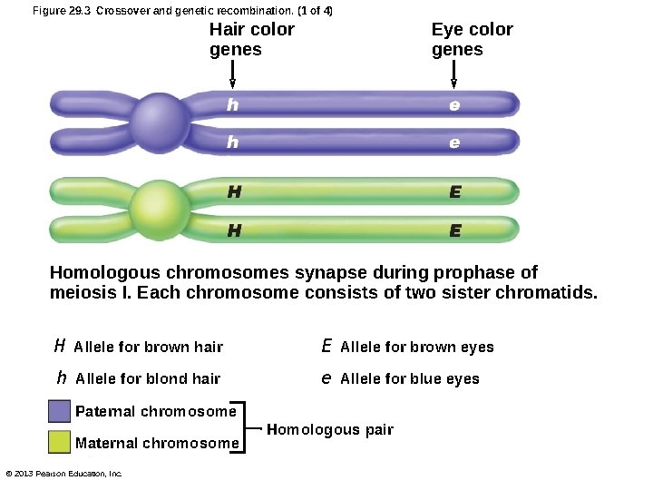
Figure 29. 3 Crossover and genetic recombination. (1 of 4) Hair color genes Eye color genes Homologous chromosomes synapse during prophase of meiosis I. Each chromosome consists of two sister chromatids. H Allele for brown hair E Allele for brown eyes h Allele for blond hair e Allele for blue eyes Paternal chromosome Maternal chromosome © 2013 Pearson Education, Inc. Homologous pair
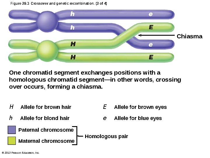
Figure 29. 3 Crossover and genetic recombination. (2 of 4) Chiasma One chromatid segment exchanges positions with a homologous chromatid segment—in other words, crossing over occurs, forming a chiasma. H Allele for brown hair E Allele for brown eyes h Allele for blond hair e Allele for blue eyes Paternal chromosome Maternal chromosome © 2013 Pearson Education, Inc. Homologous pair
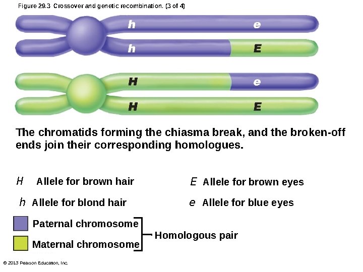
Figure 29. 3 Crossover and genetic recombination. (3 of 4) The chromatids forming the chiasma break, and the broken-off ends join their corresponding homologues. H Allele for brown hair h Allele for blond hair E Allele for brown eyes e Allele for blue eyes Paternal chromosome Maternal chromosome © 2013 Pearson Education, Inc. Homologous pair
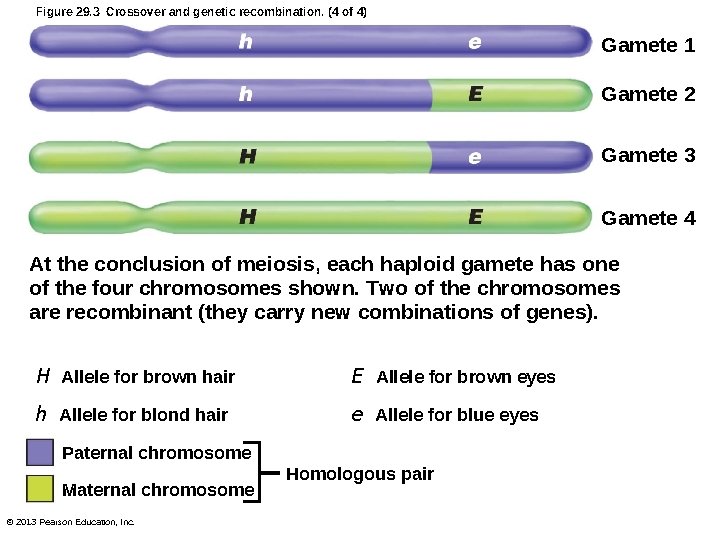
Figure 29. 3 Crossover and genetic recombination. (4 of 4) Gamete 1 Gamete 2 Gamete 3 Gamete 4 At the conclusion of meiosis, each haploid gamete has one of the four chromosomes shown. Two of the chromosomes are recombinant (they carry new combinations of genes). H Allele for brown hair E Allele for brown eyes h Allele for blond hair e Allele for blue eyes Paternal chromosome Maternal chromosome © 2013 Pearson Education, Inc. Homologous pair
Random Fertilization • Single egg fertilized by single sperm in random manner • Independent assortment and random fertilization ~ 72 trillion (8. 5 million 8. 5 million) zygote possibilities • Random fertilization increases number of possibilities exponentially © 2013 Pearson Education, Inc.
Types of Inheritance • Few phenotypes traced to single gene • Most traits determined by multiple alleles or by interaction of several gene pairs © 2013 Pearson Education, Inc.
Dominant-Recessive Inheritance • Reflects interaction of dominant and recessive alleles • Punnett square – Predicts possible gene combinations resulting from mating of parents of known genotypes – Only probability of particular genotype (phenotype) © 2013 Pearson Education, Inc.
Dominant-Recessive Inheritance • Example – Probability of genotypes from mating two heterozygous parents – Dominant allele—capital letter; recessive allele—lowercase letter – T = tongue roller and t = cannot roll tongue – TT and tt are homozygous; Tt is heterozygous © 2013 Pearson Education, Inc.
Dominant-Recessive Inheritance • Offspring: 25% TT; 50% Tt; 25% tt • Larger number of offspring greater likelihood ratios conform to predicted values • Probability of two children with same genotype – Multiply probabilities of separate events – E. g. , probability of two children who cannot roll tongue = 1/4 * 1/4 = 1/16 © 2013 Pearson Education, Inc.
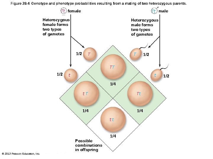
Figure 29. 4 Genotype and phenotype probabilities resulting from a mating of two heterozygous parents. Tt female Tt male Heterozygous female forms two types of gametes Heterozygous male forms two types of gametes 1/2 T T 1/2 TT 1/2 t t 1/4 t. T Tt 1/4 tt 1/4 Possible combinations in offspring © 2013 Pearson Education, Inc. 1/2
Dominant Traits • Dominant traits (for example, widow's peaks, freckles, dimples) • Dominant disorders uncommon; many lethal death before reproductive age • Huntington's disease caused by delayedaction gene – Activated ~ age 40 – Dominant gene possibly passed to offspring– 50% chance © 2013 Pearson Education, Inc.
Recessive Inheritance • Some recessive genes more desirable condition – E. g. , normal vision-recessive; astigmatism dominant • Most genetic disorders inherited as simple recessive traits – Albinism, cystic fibrosis, and Tay-Sachs disease • Heterozygotes–carriers; do not express trait but can pass to offspring © 2013 Pearson Education, Inc.
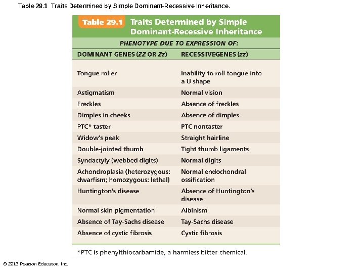
Table 29. 1 Traits Determined by Simple Dominant-Recessive Inheritance. © 2013 Pearson Education, Inc.
Incomplete Dominance • Heterozygous individuals have intermediate phenotype • Rare in humans; example-Sickling gene – SS = normal Hb made – Ss = sickle-cell trait; both aberrant and normal Hb made; can suffer sickle-cell crisis under prolonged reduction in blood O 2 – ss = sickle-cell anemia; only aberrant Hb made; more susceptible to sickle-cell crisis © 2013 Pearson Education, Inc.
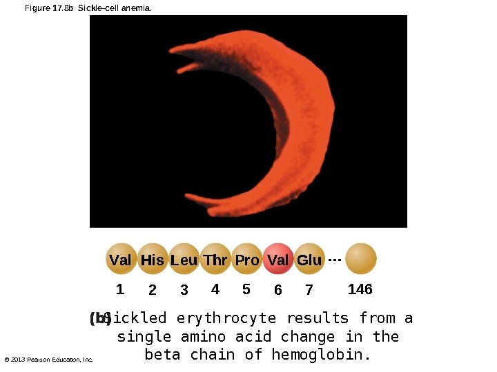
Figure 17. 8 b Sickle-cell anemia. Val His Leu Thr Pro Val Glu … 1 © 2013 Pearson Education, Inc. 2 3 4 5 6 7 146 Sickled erythrocyte results from a single amino acid change in the beta chain of hemoglobin.
Multiple-Allele Inheritance • Genes that exhibit more than two allele forms – E. g. , ABO blood grouping • Three alleles (IA, IB, i) determine ABO blood type in humans – Each person has only two – IA and IB-codominant (both expressed if present) – i - recessive © 2013 Pearson Education, Inc.
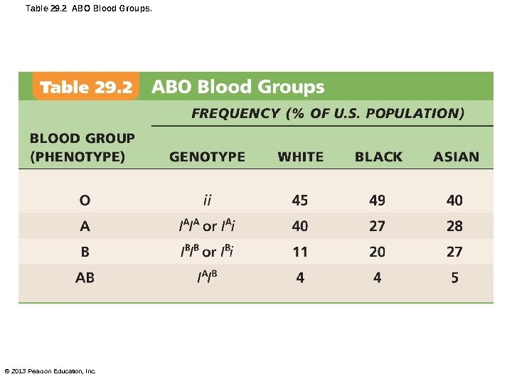
Table 29. 2 ABO Blood Groups. © 2013 Pearson Education, Inc.
Sex-Linked Inheritance • Inherited traits determined by genes on sex chromosomes • X chromosomes bear over 2500 genes (many for brain function); Y chromosomes carry about 78 genes – Few regions can participate in crossover © 2013 Pearson Education, Inc.
Sex-Linked Inheritance • X-linked genes - found only on X chromosome – X-linked recessive alleles always expressed in males • Never masked or damped in males (no Y counterpart) • Most X-linked conditions expressed in males – Passed from mothers to sons (e. g. , hemophilia or red-green color blindness); never father to sons – Passed from mothers to daughters, but females require two alleles to express © 2013 Pearson Education, Inc.
Polygene Inheritance • Traits reflecting actions of several gene pairs at different locations • Results in continuous phenotypic variation between two extremes • Examples: skin color, eye color, height, intelligence, metabolic rate © 2013 Pearson Education, Inc.
Polygene Inheritance of Skin Color • Alleles for dark skin (ABC) incompletely dominant over those for light skin (abc) • First-generation offspring of AABBCC X aabbcc cross all heterozygous (intermediate pigmentation) • Second-generation offspring have wide variation in possible pigmentations bellshaped curve © 2013 Pearson Education, Inc.
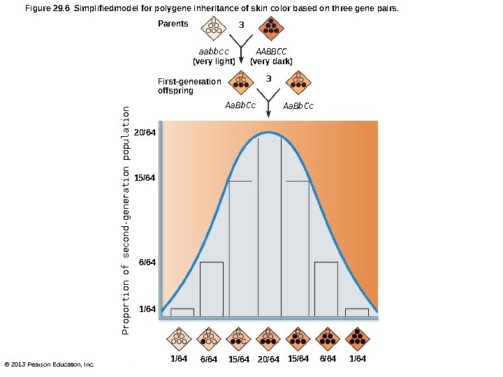
Figure 29. 6 Simplifiedmodel for polygene inheritance of skin color based on three gene pairs. Parents 3 aabbcc (very light) AABBCC (very dark) 3 Proportion of second-generation population First-generation offspring © 2013 Pearson Education, Inc. Aa. Bb. Cc 20/64 15/64 6/64 1/64 6/64 15/64 20/64 15/64 6/64 1/64
Environmental Factors in Gene Expression • Maternal factors (e. g. , drugs, pathogens) alter normal gene expression during embryonic development • E. g. , thalidomide – Embryos developed phenotypes not directed by their genes • Phenocopies-environmentally produced phenotypes; mimic conditions caused by genetic mutations © 2013 Pearson Education, Inc.
Environmental Factors in Gene Expression • Environmental factors influence gene expression after birth – Poor nutrition affects brain growth, body development, and height – Childhood hormonal deficits can lead to abnormal skeletal growth and proportions © 2013 Pearson Education, Inc.
Nontraditional Inheritance • Genetic outcomes influenced by – Small RNAs – Epigenetic marks (chemical groups attached to DNA or histone proteins) – Extranuclear inheritance (mitochondrial DNA) © 2013 Pearson Education, Inc.
Nontraditional Inheritance: Regulation of Gene Expression • Three levels of controls in human genome – First layer - protein-coding genes • Less than 2% of DNA – Second layer–small RNAs • In non-protein-coding DNA – Third layer–epigenetic marks • Stored in proteins and chemical groups that bind to DNA; and in way chromatin packaged © 2013 Pearson Education, Inc.
Small RNAs • Micro. RNAs (mi. RNAs) and short interfering RNAs (si. RNAs) – Act directly on DNA, other RNAs, or proteins – Inactivate transposons, genes that tend to replicate themselves and disable or hyperactivate other genes – Control timing of apoptosis during development – Prevent translation of another gene – Mutations linked to prostate and lung cancers, and schizophrenia © 2013 Pearson Education, Inc.
Small RNAs • Nucleotide sequences for small RNAs now determined • Extensive gene therapy research to develop RNA-interfering drugs for diseases such as • Age-related macular degeneration, Parkinson's disease, cancer, and other disorders © 2013 Pearson Education, Inc.
Epigenetic Marks • Information stored in proteins and chemical groups (e. g. , methyl and acetyl groups) bound to DNA; and in way chromatin packaged in cell • Determine whether DNA available for transcription (acetylation) or silenced (methylation) • May predispose cell to cancerous changes or devastating illness © 2013 Pearson Education, Inc.
Epigenetic Marks • Genomic imprinting – Tags certain genes with methyl group during gametogenesis as maternal or paternal; essential for normal development • Allows embryo to express only mother's or father's gene • Each new generation erases imprinting when new gametes produced © 2013 Pearson Education, Inc.
Epigenetic Marks • Mutations of imprinted genes may pathology • Same allele can have different effects depending on which parent it comes from – For example, deletions in chromosome 15 result in disorders with different symptoms • Prader-Willi syndrome if inherited from father • Angelman syndrome if inherited from mother © 2013 Pearson Education, Inc.
Extranuclear (Mitochondrial) Inheritance • Some genes (37) in mitochondrial DNA (mt. DNA) • Transmitted by mother in cytoplasm of egg • Errors in mt. DNA linked to rare disorders – Muscle disorders and neurological problems, possibly Alzheimer's and Parkinson's diseases © 2013 Pearson Education, Inc.
Genetic Screening, Counseling, and Therapy • Newborn infants routinely screened for number of genetic disorders – Congenital hip dysplasia, imperforate anus, PKU, and other metabolic disorders • Other examples – Screening adult children of parents with Huntington's disease; testing fetus of 35 yo woman for trisomy-21 (Down syndrome) © 2013 Pearson Education, Inc.
Carrier Recognition • Two ways to identify gene carriers – Pedigrees and blood tests • Pedigrees – Trace genetic trait through several generations; helps predict future © 2013 Pearson Education, Inc.
Carrier Recognition • Blood tests and DNA probes can detect presence of unexpressed recessive genes – E. g. , Tay-Sachs and cystic fibrosis genes © 2013 Pearson Education, Inc.
Fetal Testing • Used when known risk of genetic disorder; both invasive; both carry risk • Amniocentesis – Amniotic fluid withdrawn after 14 th week; fluid and cells examined for genetic abnormalities – Testing takes several weeks • Chorionic villus sampling (CVS) – Chorionic villi sampled at 8 -10 weeks; karyotyped for genetic abnormalities; testing ~ immediate © 2013 Pearson Education, Inc.
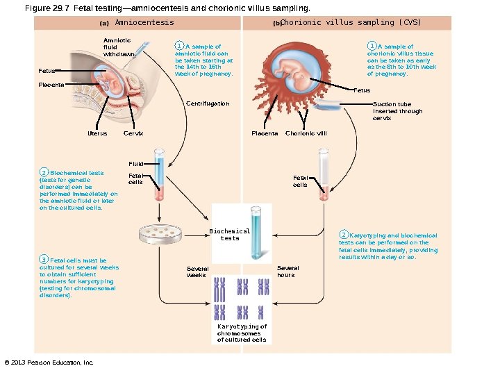
Figure 29. 7 Fetal testing—amniocentesis and chorionic villus sampling. Amniocentesis Amniotic fluid withdrawn Fetus Chorionic villus sampling (CVS) 1 A sample of amniotic fluid can be taken starting at the 14 th to 16 th week of pregnancy. 1 A sample of chorionic villus tissue can be taken as early as the 8 th to 10 th week of pregnancy. Placenta Fetus Centrifugation Uterus Suction tube inserted through cervix Placenta Cervix Chorionic villi Fluid 2 Biochemical tests (tests for genetic disorders) can be performed immediately on the amniotic fluid or later on the cultured cells. Fetal cells Biochemical tests 3 Fetal cells must be cultured for several weeks to obtain sufficient numbers for karyotyping (testing for chromosomal disorders). Several hours Several weeks Karyotyping of chromosomes of cultured cells © 2013 Pearson Education, Inc. 2 Karyotyping and biochemical tests can be performed on the fetal cells immediately, providing results within a day or so.
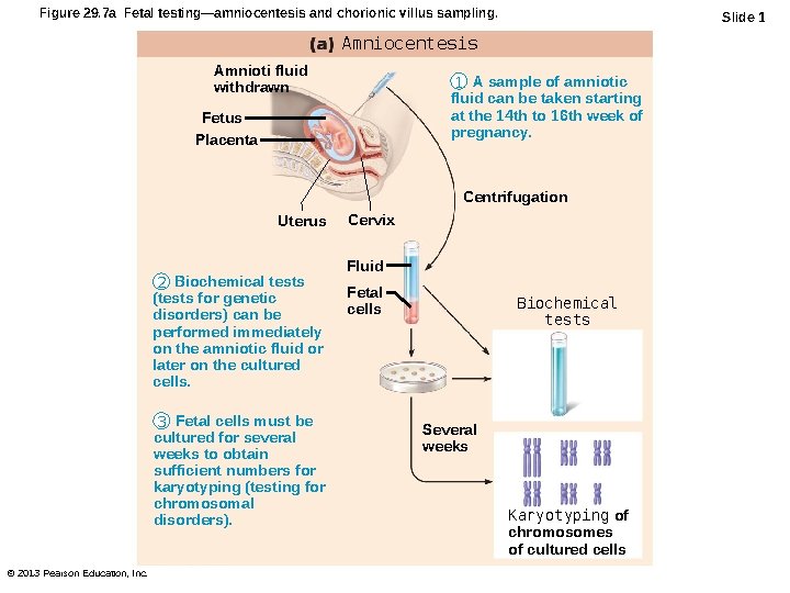
Figure 29. 7 a Fetal testing—amniocentesis and chorionic villus sampling. Slide 1 Amniocentesis Amnioti fluid withdrawn 1 A sample of amniotic fluid can be taken starting at the 14 th to 16 th week of pregnancy. Fetus Placenta Centrifugation Uterus 2 Biochemical tests (tests for genetic disorders) can be performed immediately on the amniotic fluid or later on the cultured cells. 3 Fetal cells must be cultured for several weeks to obtain sufficient numbers for karyotyping (testing for chromosomal disorders). © 2013 Pearson Education, Inc. Cervix Fluid Fetal cells Biochemical tests Several weeks Karyotyping of chromosomes of cultured cells
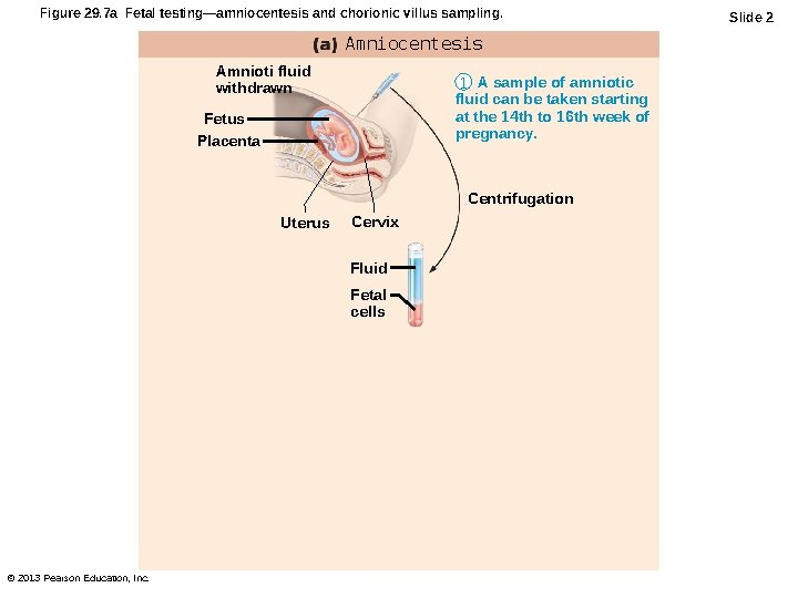
Figure 29. 7 a Fetal testing—amniocentesis and chorionic villus sampling. Amniocentesis Amnioti fluid withdrawn 1 A sample of amniotic fluid can be taken starting at the 14 th to 16 th week of pregnancy. Fetus Placenta Centrifugation Uterus Cervix Fluid Fetal cells © 2013 Pearson Education, Inc. Slide 2
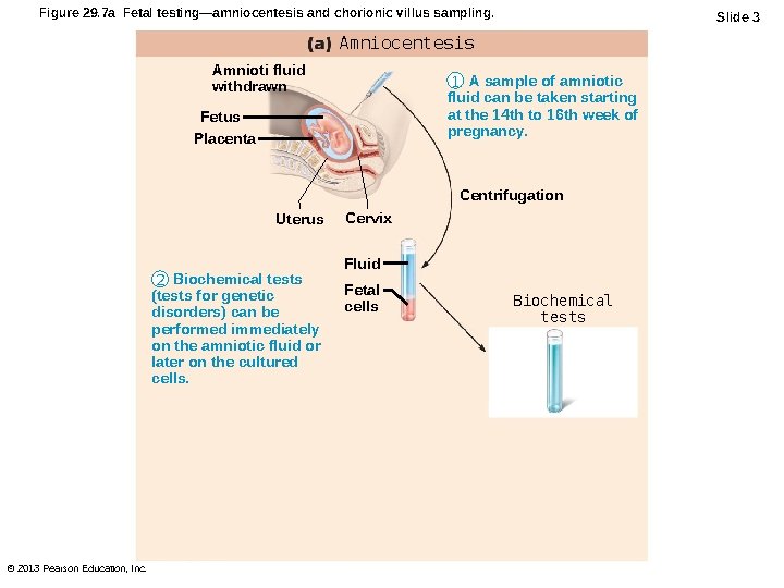
Figure 29. 7 a Fetal testing—amniocentesis and chorionic villus sampling. Slide 3 Amniocentesis Amnioti fluid withdrawn 1 A sample of amniotic fluid can be taken starting at the 14 th to 16 th week of pregnancy. Fetus Placenta Centrifugation Uterus 2 Biochemical tests (tests for genetic disorders) can be performed immediately on the amniotic fluid or later on the cultured cells. © 2013 Pearson Education, Inc. Cervix Fluid Fetal cells Biochemical tests
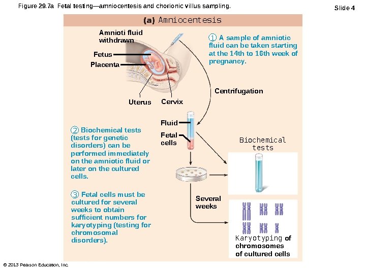
Figure 29. 7 a Fetal testing—amniocentesis and chorionic villus sampling. Slide 4 Amniocentesis Amnioti fluid withdrawn 1 A sample of amniotic fluid can be taken starting at the 14 th to 16 th week of pregnancy. Fetus Placenta Centrifugation Uterus 2 Biochemical tests (tests for genetic disorders) can be performed immediately on the amniotic fluid or later on the cultured cells. 3 Fetal cells must be cultured for several weeks to obtain sufficient numbers for karyotyping (testing for chromosomal disorders). © 2013 Pearson Education, Inc. Cervix Fluid Fetal cells Biochemical tests Several weeks Karyotyping of chromosomes of cultured cells
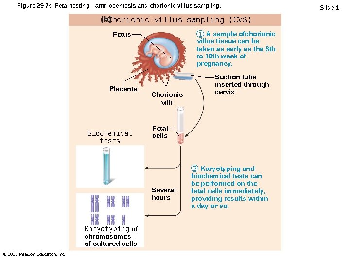
Figure 29. 7 b Fetal testing—amniocentesis and chorionic villus sampling. Chorionic villus sampling (CVS) 1 A sample ofchorionic villus tissue can be taken as early as the 8 th to 10 th week of pregnancy. Fetus Placenta Biochemical tests Chorionic villi Fetal cells Several hours Karyotyping of chromosomes of cultured cells © 2013 Pearson Education, Inc. Suction tube inserted through cervix 2 Karyotyping and biochemical tests can be performed on the fetal cells immediately, providing results within a day or so. Slide 1
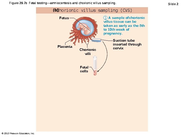
Figure 29. 7 b Fetal testing—amniocentesis and chorionic villus sampling. Chorionic villus sampling (CVS) 1 A sample ofchorionic villus tissue can be taken as early as the 8 th to 10 th week of pregnancy. Fetus Placenta Chorionic villi Fetal cells © 2013 Pearson Education, Inc. Suction tube inserted through cervix Slide 2
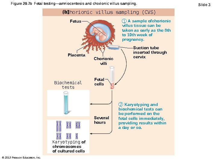
Figure 29. 7 b Fetal testing—amniocentesis and chorionic villus sampling. Chorionic villus sampling (CVS) 1 A sample ofchorionic villus tissue can be taken as early as the 8 th to 10 th week of pregnancy. Fetus Placenta Biochemical tests Chorionic villi Fetal cells Several hours Karyotyping of chromosomes of cultured cells © 2013 Pearson Education, Inc. Suction tube inserted through cervix 2 Karyotyping and biochemical tests can be performed on the fetal cells immediately, providing results within a day or so. Slide 3
Human Gene Therapy • Gene therapy may alleviate or cure disorders • Genetic engineering has potential to replace defective gene, including – Defective cells infected with genetically engineered virus containing functional gene – Inject "corrected" gene directly into cells • Prohibitively expensive; ethical, religious, societal questions © 2013 Pearson Education, Inc.
- Slides: 58