
Power Point Lecture Slides prepared by Barbara Heard
Power. Point® Lecture Slides prepared by Barbara Heard, Atlantic Cape Community Ninth Edition College Human Anatomy & Physiology CHAPTER 13 The Peripheral Nervous System and Reflex Activity: Part C © Annie Leibovitz/Contact Press Images © 2013 Pearson Education, Inc.
Spinal Nerves • 31 pairs of mixed nerves named for point of issue from spinal cord – Supply all body parts but head and part of neck – 8 cervical (C 1–C 8) – 12 thoracic (T 1–T 12) – 5 Lumbar (L 1–L 5) – 5 Sacral (S 1–S 5) – 1 Coccygeal (C 0) © 2013 Pearson Education, Inc.

Spinal Nerves • Only 7 cervical vertebrae, yet 8 pairs cervical spinal nerves – 7 exit vertebral canal superior to vertebrae for which named – 1 exits canal inferior to C 7 • Other vertebrae exit inferior to vertebra for which named © 2013 Pearson Education, Inc.
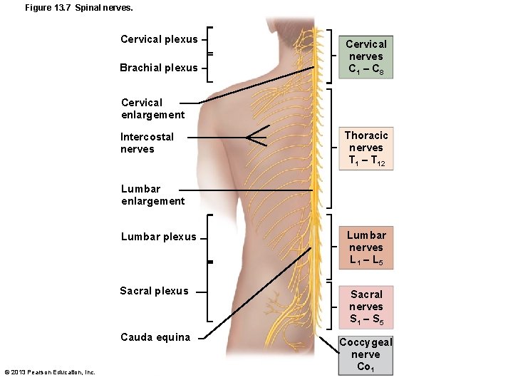
Figure 13. 7 Spinal nerves. Cervical plexus Brachial plexus Cervical nerves C 1 – C 8 Cervical enlargement Intercostal nerves Thoracic nerves T 1 – T 12 Lumbar enlargement © 2013 Pearson Education, Inc. Lumbar plexus Lumbar nerves L 1 – L 5 Sacral plexus Sacral nerves S 1 – S 5 Cauda equina Coccygeal nerve Co 1
Spinal Nerves: Roots • Each spinal nerve connects to spinal cord via two roots • Ventral roots – Contain motor (efferent) fibers from ventral horn motor neurons – Fibers innervate skeletal muscles © 2013 Pearson Education, Inc.
Spinal Nerves: Roots • Dorsal roots – Contain sensory (afferent) fibers from sensory neurons in dorsal root ganglia and conduct impulses from peripheral receptors • Dorsal and ventral roots unite to form spinal nerves, which emerge from vertebral column via intervertebral foramina © 2013 Pearson Education, Inc.
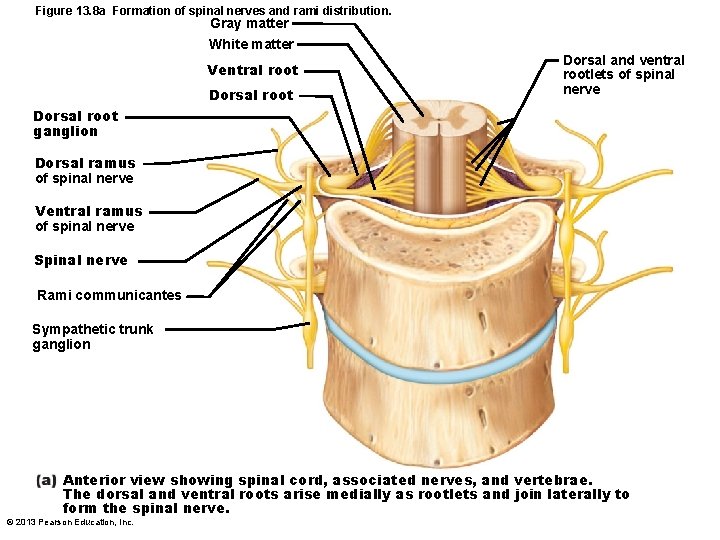
Figure 13. 8 a Formation of spinal nerves and rami distribution. Gray matter White matter Ventral root Dorsal and ventral rootlets of spinal nerve Dorsal root ganglion Dorsal ramus of spinal nerve Ventral ramus of spinal nerve Spinal nerve Rami communicantes Sympathetic trunk ganglion Anterior view showing spinal cord, associated nerves, and vertebrae. The dorsal and ventral roots arise medially as rootlets and join laterally to form the spinal nerve. © 2013 Pearson Education, Inc.
Spinal Nerves: Rami • Spinal nerves quite short (~1 -2 cm) • Each branches into mixed rami – Dorsal ramus – Ventral ramus - larger – Meningeal branch – tiny, reenters vertebral canal, innervates meninges and blood vessels – Rami communicantes (autonomic pathways) join ventral rami in thoracic region © 2013 Pearson Education, Inc.
Spinal Nerves: Rami • All ventral rami except T 2–T 12 form interlacing nerve networks called nerve plexuses (cervical, brachial, lumbar, and sacral) • Back innervated by dorsal rami via several branches • Ventral rami of T 2–T 12 as intercostal nerves supply muscles of ribs, anterolateral thorax, and abdominal wall • Spinal roots longer as move inferiorly in cord – Lumbar and sacral roots extend as cauda equina © 2013 Pearson Education, Inc.
Spinal Nerves: Plexuses • Within plexus fibers criss-cross – Each branch contains fibers from several spinal nerves – Fibers from ventral ramus go to body periphery via several routes • Each limb muscle innervated by more than one spinal nerve – Damage to one does not paralysis © 2013 Pearson Education, Inc.
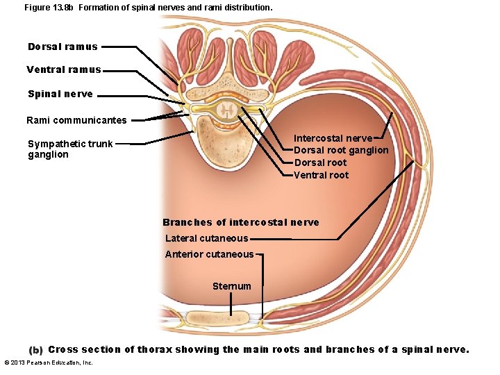
Figure 13. 8 b Formation of spinal nerves and rami distribution. Dorsal ramus Ventral ramus Spinal nerve Rami communicantes Intercostal nerve Dorsal root ganglion Dorsal root Ventral root Sympathetic trunk ganglion Branches of intercostal nerve Lateral cutaneous Anterior cutaneous Sternum Cross section of thorax showing the main roots and branches of a spinal nerve. © 2013 Pearson Education, Inc.
Cervical Plexus and the Neck • Formed by ventral rami of C 1–C 4 • Most branches form cutaneous nerves – Innervate skin of neck, ear, back of head, and shoulders – Other branches innervate neck muscles • Phrenic nerve – Major motor and sensory nerve of diaphragm (receives fibers from C 3–C 5) – Irritation hiccups © 2013 Pearson Education, Inc.
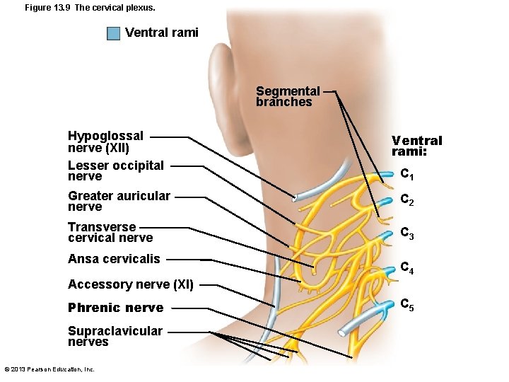
Figure 13. 9 The cervical plexus. Ventral rami Segmental branches Hypoglossal nerve (XII) Lesser occipital nerve Ventral rami: C 1 Greater auricular nerve C 2 Transverse cervical nerve C 3 Ansa cervicalis C 4 Accessory nerve (XI) Phrenic nerve Supraclavicular nerves © 2013 Pearson Education, Inc. C 5
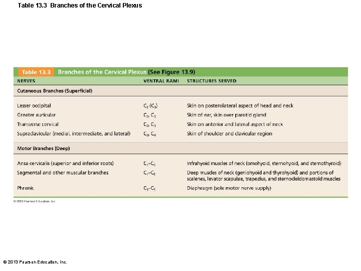
Table 13. 3 Branches of the Cervical Plexus © 2013 Pearson Education, Inc.
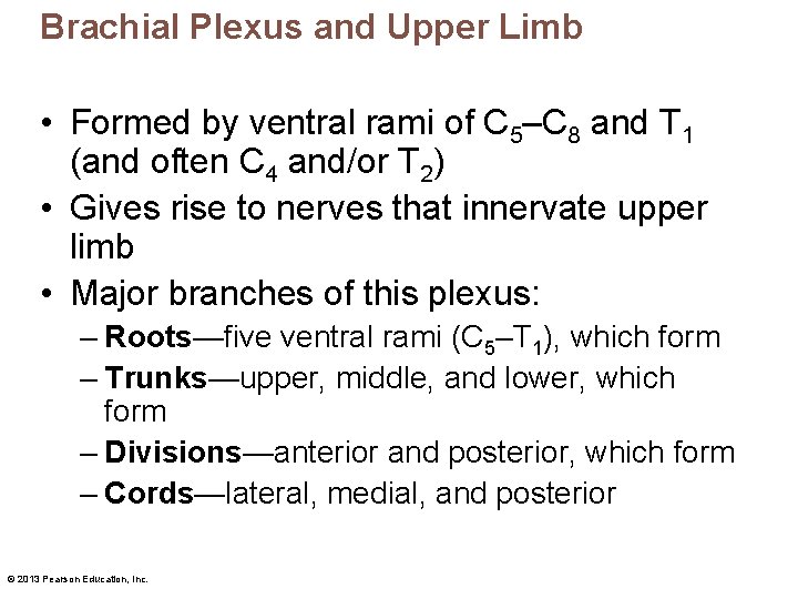
Brachial Plexus and Upper Limb • Formed by ventral rami of C 5–C 8 and T 1 (and often C 4 and/or T 2) • Gives rise to nerves that innervate upper limb • Major branches of this plexus: – Roots—five ventral rami (C 5–T 1), which form – Trunks—upper, middle, and lower, which form – Divisions—anterior and posterior, which form – Cords—lateral, medial, and posterior © 2013 Pearson Education, Inc.
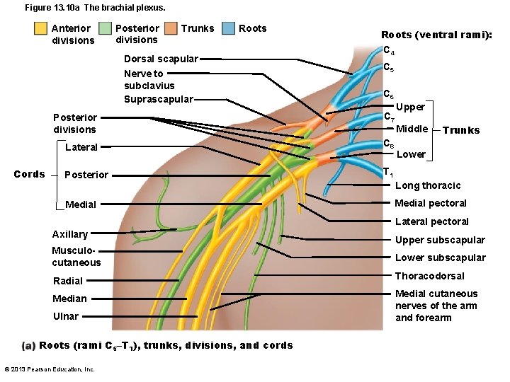
Figure 13. 10 a The brachial plexus. Anterior divisions Posterior divisions Trunks Roots Dorsal scapular Nerve to subclavius Suprascapular Cords Roots (ventral rami): C 4 C 5 C 6 Posterior divisions C 7 Lateral C 8 Posterior T 1 Medial Upper Middle Trunks Lower Long thoracic Medial pectoral Lateral pectoral Axillary Upper subscapular Musculocutaneous Lower subscapular Radial Thoracodorsal Median Ulnar Roots (rami C 5–T 1), trunks, divisions, and cords © 2013 Pearson Education, Inc. Medial cutaneous nerves of the arm and forearm
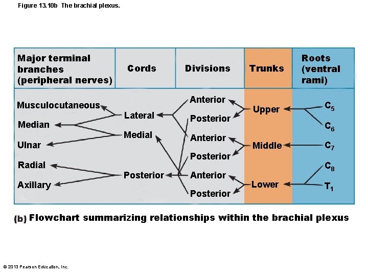
Figure 13. 10 b The brachial plexus. Major terminal branches (peripheral nerves) Cords Anterior Musculocutaneous Median Ulnar Radial Axillary Divisions Lateral Medial Posterior Anterior Trunks Upper Anterior Posterior C 5 C 6 Middle Posterior Roots (ventral rami) C 7 C 8 Lower T 1 Flowchart summarizing relationships within the brachial plexus © 2013 Pearson Education, Inc.
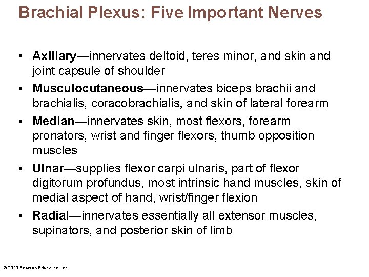
Brachial Plexus: Five Important Nerves • Axillary—innervates deltoid, teres minor, and skin and joint capsule of shoulder • Musculocutaneous—innervates biceps brachii and brachialis, coracobrachialis, and skin of lateral forearm • Median—innervates skin, most flexors, forearm pronators, wrist and finger flexors, thumb opposition muscles • Ulnar—supplies flexor carpi ulnaris, part of flexor digitorum profundus, most intrinsic hand muscles, skin of medial aspect of hand, wrist/finger flexion • Radial—innervates essentially all extensor muscles, supinators, and posterior skin of limb © 2013 Pearson Education, Inc.
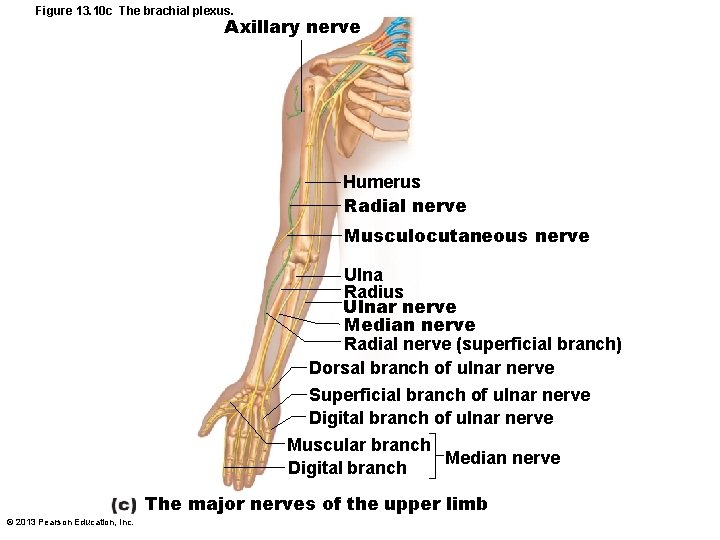
Figure 13. 10 c The brachial plexus. Axillary nerve Humerus Radial nerve Musculocutaneous nerve Ulna Radius Ulnar nerve Median nerve Radial nerve (superficial branch) Dorsal branch of ulnar nerve Superficial branch of ulnar nerve Digital branch of ulnar nerve Muscular branch Median nerve Digital branch The major nerves of the upper limb © 2013 Pearson Education, Inc.
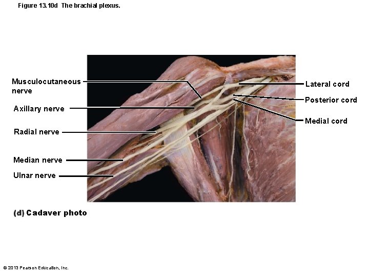
Figure 13. 10 d The brachial plexus. Musculocutaneous nerve Lateral cord Posterior cord Axillary nerve Medial cord Radial nerve Median nerve Ulnar nerve Cadaver photo © 2013 Pearson Education, Inc.
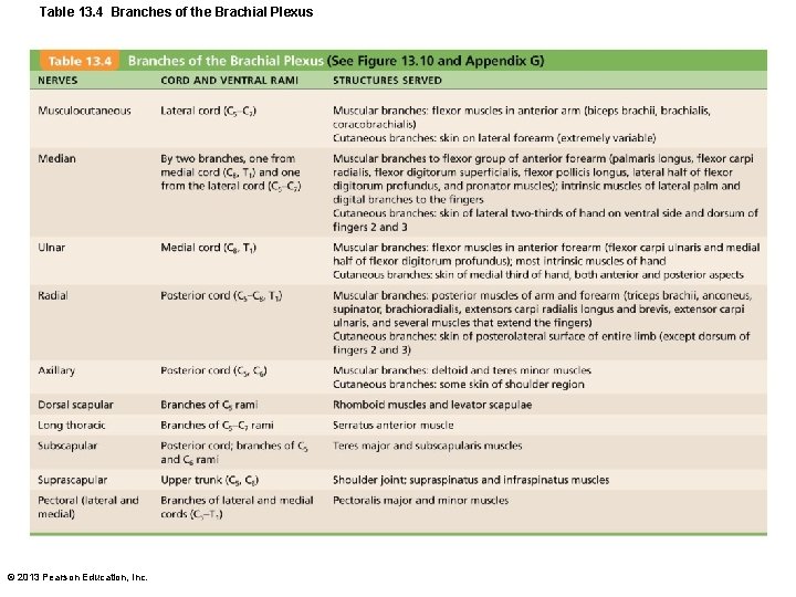
Table 13. 4 Branches of the Brachial Plexus © 2013 Pearson Education, Inc.
Lumbar Plexus • Arises from L 1–L 4 • Innervates thigh, abdominal wall, and psoas muscle • Femoral nerve—innervates quadriceps and skin of anterior thigh and medial surface of leg • Obturator nerve—passes through obturator foramen to innervate adductor muscles © 2013 Pearson Education, Inc.
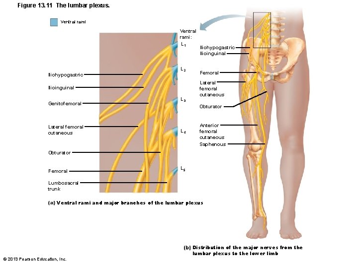
Figure 13. 11 The lumbar plexus. Ventral rami: L 1 Iliohypogastric L 2 Ilioinguinal Genitofemoral Lateral femoral cutaneous L 3 Iliohypogastric Ilioinguinal Femoral Lateral femoral cutaneous Obturator L 4 Anterior femoral cutaneous Saphenous Obturator Femoral L 5 Lumbosacral trunk Ventral rami and major branches of the lumbar plexus © 2013 Pearson Education, Inc. Distribution of the major nerves from the lumbar plexus to the lower limb
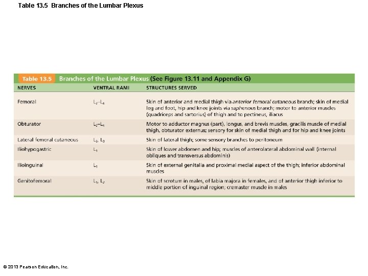
Table 13. 5 Branches of the Lumbar Plexus © 2013 Pearson Education, Inc.
Sacral Plexus • Arises from L 4–S 4 • Serves the buttock, lower limb, pelvic structures, and perineum • Sciatic nerve – Longest and thickest nerve of body – Innervates hamstring muscles, adductor magnus, and most muscles in leg and foot – Composed of two nerves: tibial and common fibular © 2013 Pearson Education, Inc.
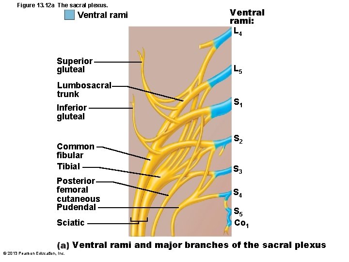
Figure 13. 12 a The sacral plexus. Ventral rami Superior gluteal Lumbosacral trunk Inferior gluteal Common fibular Tibial Posterior femoral cutaneous Pudendal Sciatic Ventral rami: L 4 L 5 S 1 S 2 S 3 S 4 S 5 Co 1 Ventral rami and major branches of the sacral plexus © 2013 Pearson Education, Inc.
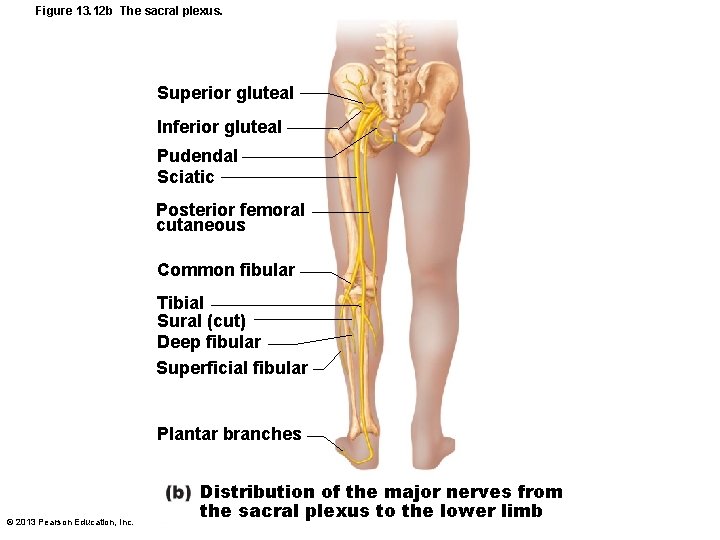
Figure 13. 12 b The sacral plexus. Superior gluteal Inferior gluteal Pudendal Sciatic Posterior femoral cutaneous Common fibular Tibial Sural (cut) Deep fibular Superficial fibular Plantar branches © 2013 Pearson Education, Inc. Distribution of the major nerves from the sacral plexus to the lower limb
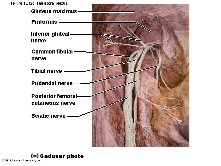
Figure 13. 12 c The sacral plexus. Gluteus maximus Piriformis Inferior gluteal nerve Common fibular nerve Tibial nerve Pudendal nerve Posterior femoral cutaneous nerve Sciatic nerve Cadaver photo © 2013 Pearson Education, Inc.
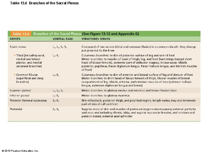
Table 13. 6 Branches of the Sacral Plexus © 2013 Pearson Education, Inc.
Anterolateral Thorax and Abdominal Wall • Ventral rami in thorax in simple segmental pattern – Form intercostal nerves that supply intercostal muscles, muscle and skin of anterolateral thorax, most abdominal wall – Give off cutaneous branches to skin along course • Dorsal rami innervate posterior body trunk © 2013 Pearson Education, Inc.
Innervation of Skin: Dermatomes • Dermatome - area of skin innervated by cutaneous branches of single spinal nerve • All spinal nerves except C 1 participate in dermatomes • Extent of spinal cord injuries ascertained by affected dermatomes • Most dermatomes overlap, so destruction of a single spinal nerve will not cause complete numbness © 2013 Pearson Education, Inc.
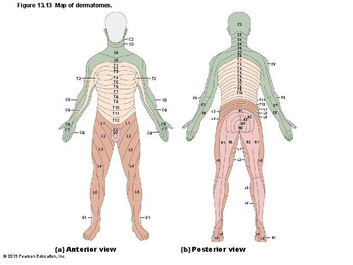
Figure 13. 13 Map of dermatomes. C 2 C 3 C 4 C 5 C 6 C 7 C 8 T 1 T 2 T 3 T 4 T 5 T 6 T 7 T 8 T 9 T 10 C 2 C 3 C 4 C 5 T 1 T 2 T 3 T 4 T 5 T 6 T 7 T 8 T 9 T 10 T 11 T 2 C 5 C 6 C 7 L 1 C 8 L 2 T 12 S 3 T 2 C 5 C 6 C 7 C 6 L 1 C 8 L 2 S 1 L 4 S 2 S 3 S 4 S 5 C 6 C 7 S 1 S 2 L 3 S 2 C 6 C 7 C 8 S 1 L 3 L 5 L 4 T 11 T 12 L 1 L 3 L 5 L 2 L 5 L 4 L 3 L 5 L 4 S 1 L 4 L 5 © 2013 Pearson Education, Inc. Anterior view Posterior view L 4 L 5 S 1
Innervation of Joints • To remember which nerves serve which synovial joint – Hilton's law: Any nerve serving a muscle that produces movement at joint also innervates joint and skin over joint © 2013 Pearson Education, Inc.
- Slides: 33