
Power Point Lecture Slide Presentation by Patty BostwickTaylor
Power. Point® Lecture Slide Presentation by Patty Bostwick-Taylor, Florence-Darlington Technical College Cells and Tissues 3 PART C Copyright © 2009 Pearson Education, Inc. , publishing as Benjamin Cummings
Protein Synthesis § Gene—DNA segment that carries a blueprint for building one protein § Proteins have many functions § Building materials for cells § Act as enzymes (biological catalysts) § RNA is essential for protein synthesis Copyright © 2009 Pearson Education, Inc. , publishing as Benjamin Cummings
Role of RNA § Transfer RNA (t. RNA) § Transfers appropriate amino acids to the ribosome for building the protein § Ribosomal RNA (r. RNA) § Helps form the ribosomes where proteins are built § Messenger RNA (m. RNA) § Carries the instructions for building a protein from the nucleus to the ribosome Copyright © 2009 Pearson Education, Inc. , publishing as Benjamin Cummings
Transcription and Translation § Transcription § Transfer of information from DNA’s base sequence to the complimentary base sequence of m. RNA § Three-base sequences on m. RNA are called codons Copyright © 2009 Pearson Education, Inc. , publishing as Benjamin Cummings
Transcription and Translation § Base sequence of nucleic acid is translated to an amino acid sequence § Amino acids are the building blocks of proteins Copyright © 2009 Pearson Education, Inc. , publishing as Benjamin Cummings
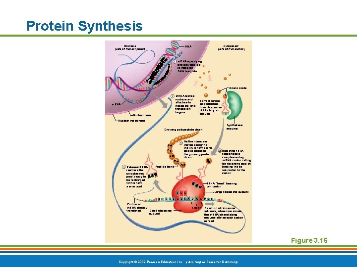
Protein Synthesis Nucleus (site of transcription) Cytoplasm (site of translation) DNA m. RNA specifying one polypeptide is made on DNA template Amino acids m. RNA leaves nucleus and attaches to ribosome, and translation begins m. RNA Nuclear pore Correct amino acid attached to each species of t. RNA by an enzyme Nuclear membrane Synthetase enzyme Growing polypeptide chain As the ribosome moves along the m. RNA, a new amino acid is added to the growing protein chain Met Gly Ser Phe Released t. RNA reenters the cytoplasmic pool, ready to be recharged with a new amino acid Peptide bond Ala Incoming t. RNA recognizes a complementary m. RNA codon calling for its amino acid by binding via its anticodon to the codon t. RNA “head” bearing anticodon Large ribosomal subunit C G G AU U U C G C C A U A G U C C Portion of m. RNA already Small ribosomal translated subunit Codon Direction of ribosome advance; ribosome moves the m. RNA strand along sequentially as each codon is read Figure 3. 16 Copyright © 2009 Pearson Education, Inc. , publishing as Benjamin Cummings
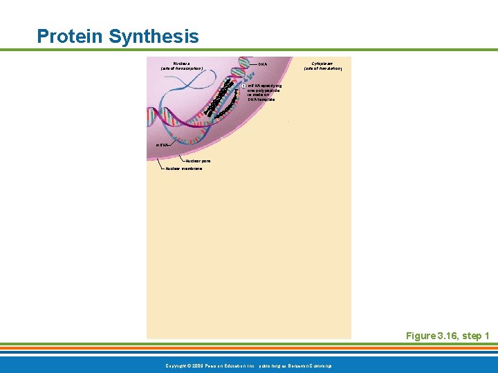
Protein Synthesis Nucleus (site of transcription) DNA Cytoplasm (site of translation) m. RNA specifying one polypeptide is made on DNA template m. RNA Nuclear pore Nuclear membrane Figure 3. 16, step 1 Copyright © 2009 Pearson Education, Inc. , publishing as Benjamin Cummings
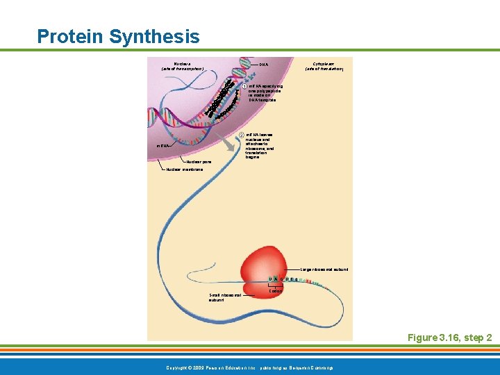
Protein Synthesis Nucleus (site of transcription) Cytoplasm (site of translation) DNA m. RNA specifying one polypeptide is made on DNA template m. RNA Nuclear pore m. RNA leaves nucleus and attaches to ribosome, and translation begins Nuclear membrane Large ribosomal subunit U A G U CC Small ribosomal subunit Codon Figure 3. 16, step 2 Copyright © 2009 Pearson Education, Inc. , publishing as Benjamin Cummings
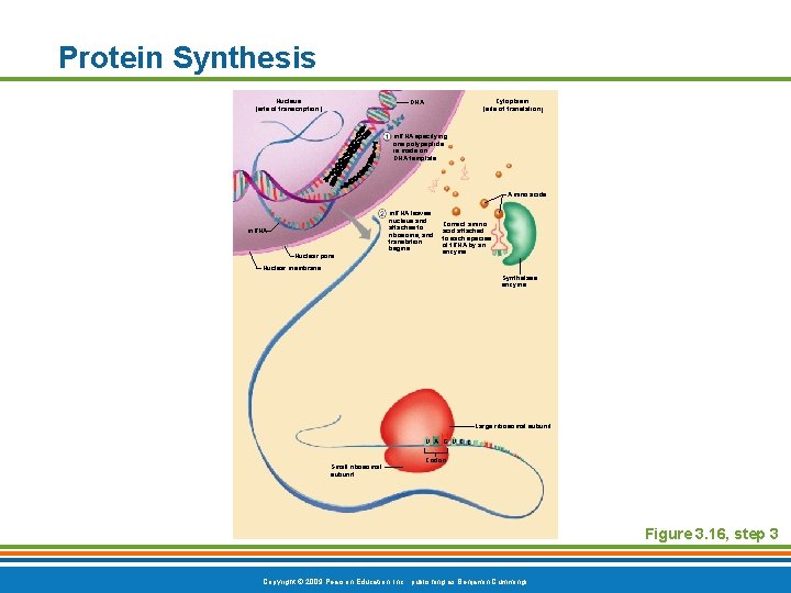
Protein Synthesis Nucleus (site of transcription) Cytoplasm (site of translation) DNA m. RNA specifying one polypeptide is made on DNA template Amino acids m. RNA Nuclear pore m. RNA leaves nucleus and attaches to ribosome, and translation begins Correct amino acid attached to each species of t. RNA by an enzyme Nuclear membrane Synthetase enzyme Large ribosomal subunit U A G U CC Small ribosomal subunit Codon Figure 3. 16, step 3 Copyright © 2009 Pearson Education, Inc. , publishing as Benjamin Cummings
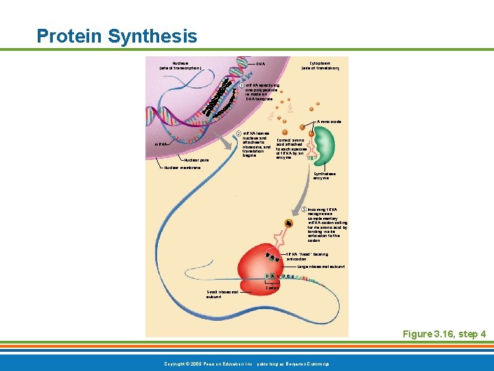
Protein Synthesis Nucleus (site of transcription) Cytoplasm (site of translation) DNA m. RNA specifying one polypeptide is made on DNA template Amino acids m. RNA Nuclear pore m. RNA leaves nucleus and attaches to ribosome, and translation begins Correct amino acid attached to each species of t. RNA by an enzyme Nuclear membrane Synthetase enzyme Incoming t. RNA recognizes a complementary m. RNA codon calling for its amino acid by binding via its anticodon to the codon t. RNA “head” bearing anticodon Large ribosomal subunit U A G U CC Small ribosomal subunit Codon Figure 3. 16, step 4 Copyright © 2009 Pearson Education, Inc. , publishing as Benjamin Cummings
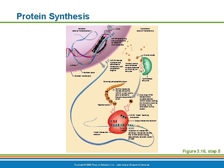
Protein Synthesis Nucleus (site of transcription) Cytoplasm (site of translation) DNA m. RNA specifying one polypeptide is made on DNA template Amino acids m. RNA leaves nucleus and attaches to ribosome, and translation begins m. RNA Nuclear pore Correct amino acid attached to each species of t. RNA by an enzyme Nuclear membrane Synthetase enzyme Growing polypeptide chain As the ribosome moves along the m. RNA, a new amino acid is added to the growing protein chain Met Gly Ser Phe Peptide bond Ala Incoming t. RNA recognizes a complementary m. RNA codon calling for its amino acid by binding via its anticodon to the codon t. RNA “head” bearing anticodon Large ribosomal subunit C G G G C C A U A G U CC Small ribosomal subunit Codon Direction of ribosome advance; ribosome moves the m. RNA strand along sequentially as each codon is read Figure 3. 16, step 5 Copyright © 2009 Pearson Education, Inc. , publishing as Benjamin Cummings
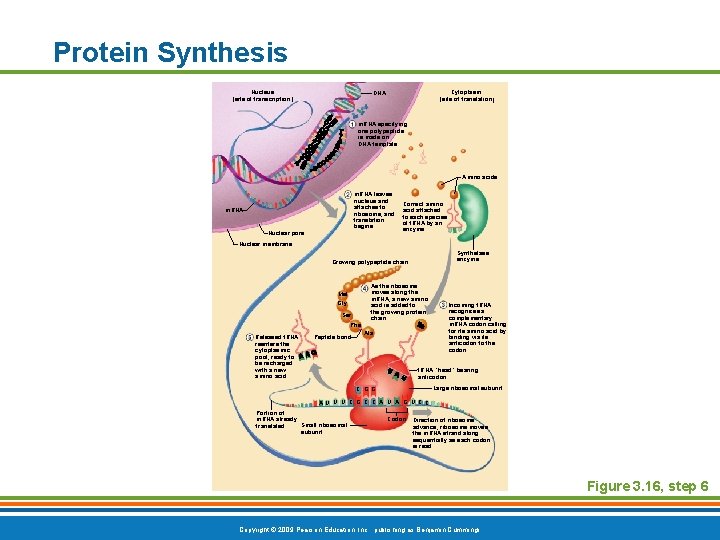
Protein Synthesis Nucleus (site of transcription) Cytoplasm (site of translation) DNA m. RNA specifying one polypeptide is made on DNA template Amino acids m. RNA leaves nucleus and attaches to ribosome, and translation begins m. RNA Nuclear pore Correct amino acid attached to each species of t. RNA by an enzyme Nuclear membrane Synthetase enzyme Growing polypeptide chain As the ribosome moves along the m. RNA, a new amino acid is added to the growing protein chain Met Gly Ser Phe Released t. RNA reenters the cytoplasmic pool, ready to be recharged with a new amino acid Peptide bond Ala Incoming t. RNA recognizes a complementary m. RNA codon calling for its amino acid by binding via its anticodon to the codon t. RNA “head” bearing anticodon Large ribosomal subunit C G G AU U U C G C C A U A G U CC Portion of m. RNA already Small ribosomal translated subunit Codon Direction of ribosome advance; ribosome moves the m. RNA strand along sequentially as each codon is read Figure 3. 16, step 6 Copyright © 2009 Pearson Education, Inc. , publishing as Benjamin Cummings
Body Tissues § Groups of cells with similar structure and function § Four primary types § Epithelial tissue (epithelium) § Connective tissue § Muscle tissue § Nervous tissue Copyright © 2009 Pearson Education, Inc. , publishing as Benjamin Cummings
Epithelial Tissues § Locations § Body coverings § Body linings § Glandular tissue § Functions § Protection § Absorption § Filtration § Secretion Copyright © 2009 Pearson Education, Inc. , publishing as Benjamin Cummings
Epithelium Characteristics § Cells fit closely together and often form sheets § The apical surface is the free surface of the tissue § The lower surface of the epithelium rests on a basement membrane § Avascular (no blood supply) § Regenerate easily if well nourished Copyright © 2009 Pearson Education, Inc. , publishing as Benjamin Cummings
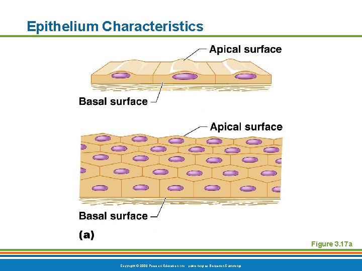
Epithelium Characteristics Figure 3. 17 a Copyright © 2009 Pearson Education, Inc. , publishing as Benjamin Cummings
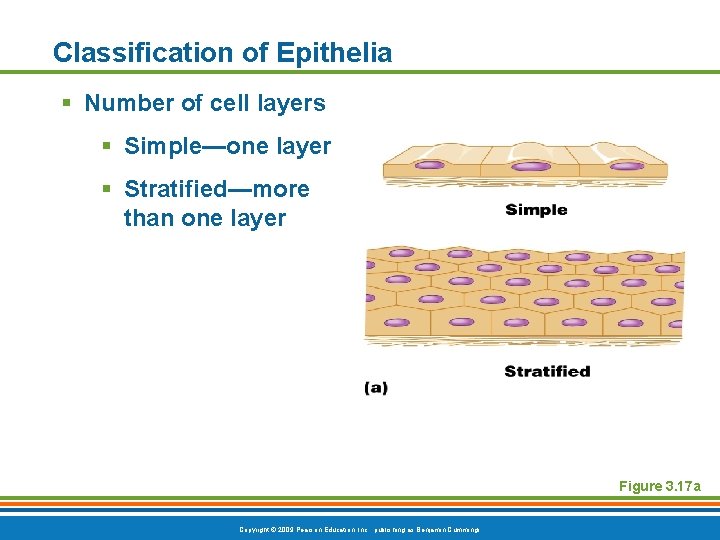
Classification of Epithelia § Number of cell layers § Simple—one layer § Stratified—more than one layer Figure 3. 17 a Copyright © 2009 Pearson Education, Inc. , publishing as Benjamin Cummings
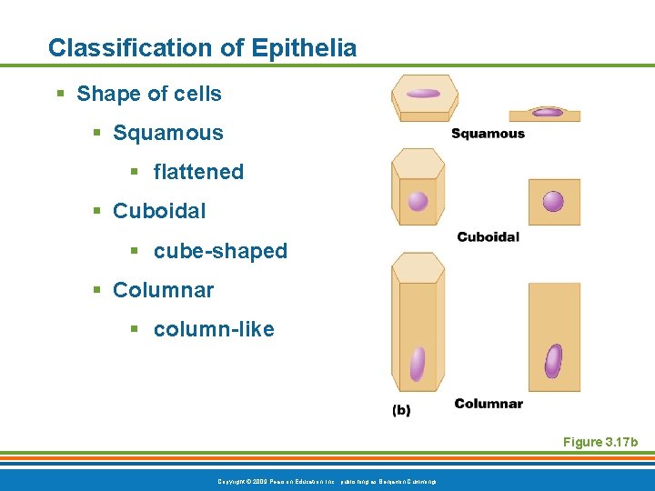
Classification of Epithelia § Shape of cells § Squamous § flattened § Cuboidal § cube-shaped § Columnar § column-like Figure 3. 17 b Copyright © 2009 Pearson Education, Inc. , publishing as Benjamin Cummings
Simple Epithelia § Simple squamous § Single layer of flat cells § Usually forms membranes § Lines body cavities § Lines lungs and capillaries Copyright © 2009 Pearson Education, Inc. , publishing as Benjamin Cummings
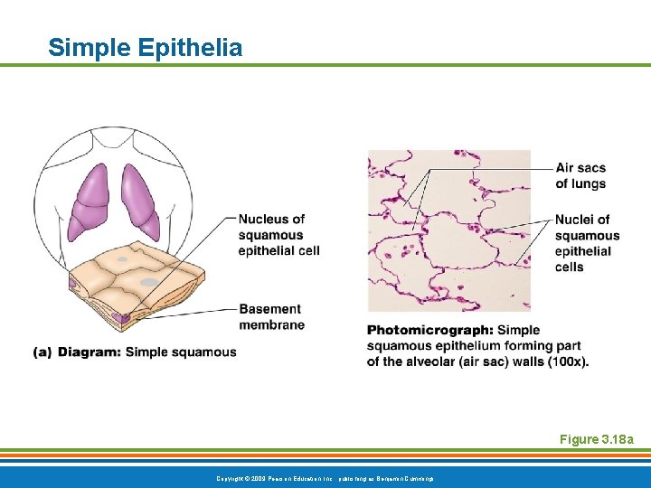
Simple Epithelia Figure 3. 18 a Copyright © 2009 Pearson Education, Inc. , publishing as Benjamin Cummings
Simple Epithelia § Simple cuboidal § Single layer of cube-like cells § Common in glands and their ducts § Forms walls of kidney tubules § Covers the ovaries Copyright © 2009 Pearson Education, Inc. , publishing as Benjamin Cummings
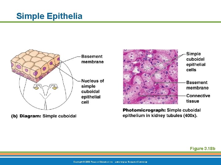
Simple Epithelia Figure 3. 18 b Copyright © 2009 Pearson Education, Inc. , publishing as Benjamin Cummings
Simple Epithelia § Simple columnar § Single layer of tall cells § Often includes mucus-producing goblet cells § Lines digestive tract Copyright © 2009 Pearson Education, Inc. , publishing as Benjamin Cummings
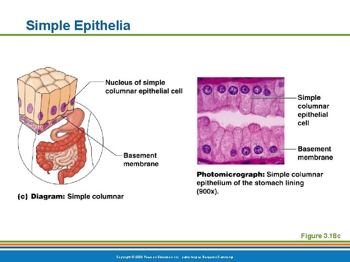
Simple Epithelia Figure 3. 18 c Copyright © 2009 Pearson Education, Inc. , publishing as Benjamin Cummings
Simple Epithelia § Pseudostratified columnar § Single layer, but some cells are shorter than others § Often looks like a double layer of cells § Sometimes ciliated, such as in the respiratory tract § May function in absorption or secretion Copyright © 2009 Pearson Education, Inc. , publishing as Benjamin Cummings
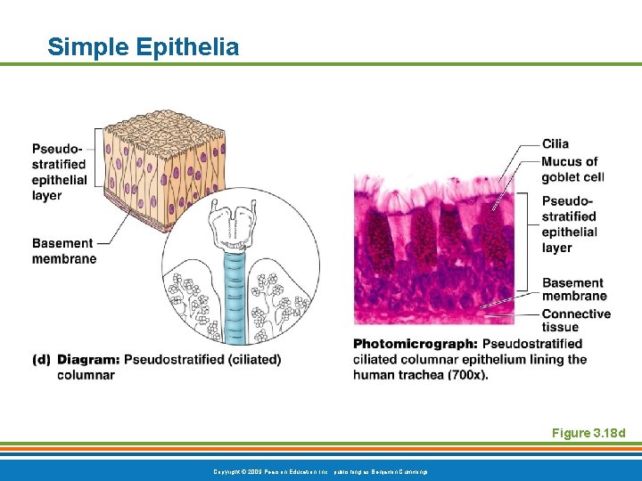
Simple Epithelia Figure 3. 18 d Copyright © 2009 Pearson Education, Inc. , publishing as Benjamin Cummings
Stratified Epithelia § Stratified squamous § Cells at the apical surface are flattened § Found as a protective covering where friction is common § Locations § Skin § Mouth § Esophagus Copyright © 2009 Pearson Education, Inc. , publishing as Benjamin Cummings
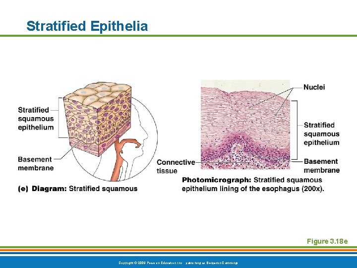
Stratified Epithelia Figure 3. 18 e Copyright © 2009 Pearson Education, Inc. , publishing as Benjamin Cummings
Stratified Epithelia § Stratified cuboidal—two layers of cuboidal cells § Stratified columnar—surface cells are columnar, cells underneath vary in size and shape § Stratified cuboidal and columnar § Rare in human body § Found mainly in ducts of large glands Copyright © 2009 Pearson Education, Inc. , publishing as Benjamin Cummings
Stratified Epithelia § Transitional epithelium § Shape of cells depends upon the amount of stretching § Lines organs of the urinary system Copyright © 2009 Pearson Education, Inc. , publishing as Benjamin Cummings
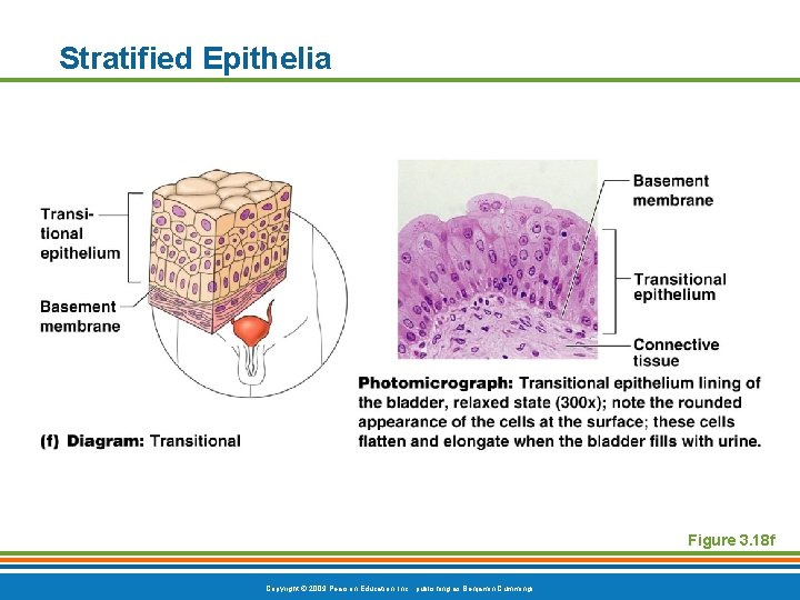
Stratified Epithelia Figure 3. 18 f Copyright © 2009 Pearson Education, Inc. , publishing as Benjamin Cummings
Glandular Epithelium § Gland § One or more cells responsible for secreting a particular product Copyright © 2009 Pearson Education, Inc. , publishing as Benjamin Cummings
Glandular Epithelium § Two major gland types § Endocrine gland § Ductless since secretions diffuse into blood vessels § All secretions are hormones § Exocrine gland § Secretions empty through ducts to the epithelial surface § Include sweat and oil glands Copyright © 2009 Pearson Education, Inc. , publishing as Benjamin Cummings
- Slides: 33