
Power Point Lecture Presentations prepared by Bradley W

Power. Point® Lecture Presentations prepared by Bradley W. Christian, Mc. Lennan Community College CHAPTER 26 Microbial Diseases of the Urinary and Reproductive Systems © 2016 Pearson Education, Inc.
© 2016 Pearson Education, Inc.
Structure and Function of the Urinary System Learning Objective 26 -1 List the antimicrobial features of the urinary system. © 2016 Pearson Education, Inc.
Structure and Function of the Urinary System • Urinary system • • Two kidneys Two ureters One urinary bladder One urethra • Infection prevented by: • Valves that prevent backflow to the kidneys • Acidity of urine • Mechanical flushing © 2016 Pearson Education, Inc.
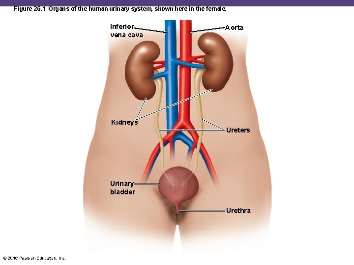
Figure 26. 1 Organs of the human urinary system, shown here in the female. Inferior vena cava Aorta Kidneys Ureters Urinary bladder Urethra © 2016 Pearson Education, Inc.
Check Your Understanding ü Does the p. H of urine facilitate the growth of most bacteria? 26 -1 © 2016 Pearson Education, Inc.
Structure and Function of the Reproductive Systems Learning Objective 26 -2 Identify the portals of entry for microbes into the female and male reproductive systems. © 2016 Pearson Education, Inc.
Structure and Function of the Reproductive Systems • Female reproductive system • • • Two ovaries Two uterine (fallopian) tubes The uterus, including the cervix The vagina External genitals (vulva) © 2016 Pearson Education, Inc.
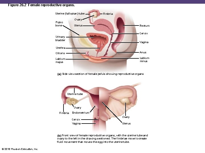
Figure 26. 2 Female reproductive organs. Uterine (fallopian) tube Fimbria Ovary Pubic bone Uterus Rectum Cervix Urinary bladder Vagina Urethra Clitoris Anus Labium majus Labium minus Side view section of female pelvis showing reproductive organs Uterine tube Ovary Fimbria Endometrium Cervix Vagina Ovary Uterus Front view of female reproductive organs, with the uterine tube and ovary to the left in the drawing sectioned. The fimbriae move to create fluid movement that moves the egg into the uterine tube. © 2016 Pearson Education, Inc.
Structure and Function of the Reproductive Systems • Male reproductive system • Two testes • System of ducts • • Epididymis Ductus (vas) deferens Ejaculatory duct Urethra • Accessory glands • Penis © 2016 Pearson Education, Inc.
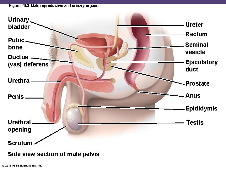
Figure 26. 3 Male reproductive and urinary organs. Urinary bladder Pubic bone Ductus (vas) deferens Ureter Rectum Seminal vesicle Ejaculatory duct Urethra Prostate Penis Anus Epididymis Urethral opening Scrotum Side view section of male pelvis © 2016 Pearson Education, Inc. Testis
Check Your Understanding ü Look at Figure 26. 2. Is a microbe entering the female reproductive system (the uterus, etc. ) necessarily also entering the bladder, causing cystitis? 26 -2 © 2016 Pearson Education, Inc.
Normal Microbiota of the Urinary and Reproductive Systems Learning Objective 26 -3 Describe the normal microbiota of the upper urinary tract, the male urethra, and the female urethra and vagina. © 2016 Pearson Education, Inc.
Normal Microbiota of the Urinary and Reproductive Systems • Urinary bladder and upper urinary tract are sterile • Predominant microbes of the vagina: • Lactobacilli • Produce H 2 O 2 and lactic acid • Growth promoted by estrogen • Streptococci, anaerobes, some gram-negatives • Candida albicans yeast • Male urethra is usually sterile © 2016 Pearson Education, Inc.
Check Your Understanding ü What is the association between estrogens and the microbiota of the vagina? 26 -3 © 2016 Pearson Education, Inc.
Bacterial Diseases of the Urinary System Learning Objectives 26 -4 Describe the modes of transmission for urinary and reproductive system infections. 26 -5 List the microorganisms that cause cystitis, pyelonephritis, and leptospirosis, and name the predisposing factors for these diseases. © 2016 Pearson Education, Inc.
Bacterial Diseases of the Urinary System • Urethritis • An inflammation of the urethra • Cystitis • An inflammation of the urinary bladder • Ureteritis • Infection of the ureters • Pyelonephritis • An inflammation of one or both kidneys • 7 million urinary tract infections annually • Most due to Escherichia coli © 2016 Pearson Education, Inc.
Cystitis • Commonly caused by E. coli; also Staphylococcus saprophyticus • Dysuria (difficult or painful urination); pyuria • Eight times more common in women than men • Due to the short length of the urethra • Diagnosis: >100 CFU/ml of potential pathogens and a positive leukocyte esterase (LE) test • Treatment with trimethoprim-sulfamethexazole © 2016 Pearson Education, Inc.
Pyelonephritis • • 75% of cases caused by E. coli Fever and back or flank pain Generally results in bacteremia Can form scar tissue in kidneys and become lifethreatening • Diagnosis: >10, 000 CFU/ml and a positive LE test • Treatment with cephalosporin © 2016 Pearson Education, Inc.
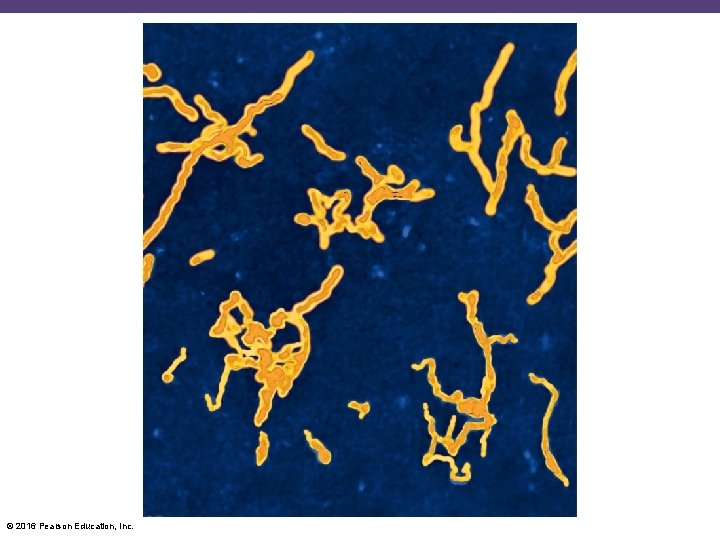
Leptospirosis • Caused by Leptospira interrogans • Fine spiral; stains poorly; obligate aerobe • Transmitted by skin/mucosal contact from urinecontaminated water from domestic or wild animals • Headaches, muscular aches, fever • Kidney failure (Weil's disease) • Pulmonary hemorrhagic syndrome • Diagnosed with a rapid serological test • Treatment with doxycycline © 2016 Pearson Education, Inc.
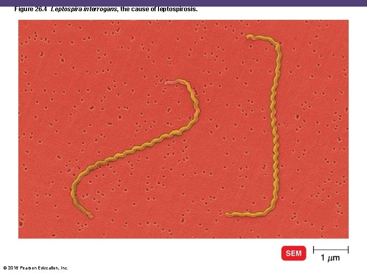
Figure 26. 4 Leptospira interrogans, the cause of leptospirosis. © 2016 Pearson Education, Inc.
Diseases in Focus: Bacterial Diseases of the Urinary System • A 20 -year-old woman feels a stinging sensation when urinating and feels an urgent need to urinate, even if very little urine is excreted. Lactosefermenting, gram-negative rods are cultured from her urine. • Can you identify infections that could cause these symptoms? © 2016 Pearson Education, Inc.
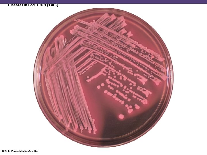
Diseases in Focus 26. 1 (1 of 2) © 2016 Pearson Education, Inc.
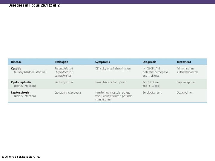
Diseases in Focus 26. 1 (2 of 2) © 2016 Pearson Education, Inc.
Check Your Understanding ü Why is urethritis, an infection of the urethra, frequently preliminary to further infections of the urinary tract? 26 -4 ü Why is E. coli the most common cause of cystitis, especially in females? 26 -5 © 2016 Pearson Education, Inc.
Bacterial Diseases of the Reproductive Systems Learning Objective 26 -6 List the causative agents, symptoms, methods of diagnosis, and treatments for gonorrhea, nongonococcal urethritis (NGU), pelvic inflammatory disease (PID), syphilis, lymphogranuloma venereum (LGV), chancroid, and bacterial vaginosis. © 2016 Pearson Education, Inc.
Bacterial Diseases of the Reproductive Systems • Sexually transmitted diseases (STDs) • • • Also known as sexually transmitted infections (STIs) Often no signs or symptoms Over 30 types of infections 15 million new cases in the United States annually Treatment with antibiotics and prevented with condoms © 2016 Pearson Education, Inc.
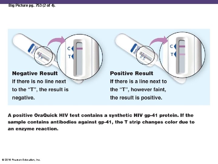
Big Picture: STI Home Test Kits • Collect samples at home and mail to a lab • Screens for chlamydia, gonorrhea, and trichomoniasis • Results in 1– 2 weeks • Positive tests receive referrals to clinics • Test for HIV • Ora. Quick: oral test • Test for urinary tract infections • Uritest dipstick test © 2016 Pearson Education, Inc.
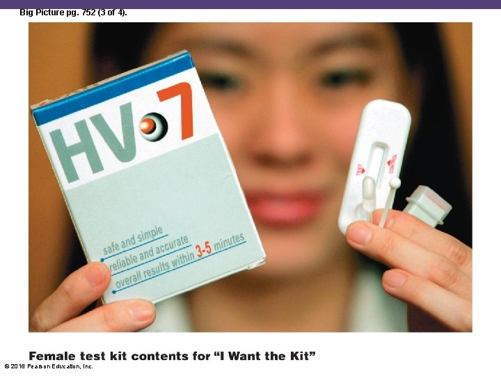
Big Picture pg. 752 (3 of 4). © 2016 Pearson Education, Inc.
Big Picture: STI Home Test Kits • Pros of at-home testing • More cases are diagnosed • Better access for patients • Quicker treatment • Cons of home-testing • Cost • Privacy • Not all home test kits are equally accurate © 2016 Pearson Education, Inc.
Big Picture pg. 753 (2 of 4). © 2016 Pearson Education, Inc.
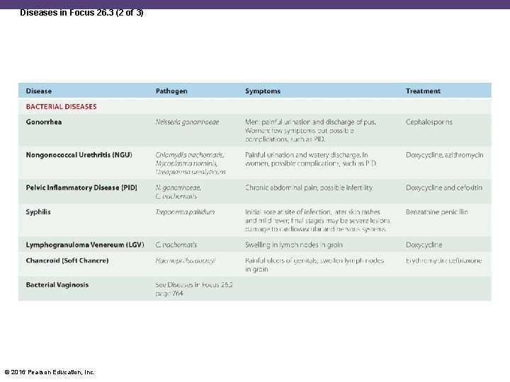
Gonorrhea • Caused by Neisseria gonorrhoeae • Gram-negative diplococcus • 300, 000 cases in the United States annually • Attaches to the epithelial mucosa by the fimbriae • Invades the spaces between the columnar epithelial cells • Causes inflammation • Forms pus • Pharyngeal gonorrhea and anal gonorrhea © 2016 Pearson Education, Inc.
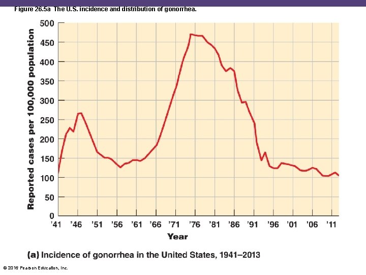
Figure 26. 5 a The U. S. incidence and distribution of gonorrhea. © 2016 Pearson Education, Inc.
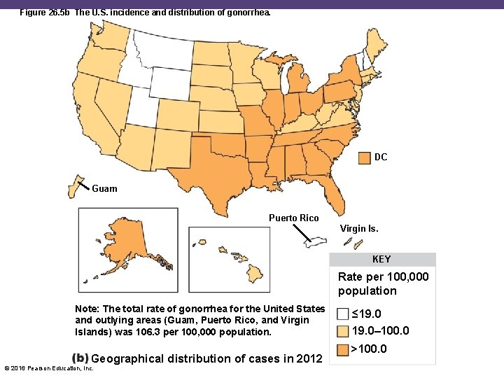
Figure 26. 5 b The U. S. incidence and distribution of gonorrhea. DC Guam Puerto Rico Virgin Is. KEY Rate per 100, 000 population Note: The total rate of gonorrhea for the United States and outlying areas (Guam, Puerto Rico, and Virgin Islands) was 106. 3 per 100, 000 population. Geographical distribution of cases in 2012 © 2016 Pearson Education, Inc. ≤ 19. 0– 100. 0 >100. 0
Gonorrhea • Symptoms • Men: painful urination and discharge of pus; epididymitis • Women: fewer symptoms; pelvic inflammatory disease • If left untreated, may disseminate and become systemic • Endocarditis • Meningitis • Arthritis • Ophthalmia neonatorum: infant blindness due to a gonorrheal infection of the eyes © 2016 Pearson Education, Inc.
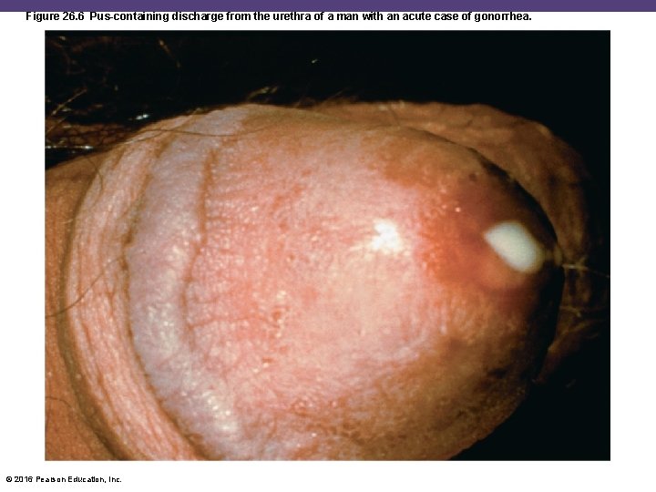
Figure 26. 6 Pus-containing discharge from the urethra of a man with an acute case of gonorrhea. © 2016 Pearson Education, Inc.
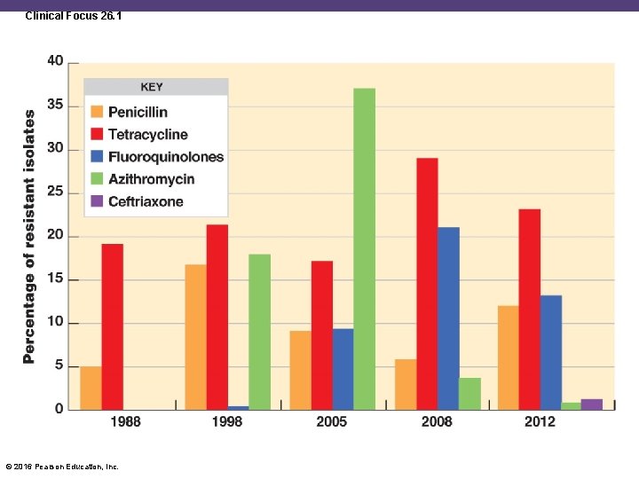
Gonorrhea • No adaptive immunity • Antigenic variability • Opa proteins bind to T cell receptors, preventing activation and immunological memory • Diagnosis with Gram stain, ELISA, or monoclonal antibodies • Treatment first with cephalosporins • Fluoroquinolones not recommended due to resistance © 2016 Pearson Education, Inc.
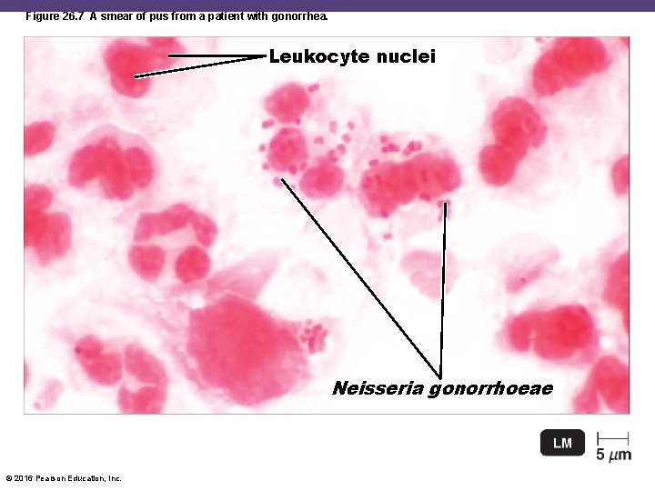
Figure 26. 7 A smear of pus from a patient with gonorrhea. Leukocyte nuclei Neisseria gonorrhoeae © 2016 Pearson Education, Inc.
Clinical Focus 26. 1 © 2016 Pearson Education, Inc.
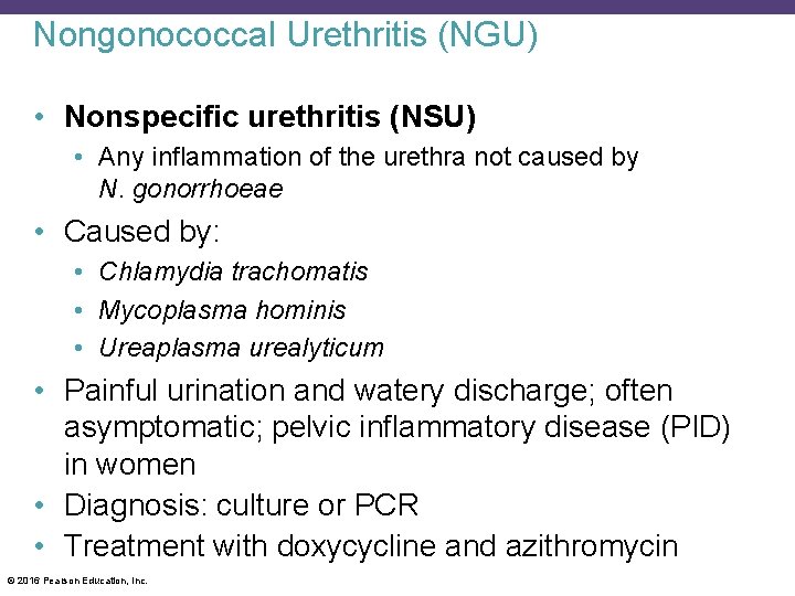
Nongonococcal Urethritis (NGU) • Nonspecific urethritis (NSU) • Any inflammation of the urethra not caused by N. gonorrhoeae • Caused by: • Chlamydia trachomatis • Mycoplasma hominis • Ureaplasma urealyticum • Painful urination and watery discharge; often asymptomatic; pelvic inflammatory disease (PID) in women • Diagnosis: culture or PCR • Treatment with doxycycline and azithromycin © 2016 Pearson Education, Inc.
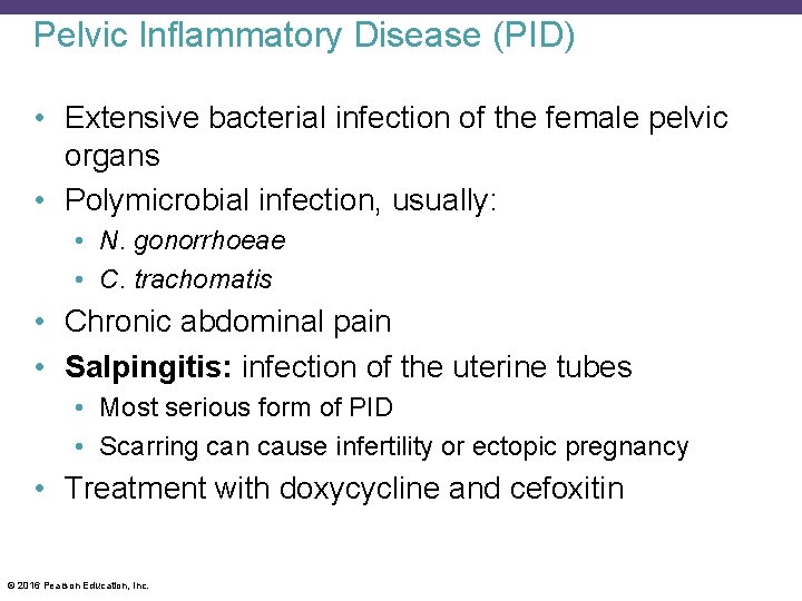
Pelvic Inflammatory Disease (PID) • Extensive bacterial infection of the female pelvic organs • Polymicrobial infection, usually: • N. gonorrhoeae • C. trachomatis • Chronic abdominal pain • Salpingitis: infection of the uterine tubes • Most serious form of PID • Scarring can cause infertility or ectopic pregnancy • Treatment with doxycycline and cefoxitin © 2016 Pearson Education, Inc.
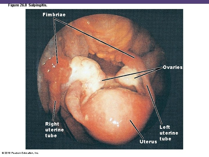
Figure 26. 8 Salpingitis. Fimbriae Ovaries Right uterine tube © 2016 Pearson Education, Inc. Uterus Left uterine tube
Syphilis • Caused by Treponema pallidum • Gram-negative spirochete • Grows slowly in cell culture • Invades the mucosa or through skin breaks and enters the bloodstream • Induces an inflammatory response • Some strains cause yaws • Skin disease is not sexually transmitted • Stable incidence in the United States © 2016 Pearson Education, Inc.
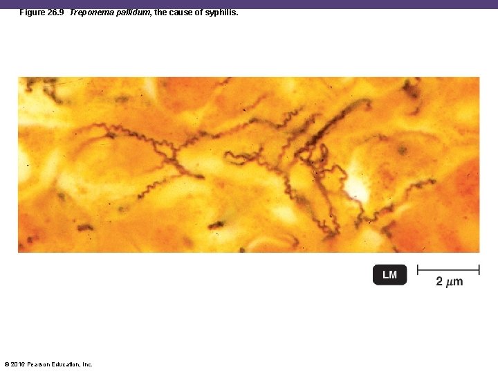
Figure 26. 9 Treponema pallidum, the cause of syphilis. © 2016 Pearson Education, Inc.
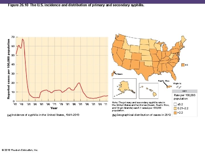
Figure 26. 10 The U. S. incidence and distribution of primary and secondary syphilis. DC Guam Puerto Rico Virgin Is. KEY Rate per 100, 000 population Note: The primary and secondary syphilis rate in the United States and territories (Guam, Puerto Rico, and Virgin Islands) was 5. 1 cases per 100, 000 population. Incidence of syphilis in the United States, 1941– 2013 © 2016 Pearson Education, Inc. Geographical distribution of cases in 2012 ≤ 0. 21– 2. 2 >2. 2
Syphilis • Primary stage • Chancre at the site of infection about 3 weeks after exposure • Painless and highly infectious • Disappears after 2 weeks • Secondary stage • Skin and mucosal rashes, especially on the palms and soles • Due to an inflammatory response • Latent period • No symptoms © 2016 Pearson Education, Inc.
Syphilis • Tertiary stage • Appear years after latency • Due to cell-mediated immune reactions • Gummatous syphilis: gummas on many organs • Cardiovascular syphilis: weakens the aorta • Neurosyphilis: affects the CNS; dementia • Congenital: neurological damage to the fetus © 2016 Pearson Education, Inc.
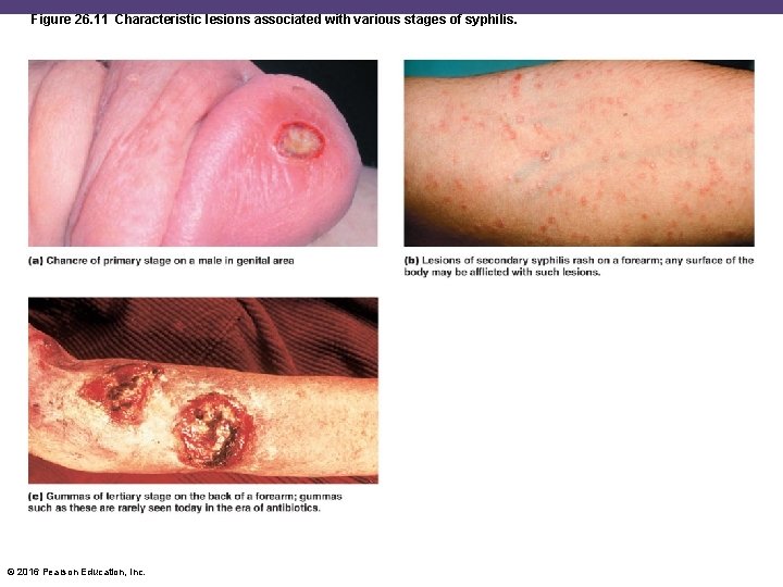
Figure 26. 11 Characteristic lesions associated with various stages of syphilis. © 2016 Pearson Education, Inc.
Syphilis • Microscopic tests • Direct fluorescent-antibody test (DFA-TP) with monoclonal antibodies • Nontreponemal serological tests • Slide agglutination VDRL test • Rapid plasma reagin (RPR) test • Treponemal-type serological tests • Enzyme immunoassay (EIA) • Fluorescent treponemal antibody absorption test (FTA-ABS) • Treatment with benzathine penicillin © 2016 Pearson Education, Inc.
Lymphogranuloma Venereum (LGV) • Caused by C. trachomatis • Infects the lymphoid tissue • Regional lymph nodes become enlarged and tender • Discharge of pus and scarring • Diagnosis with blood test for antibodies to the organism • Treatment with doxycycline © 2016 Pearson Education, Inc.
Chancroid (Soft Chancre) • Caused by Haemophilus ducreyi • Gram-negative rod • Associated with drug use • Painful ulcers of the genitals and swollen lymph nodes in the groin • Factors in the sexual transmission of HIV • Treatment with azithromycin or ceftriaxone © 2016 Pearson Education, Inc.
Bacterial Vaginosis • Caused by Gardnerella vaginalis • Pleomorphic gram-negative rod • Vaginitis: inflammation of the vagina due to infection • Vaginosis: no sign of inflammation • p. H above 4. 5, fishy odor, copious frothy discharge • Clue cells • Sloughed-off vaginal epithelial cells covered with a biofilm of G. vaginalis • Treatment with metronidazole © 2016 Pearson Education, Inc.
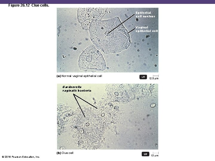
Figure 26. 12 Clue cells. Epithelial cell nucleus Vaginal epithelial cell Normal vaginal epithelial cell Gardnerella vaginalis bacteria Clue cell © 2016 Pearson Education, Inc.
Check Your Understanding ü Why is the disease condition of the female reproductive system, principally featuring growth of Gardnerella vaginalis, termed vaginosis rather than vaginitis? 26 -6 © 2016 Pearson Education, Inc.
Viral Diseases of the Reproductive Systems Learning Objective 26 -7 Discuss the epidemiology of genital herpes and genital warts. © 2016 Pearson Education, Inc.
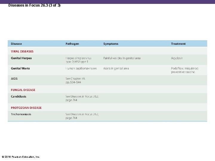
Genital Herpes • Caused by herpes simplex virus type 2 (HSV– 2) • In the United States, 1 in 4 over age 30 are infected • Painful vesicles on the genitals; painful urination • Heals within 2 weeks • Recurrences from viruses latent in nerve cells • Due to menstruation, emotional stress, or illness • Diagnosis via culture or PCR • No cure; suppression and management with acyclovir, famciclovir, and valacyclovir © 2016 Pearson Education, Inc.
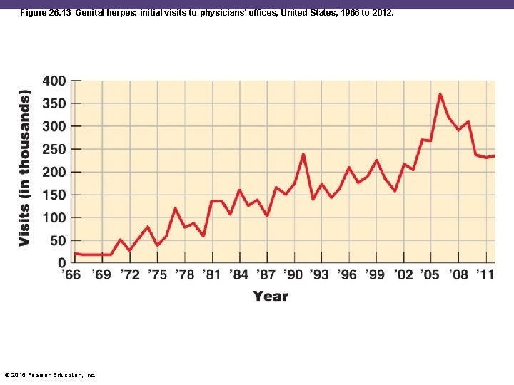
Figure 26. 13 Genital herpes: initial visits to physicians' offices, United States, 1966 to 2012. © 2016 Pearson Education, Inc.
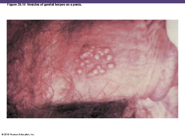
Figure 26. 14 Vesicles of genital herpes on a penis. © 2016 Pearson Education, Inc.
Neonatal Herpes • Herpesvirus crosses the placental barrier and infects the fetus • Damages the CNS, developmental delays, blindness, hearing loss • Survival rate of 40% • Newborns infected from HSV exposure during delivery • Diagnosed by PCR tests and fluorescent antibody tests • Treatment with intravenous acyclovir © 2016 Pearson Education, Inc.
Genital Warts • Also known as condyloma acuminata • Caused by human papillomaviruses • 25% of 14 - to 59 -year-old women in the United States are infected • Visible warts caused by serotypes 6 and 11 • Serotypes 16 and 18 cause cervical cancer • Kills 4000 women in the United States annually • Treatment via removal of warts; podofilox and imiquimod • Prevention with the Gardasil and Cervarix vaccines © 2016 Pearson Education, Inc.
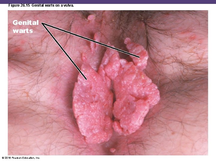
Figure 26. 15 Genital warts on a vulva. Genital warts © 2016 Pearson Education, Inc.
Check Your Understanding ü Both genital herpes and genital warts are caused by viruses; which one is the greater danger to a pregnancy? 26 -7 © 2016 Pearson Education, Inc.
Fungal Disease of the Reproductive Systems Learning Objective 26 -8 Discuss the epidemiology of candidiasis. © 2016 Pearson Education, Inc.
Candidiasis • Caused by C. albicans • Grows on the mucosa of the mouth, the intestinal tract, and the genitourinary tract • Due to opportunistic overgrowth caused by: • Antibiotic use • Diabetes • Hormones • • Oral candidiasis: thrush Vulvovaginal candidiasis: vaginitis Yeasty, thick, yellow discharge Treatment with clotrimazole or fluconazole © 2016 Pearson Education, Inc.
Check Your Understanding ü What changes in the vaginal bacterial microbiota tend also to favor the growth of the yeast Canidida albicans? 26 -8 © 2016 Pearson Education, Inc.
Protozoan Disease of the Reproductive Systems Learning Objectives 26 -9 Discuss the epidemiology of trichomoniasis. 26 -10 List reproductive system diseases that can cause congenital and neonatal infections, and explain how these infections can be prevented. © 2016 Pearson Education, Inc.
Trichomoniasis • Caused by Trichomonas vaginalis • Normal inhabitant of the vagina and urethra • Grows when normal acidity of the vagina is disturbed • Irritation and a profuse foul, greenish yellow frothy discharge • Diagnosis with microscopic identification or a DNA probe • Treatment with metronidazole © 2016 Pearson Education, Inc.
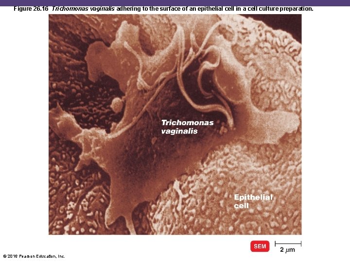
Figure 26. 16 Trichomonas vaginalis adhering to the surface of an epithelial cell in a cell culture preparation. © 2016 Pearson Education, Inc.
The TORCH Panel of Tests • Panel of tests that screens for antibodies to infections in pregnant women • Toxoplasmosis • Other (such as syphilis, hepatitis B, enterovirus, Epstein-Barr virus, varicella-zoster virus) • Rubella • Cytomegalovirus • Herpes simplex virus © 2016 Pearson Education, Inc.
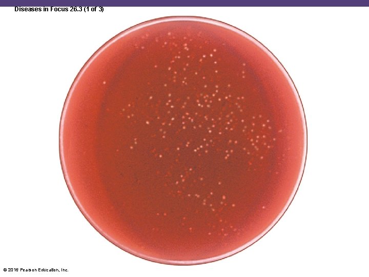
Diseases in Focus: Microbial Diseases of the Reproductive System • A 26 -year-old woman has abdominal pain, painful urination, and fever. Cultures grown in a high-CO 2 environment reveal gram-negative diplococci. • Can you identify infections that could cause these symptoms? © 2016 Pearson Education, Inc.
Diseases in Focus 26. 3 (1 of 3) © 2016 Pearson Education, Inc.
Diseases in Focus 26. 3 (2 of 3) © 2016 Pearson Education, Inc.
Diseases in Focus 26. 3 (3 of 3) © 2016 Pearson Education, Inc.
Check Your Understanding ü What are the symptoms of the presence of Trichomonas vaginalis in the male reproductive system? 26 -9 ü What is the intent of the TORCH panel of tests? 26 -10 © 2016 Pearson Education, Inc.
- Slides: 74