
Power Point Handout Lab 11 Anterior Forearm Palmar
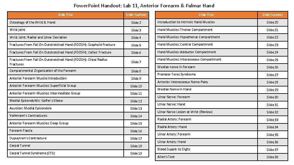
Power. Point Handout: Lab 11, Anterior Forearm & Palmar Hand Slide Title Slide Number Osteology of the Wrist & Hand Slide 2 Introduction to Intrinsic Hand Muscles Slide 20 Wrist joint Slide 3 Hand Muscles: Thenar Compartment Slide 21 Wrist Joint: Radial and Ulnar Deviation Slide 4 Hand Muscles: Hypothenar Compartment Slide 22 Fractures From Fall On Outstretched Hand (FOOSH): Scaphoid Fracture Slide 5 Hand Muscles: Central Compartment Slide 23 Fractures From Fall On Outstretched Hand (FOOSH): Colles’ Fracture Slide 6 Hand Muscles: Adductor Compartment Slide 24 Fractures From Fall On Outstretched Hand (FOOSH): Distal Radius Fractures Slide 7 Hand Muscles: Interosseous Compartment Slide 25 Compartmental Organization of the Forearm Slide 8 Median nerve in Forearm Slide 26 Anterior Forearm Muscle Introduction Slide 9 Pronator Teres Syndrome Slide 27 Anterior Forearm Muscles: Superficial Group Slide 10 Anterior Interosseous Nerve Palsy Slide 28 Anterior Forearm Muscles: Intermediate Group Slide 11 Median Nerve in Hand Slide 29 Medial Epicondylitis: Golfer’s Elbow Slide 12 Ulnar Nerve: Forearm Slide 30 Avulsion: Medial Epicondyle Slide 13 Ulnar Nerve: Hand Slide 31 Volkmann’s Contractures Slide 14 Ulnar Nerve Lesion at Wrist (Review) Slide 32 Anterior Forearm Muscles: Deep Group Slide 15 Radial Artery: Forearm Slide 33 Forearm Fascia Slide 16 Radial Artery: Hand Slide 34 Dupuytren's Contracture Slide 17 Ulnar Artery: Forearm Slide 35 Carpal Tunnel Slide 18 Ulnar Artery: Hand Slide 36 Carpal Tunnel Syndrome (CTS) Slide 19 Blood Supply to Digits Slide 37 Allen’s Test Slide 38
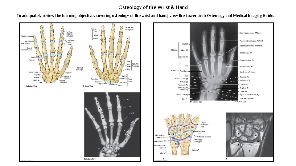
Osteology of the Wrist & Hand To adequately review the learning objectives covering osteology of the wrist and hand, view the Lower Limb Osteology and Medical Imaging Guide.
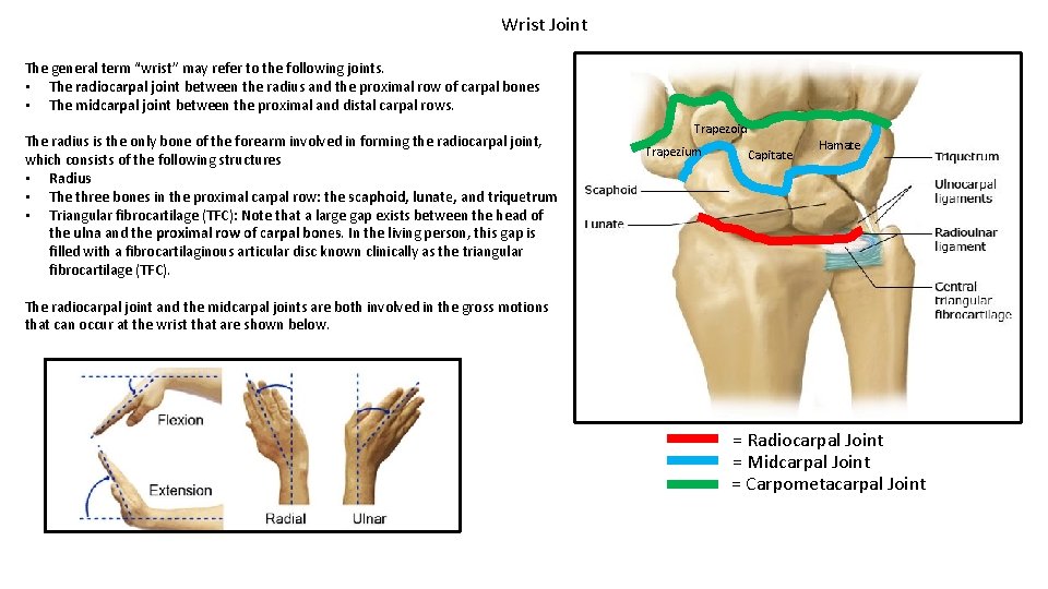
Wrist Joint The general term “wrist” may refer to the following joints. • The radiocarpal joint between the radius and the proximal row of carpal bones • The midcarpal joint between the proximal and distal carpal rows. The radius is the only bone of the forearm involved in forming the radiocarpal joint, which consists of the following structures • Radius • The three bones in the proximal carpal row: the scaphoid, lunate, and triquetrum • Triangular fibrocartilage (TFC): Note that a large gap exists between the head of the ulna and the proximal row of carpal bones. In the living person, this gap is filled with a fibrocartilaginous articular disc known clinically as the triangular fibrocartilage (TFC). Trapezoid Trapezium Capitate Hamate The radiocarpal joint and the midcarpal joints are both involved in the gross motions that can occur at the wrist that are shown below. = Radiocarpal Joint = Midcarpal Joint = Carpometacarpal Joint
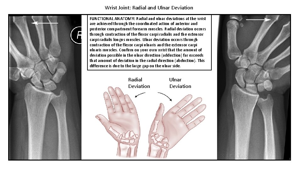
Wrist Joint: Radial and Ulnar Deviation FUNCTIONAL ANATOMY: Radial and ulnar deviations at the wrist are achieved through the coordinated action of anterior and posterior compartment forearm muscles. Radial deviation occurs through contraction of the flexor carpi radialis and the extensor carpi radialis longus muscles. Ulnar deviation occurs through contraction of the flexor carpi ulnaris and the extensor carpi ulnaris muscles. Confirm on your own wrist that the amount of deviation possible in the ulnar direction (adduction) far exceeds that amount of deviation in the radial direction (abduction). This difference is due to the large gap on the ulnar side. Radial Deviation Ulnar Deviation

Fractures From Fall On Outstretched Hand (FOOSH): Scaphoid Fracture CLINICAL ANATOMY: Scaphoid fractures account for 70 -80% of all carpal bone fractures. Although they occur essentially at any age, adolescents and young adults are most commonly affected. Older patients falling in a similar manner are more likely to sustain a distal radial fracture (usually a Colles’ fracture: next slide). Symptoms generally include pain at the base of the thumb in the region of the anatomic snuffbox, which is worse with use of the hand. In addition, edema can occur in the region of the snuffbox. (The anatomical snuffbox will be covered in more detail when we study the posterior forearm in the next unit. )

Fractures From Fall On Outstretched Hand (FOOSH): Colles’ Fracture CLINICAL ANATOMY: A Colles’ Fracture is a complete fracture of the radius close to the wrist, which typically originates from a fall on the outstretched hand in an adult. The fracture results in a dorsal and radial displacement of the distal fragment, and disturbance of the radial-ulnar articulation. The displacement puts the wrist into a position that is sometimes called a ”dinner fork” deformity.
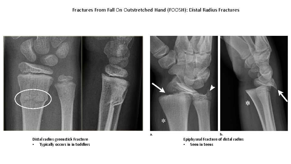
Fractures From Fall On Outstretched Hand (FOOSH): Distal Radius Fractures Distal radius greenstick Fracture • Typically occurs in in toddlers Epiphyseal Fracture of distal radius • Seen in teens
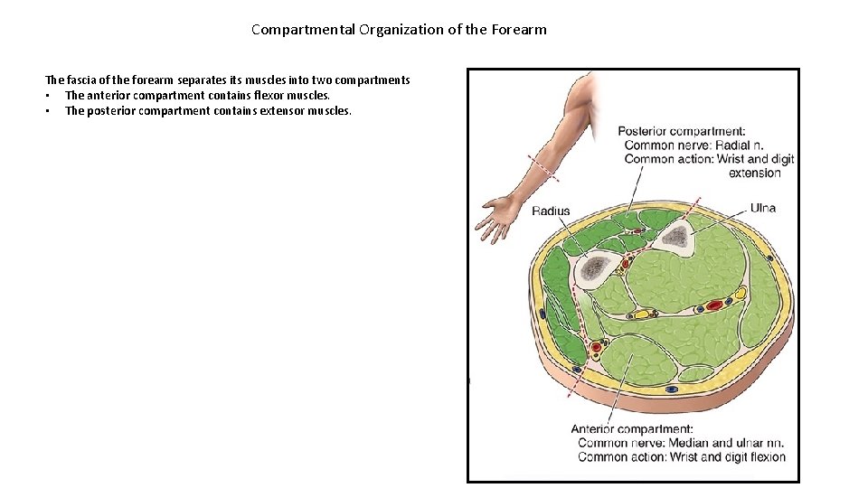
Compartmental Organization of the Forearm The fascia of the forearm separates its muscles into two compartments • The anterior compartment contains flexor muscles. • The posterior compartment contains extensor muscles.
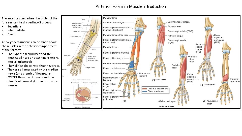
Anterior Forearm Muscle Introduction The anterior compartment muscles of the forearm can be divided into 3 groups. • Superficial • Intermediate • Deep A few generalizations can be made about the muscles in the anterior compartment of the forearm. • The superficial and Intermediate muscles all have an attachment on the medial epicondyle. • They all flex the joint(s) that they cross. • They are all innervated by the median nerve (or a branch of the median), EXCEPT flexor carpi ulnaris and the ulnar ½ of flexor digitorum profundus muscle.
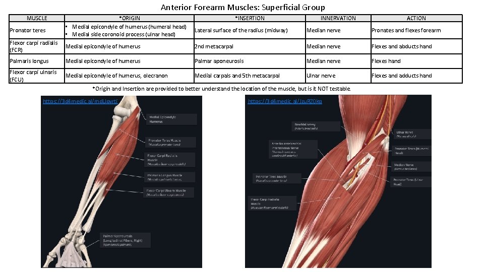
Anterior Forearm Muscles: Superficial Group MUSCLE Pronator teres *ORIGIN • Medial epicondyle of humerus (humeral head) • Medial side coronoid process (ulnar head) *INSERTION INNERVATION ACTION Lateral surface of the radius (midway) Median nerve Pronates and flexes forearm Flexor carpi radialis (FCR) Medial epicondyle of humerus 2 nd metacarpal Median nerve Flexes and abducts hand Palmaris longus Medial epicondyle of humerus Palmar aponeurosis Median nerve Flexes hand Flexor carpi ulnaris (FCU) Medial epicondyle of humerus, olecranon Medial carpals and 5 th metacarpal Ulnar nerve Flexes and adducts hand *Origin and Insertion are provided to better understand the location of the muscle, but is it NOT testable. https: //3 d 4 medic. al/md. Upvrti https: //3 d 4 medic. al/Jzu. RZ 0 kq
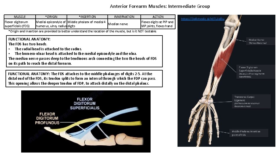
Anterior Forearm Muscles: Intermediate Group MUSCLE Flexor digitorum superficialis (FDS) *ORIGIN *INSERTION INNERVATION Medial epicondyle of Middle phalanx of medial 4 Median nerve humerus, ulna, radius digits ACTION Flexes digits at PIP and MP joints, flexes hand *Origin and Insertion are provided to better understand the location of the muscle, but is it NOT testable. FUNCTIONAL ANATOMY: The FDS has two heads. • The radial head is attached to the radius. • The humero-ulnar head is attached to the medial epicondyle and the ulna. The median nerve passes deep to the tendinous arch connecting the two the heads of FDS on its path to reach the distal forearm. FUNCTIONAL ANATOMY: The FDS attaches to the middle phalanges of digits 2 -5. At the distal end of the FDS, its tendon splits to form an interval through which the FDP can pass. This opening allows the deeper tendon of FDP, to attach distally on the distal phalanx. https: //3 d 4 medic. al/k. ETUjd. Bu
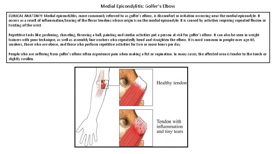
Medial Epicondylitis: Golfer’s Elbow CLINICAL ANATOMY: Medial epicondylitis, more commonly referred to as golfer’s elbow, is discomfort or irritation occurring near the medial epicondyle. It occurs as a result of inflammation/tearing of the flexor tendons whose origin is on the medial epicondyle. It is caused by activities requiring repeated flexion or twisting of the wrist. Repetitive tasks like gardening, shoveling, throwing a ball, painting and similar activities put a person at risk for golfer’s elbow. It can also be seen in weight trainers with poor technique, as well as assembly line workers who repeatedly bend and straighten the elbow. It is most common in people over age 40, smokers, those who are obese, and those who perform repetitive activities for two or more hours per day. People who are suffering from golfer’s elbow often experience pain when making a fist or supination. In many cases, the affected area is tender to the touch or slightly swollen.
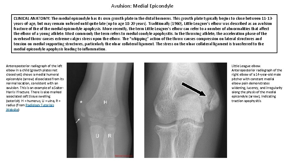
Avulsion: Medial Epicondyle CLINICAL ANATOMY: The medial epicondyle has its own growth plate in the distal humerus. This growth plate typically begins to close between 11 -13 years-of-age, but may remain unfused until quite late (up to age 18 -20 years). Traditionally (1960), Little Leaguer’s elbow was described as an avulsion fracture of the medial epicondyle apophysis. More recently, the term Little Leaguer's elbow can refer to a number of abnormalities that affect the elbow of a young athlete. Most commonly the term refers to medial condyle apophysitis. In the throwing athlete, the acceleration phase of the overhead throw causes extreme valgus stress upon the elbow. The “whipping” action of the throw causes compression on lateral structures and tension on medial supporting structures, particularly the ulnar collateral ligament. The stress on the ulnar collateral ligament is transferred to the medial epicondyle apophysis leading to inflammation. Anteroposterior radiograph of the left elbow in a child (growth plates not closed yet) shows a medial humeral epicondyle (arrow) dissociated from its normal location, consistent with an avulsion. This is an example of a Salter. Harris I fracture. There is also marked associated soft tissue swelling (asterisk). H = humerus, U = ulna, R = radius (From Radiology Tutorials Website) Little League elbow. Anteroposterior radiograph of the right elbow of a 14 -year-old male pitcher with constant medial elbow pain demonstrates widening, lucency, and irregularity along the physis of the medial epicondyle (arrow), indicating traction apophysitis.
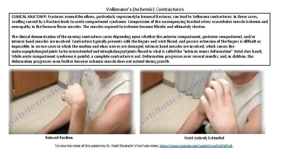
Volkmann’s (Ischemic) Contractures CLINICAL ANATOMY: Fractures around the elbow, particularly supracondylar humeral fractures, can lead to Volkmann contractures. In these cases, swelling caused by a fracture leads to acute compartment syndrome. Compression of the accompanying brachial artery exacerbates muscle ischemia and neuropathy in the forearm flexor muscles. The muscles exposed to ischemia become fibrotic and ultimately shorten. The clinical demonstration of the ensuing contracture varies depending upon whether the anterior compartment, posterior compartment, and/or intrinsic hand muscles are involved. Contracture typically presents with the fingers and wrist flexed, and passive extension of the fingers is difficult or impossible. In severe cases in which the median and ulnar nerves are damaged, intrinsic hand muscles are involved, which causes the metacarpophalangeal joints to be overextended and interphalangeal joints flexed in what is called the "intrinsic minus deformation” (total claw hand). While acute compartment syndrome is painful, a complete contracture is not. Deformation progresses over several months, and, in children, the deformation progresses even further because ischemic muscle does not extend during growth. Relaxed Position Wrist Actively Extended To view the video of this patient by Dr. Nabil Ebraheim’s You. Tube video: https: //www. youtube. com/watch? v=zx 7 q. F 2 Wfrg 4
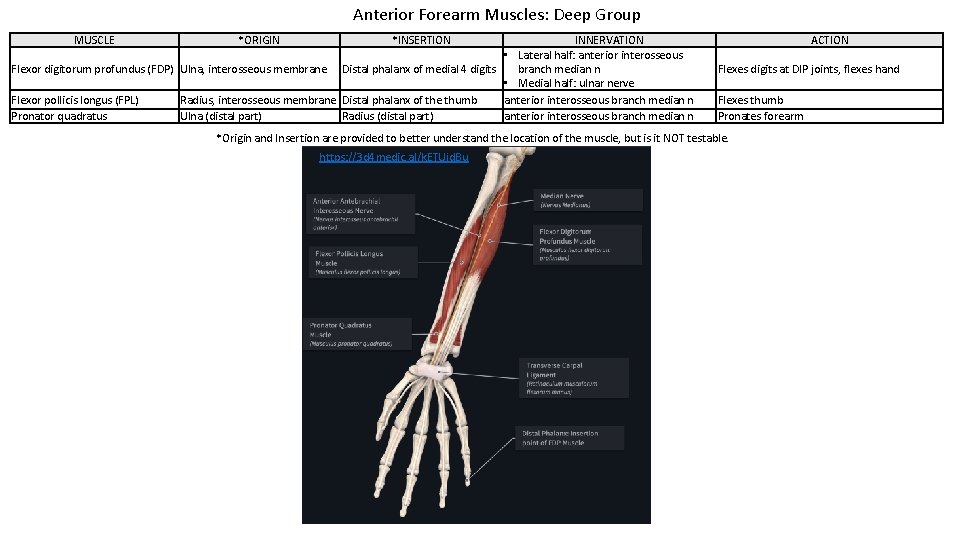
Anterior Forearm Muscles: Deep Group MUSCLE *ORIGIN *INSERTION INNERVATION • Lateral half: anterior interosseous Flexor digitorum profundus (FDP) Ulna, interosseous membrane Distal phalanx of medial 4 digits branch median n • Medial half: ulnar nerve Flexor pollicis longus (FPL) Radius, interosseous membrane Distal phalanx of the thumb anterior interosseous branch median n Pronator quadratus Ulna (distal part) Radius (distal part) anterior interosseous branch median n ACTION Flexes digits at DIP joints, flexes hand Flexes thumb Pronates forearm *Origin and Insertion are provided to better understand the location of the muscle, but is it NOT testable. https: //3 d 4 medic. al/k. ETUjd. Bu
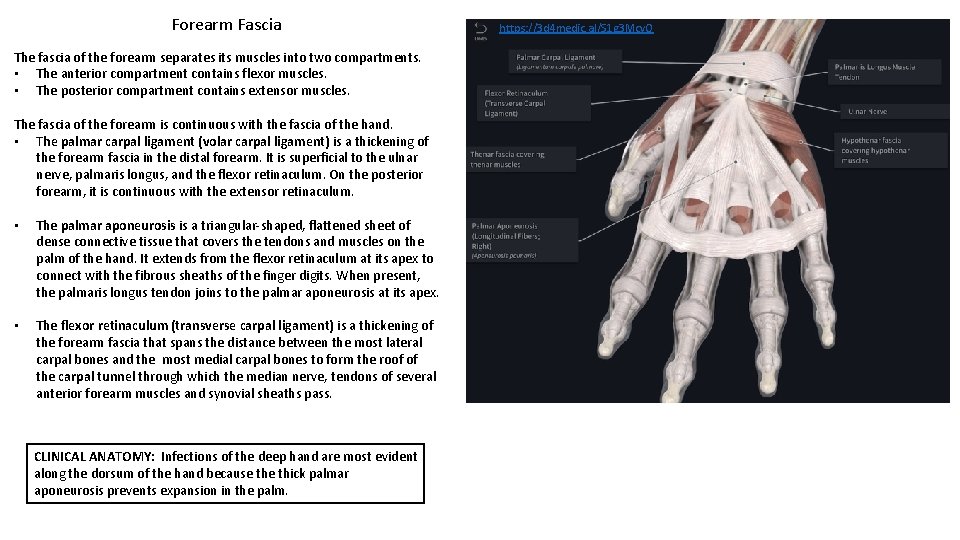
Forearm Fascia The fascia of the forearm separates its muscles into two compartments. • The anterior compartment contains flexor muscles. • The posterior compartment contains extensor muscles. The fascia of the forearm is continuous with the fascia of the hand. • The palmar carpal ligament (volar carpal ligament) is a thickening of the forearm fascia in the distal forearm. It is superficial to the ulnar nerve, palmaris longus, and the flexor retinaculum. On the posterior forearm, it is continuous with the extensor retinaculum. • The palmar aponeurosis is a triangular-shaped, flattened sheet of dense connective tissue that covers the tendons and muscles on the palm of the hand. It extends from the flexor retinaculum at its apex to connect with the fibrous sheaths of the finger digits. When present, the palmaris longus tendon joins to the palmar aponeurosis at its apex. • The flexor retinaculum (transverse carpal ligament) is a thickening of the forearm fascia that spans the distance between the most lateral carpal bones and the most medial carpal bones to form the roof of the carpal tunnel through which the median nerve, tendons of several anterior forearm muscles and synovial sheaths pass. CLINICAL ANATOMY: Infections of the deep hand are most evident along the dorsum of the hand because thick palmar aponeurosis prevents expansion in the palm. https: //3 d 4 medic. al/S 1 g 3 Mcv. O
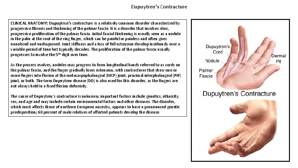
Dupuytren's Contracture CLINICAL ANATOMY: Dupuytren's contracture is a relatively common disorder characterized by progressive fibrosis and thickening of the palmar fascia. It is a disorder that involves slow, progressive proliferation of the palmar fascia. Initial fascial thickening is usually seen as a nodule in the palm at the root of the ring finger, which can be painful or painless and often goes unnoticed and undiagnosed. Joint stiffness and a loss of full extension develop insidiously over a variable period of time but typically decades. The proliferation of the palmar fascia usually progresses to involve the 5 th digit over time. As the process evolves, nodules may progress to form longitudinal bands referred to as cords on the palmar fascia, and the finger gradually loses extension, with contractures that draw one or more fingers into flexion at the metacarpophalangeal (MCP) joint, proximal interphalangeal (PIP) joint, or both. The term Dupuytren disease (DD) is also used for this disorder, as the fingers are not always held in a fixed flexion deformity. The cause of Dupuytren's contracture is unknown; important factors include genetics, ethnicity, sex, and age and may include certain environmental factors and other diseases. The disorder, which most affects those of northern European ancestry, appears to have a pronounced genetic predisposition; 68 percent of male relatives of affected patients develop the disease.
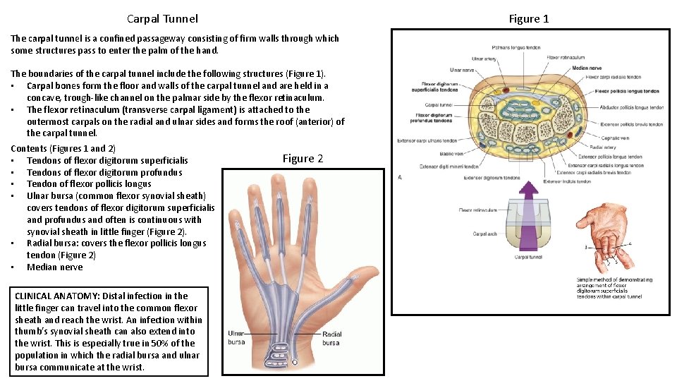
Figure 1 Carpal Tunnel The carpal tunnel is a confined passageway consisting of firm walls through which some structures pass to enter the palm of the hand. The boundaries of the carpal tunnel include the following structures (Figure 1). • Carpal bones form the floor and walls of the carpal tunnel and are held in a concave, trough-like channel on the palmar side by the flexor retinaculum. • The flexor retinaculum (transverse carpal ligament) is attached to the outermost carpals on the radial and ulnar sides and forms the roof (anterior) of the carpal tunnel. Contents (Figures 1 and 2) • Tendons of flexor digitorum superficialis • Tendons of flexor digitorum profundus • Tendon of flexor pollicis longus • Ulnar bursa (common flexor synovial sheath) covers tendons of flexor digitorum superficialis and profundus and often is continuous with synovial sheath in little finger (Figure 2). • Radial bursa: covers the flexor pollicis longus tendon (Figure 2) • Median nerve CLINICAL ANATOMY: Distal infection in the little finger can travel into the common flexor sheath and reach the wrist. An infection within thumb’s synovial sheath can also extend into the wrist. This is especially true in 50% of the population in which the radial bursa and ulnar bursa communicate at the wrist. Figure 2
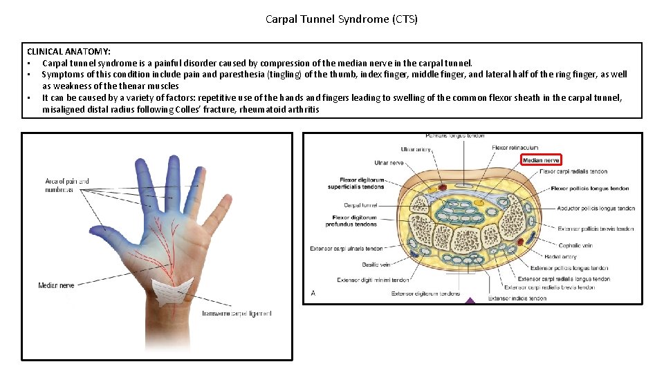
Carpal Tunnel Syndrome (CTS) CLINICAL ANATOMY: • Carpal tunnel syndrome is a painful disorder caused by compression of the median nerve in the carpal tunnel. • Symptoms of this condition include pain and paresthesia (tingling) of the thumb, index finger, middle finger, and lateral half of the ring finger, as well as weakness of thenar muscles • It can be caused by a variety of factors: repetitive use of the hands and fingers leading to swelling of the common flexor sheath in the carpal tunnel, misaligned distal radius following Colles’ fracture, rheumatoid arthritis
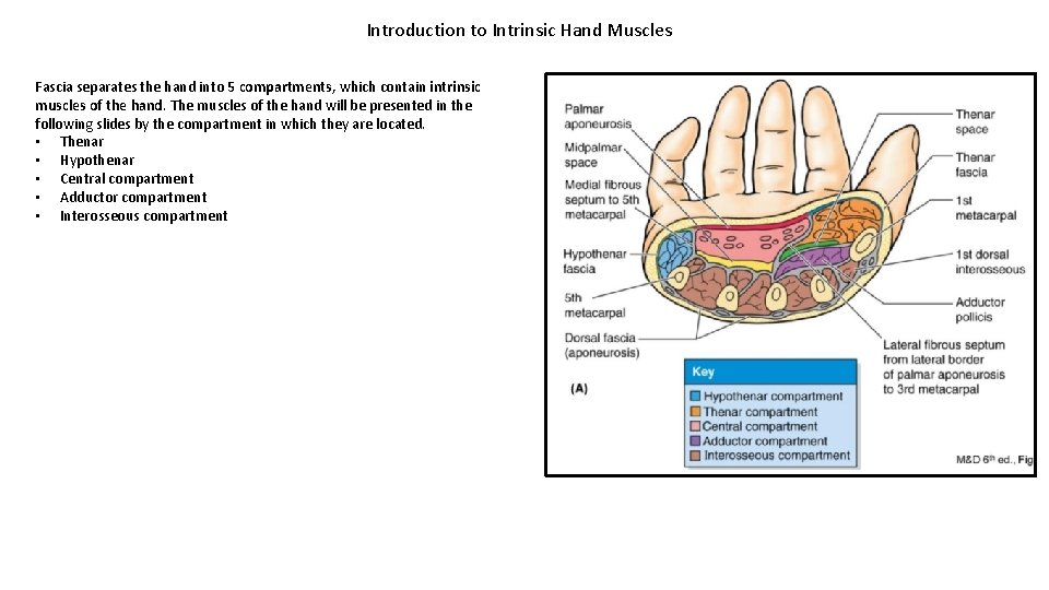
Introduction to Intrinsic Hand Muscles Fascia separates the hand into 5 compartments, which contain intrinsic muscles of the hand. The muscles of the hand will be presented in the following slides by the compartment in which they are located. • Thenar • Hypothenar • Central compartment • Adductor compartment • Interosseous compartment
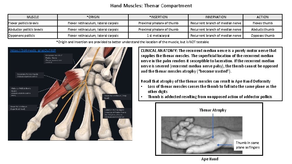
Hand Muscles: Thenar Compartment MUSCLE *ORIGIN *INSERTION INNERVATION ACTION Flexor pollicis brevis Flexor retinaculum, lateral carpals Proximal phalanx of thumb Recurrent branch of median nerve Flexes thumb Abductor pollicis brevis Flexor retinaculum, lateral carpals Proximal phalanx of thumb Recurrent branch of median nerve Abducts thumb Opponens pollicis Flexor retinaculum, lateral carpals 1 st metacarpal Recurrent branch of median nerve Opposes thumb *Origin and Insertion are provided to better understand the location of the muscle, but is NOT testable. https: //3 d 4 medic. al/gc. Qu. T 4 t. P CLINICAL ANATOMY: The recurrent median nerve is a purely motor nerve that supplies thenar muscles. The superficial location of the recurrent median nerve in the palm renders it susceptible to laceration. If the recurrent median nerve is severed (recurrent median nerve palsy), the thumb cannot be opposed and thenar muscles atrophy (“become wasted”). Recall that atrophy of thenar muscles can result in Ape Hand Deformity • Loss of thenar muscles causes the thumb to fall into the same plane as the other digits • Thumb is adducted resulting from unopposed action of adductor pollicis Thenar Atrophy Thumb in same plane as fingers Ape Hand
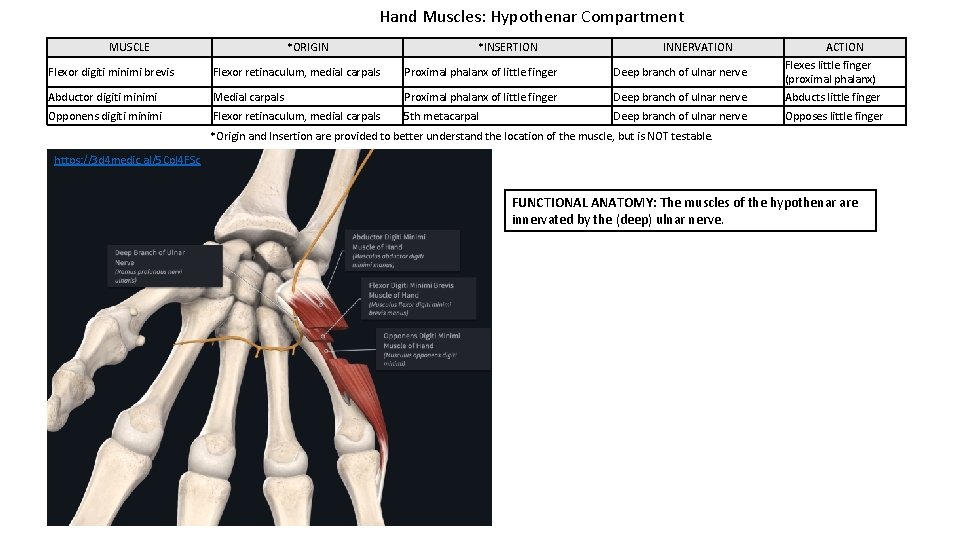
Hand Muscles: Hypothenar Compartment MUSCLE *ORIGIN *INSERTION INNERVATION Flexor digiti minimi brevis Flexor retinaculum, medial carpals Proximal phalanx of little finger Deep branch of ulnar nerve Abductor digiti minimi Medial carpals Proximal phalanx of little finger Deep branch of ulnar nerve ACTION Flexes little finger (proximal phalanx) Abducts little finger Opponens digiti minimi Flexor retinaculum, medial carpals 5 th metacarpal Deep branch of ulnar nerve Opposes little finger *Origin and Insertion are provided to better understand the location of the muscle, but is NOT testable. https: //3 d 4 medic. al/5 Cpl 4 FSc FUNCTIONAL ANATOMY: The muscles of the hypothenar are innervated by the (deep) ulnar nerve.
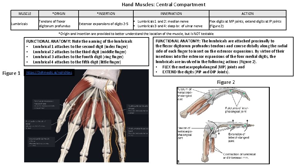
Hand Muscles: Central Compartment MUSCLE Lumbricals *ORIGIN Tendons of flexor digitorum profundus *INSERTION Extensor expansions of digits 2 -5 INNERVATION • Lumbricals 1 and 2: median nerve • Lumbricals 3 and 4: deep br. of ulnar nerve ACTION Flex digits at MP joints, extend digits at IP joints (Figure 2) *Origin and Insertion are provided to better understand the location of the muscle, but is NOT testable. FUNCTIONAL ANATOMY: Note the naming of the lumbricals • Lumbrical 1 attaches to the second digit (index finger) • Lumbrical 2 attaches to the third digit (middle finger) • Lumbrical 3 attaches to the fourth digit (ring finger) • Lumbrical 4 attaches to the fifth digit (little finger) Figure 1 https: //3 d 4 medic. al/na. Yy 9 bxc FUNCTIONAL ANATOMY: The lumbricals are attached proximally to the flexor digitorum profundus tendons and course distally along the radial side of each finger to insert on the extensor expansions. By virtue of their insertions into the extensor expansions of the four medial digits, the lumbricals are involved in the following actions (Figure 2). • FLEX the metacarpophalangeal (MP) joints and • EXTEND the digits (PIP and DIP Joints). Figure 2
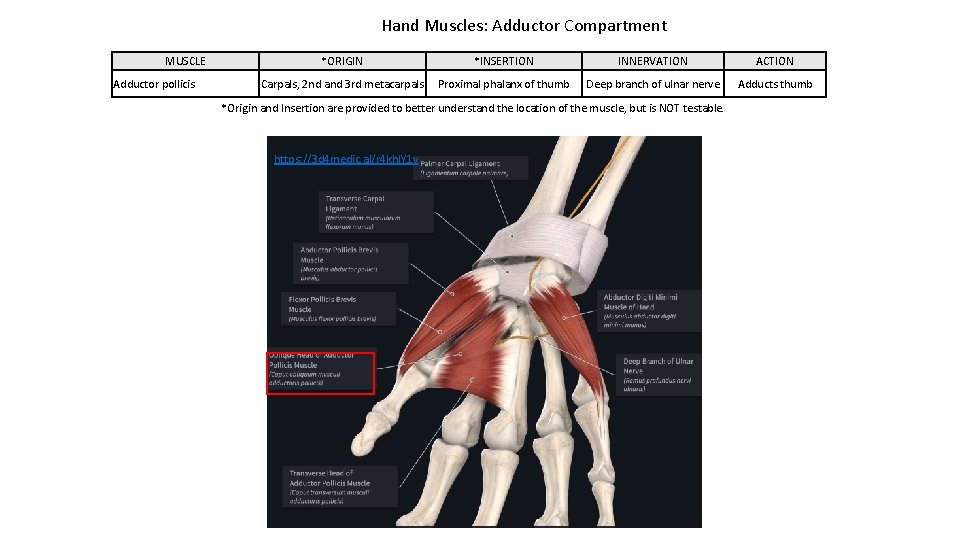
Hand Muscles: Adductor Compartment MUSCLE Adductor pollicis *ORIGIN *INSERTION INNERVATION ACTION Carpals, 2 nd and 3 rd metacarpals Proximal phalanx of thumb Deep branch of ulnar nerve Adducts thumb *Origin and Insertion are provided to better understand the location of the muscle, but is NOT testable. https: //3 d 4 medic. al/r 4 khl. Y 1 y
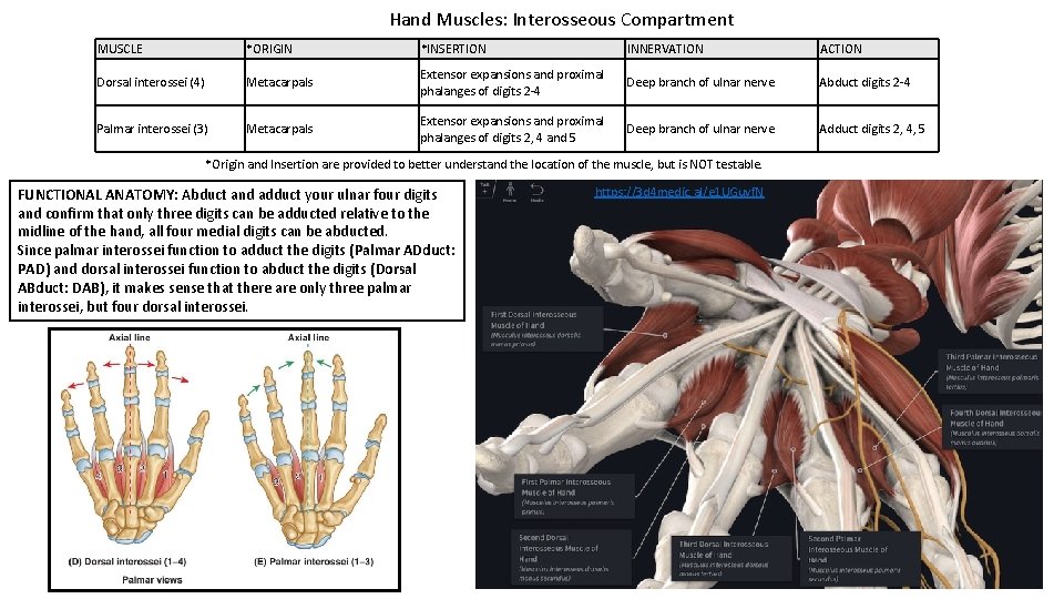
Hand Muscles: Interosseous Compartment MUSCLE *ORIGIN *INSERTION INNERVATION ACTION Dorsal interossei (4) Metacarpals Extensor expansions and proximal phalanges of digits 2 -4 Deep branch of ulnar nerve Abduct digits 2 -4 Palmar interossei (3) Metacarpals Extensor expansions and proximal phalanges of digits 2, 4 and 5 Deep branch of ulnar nerve Adduct digits 2, 4, 5 *Origin and Insertion are provided to better understand the location of the muscle, but is NOT testable. FUNCTIONAL ANATOMY: Abduct and adduct your ulnar four digits and confirm that only three digits can be adducted relative to the midline of the hand, all four medial digits can be abducted. Since palmar interossei function to adduct the digits (Palmar ADduct: PAD) and dorsal interossei function to abduct the digits (Dorsal ABduct: DAB), it makes sense that there are only three palmar interossei, but four dorsal interossei. https: //3 d 4 medic. al/e 1 UGuvf. N
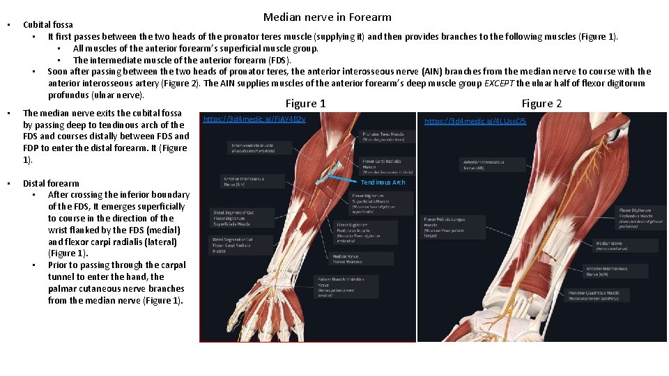
Median nerve in Forearm • Cubital fossa • It first passes between the two heads of the pronator teres muscle (supplying it) and then provides branches to the following muscles (Figure 1). • All muscles of the anterior forearm’s superficial muscle group. • The intermediate muscle of the anterior forearm (FDS). • Soon after passing between the two heads of pronator teres, the anterior interosseous nerve (AIN) branches from the median nerve to course with the anterior interosseous artery (Figure 2). The AIN supplies muscles of the anterior forearm’s deep muscle group EXCEPT the ulnar half of flexor digitorum profundus (ulnar nerve). • The median nerve exits the cubital fossa by passing deep to tendinous arch of the FDS and courses distally between FDS and FDP to enter the distal forearm. It (Figure 1). • Distal forearm • After crossing the inferior boundary of the FDS, It emerges superficially to course in the direction of the wrist flanked by the FDS (medial) and flexor carpi radialis (lateral) (Figure 1). • Prior to passing through the carpal tunnel to enter the hand, the palmar cutaneous nerve branches from the median nerve (Figure 1). Figure 1 Figure 2 https: //3 d 4 medic. al/Fi. AY 4 B 2 v https: //3 d 4 medic. al/4 LUss. Ci 5 Tendinous Arch
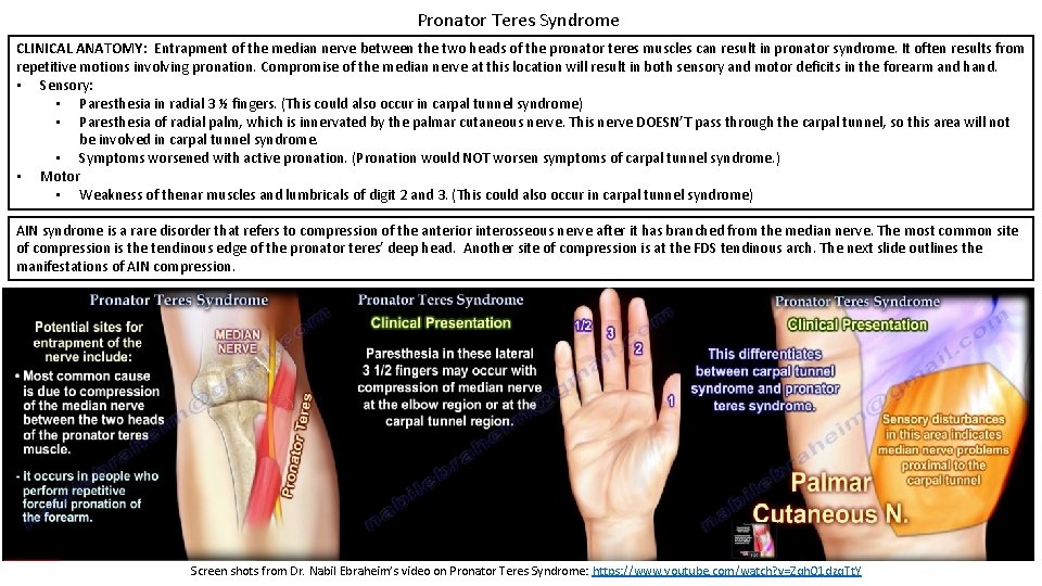
Pronator Teres Syndrome CLINICAL ANATOMY: Entrapment of the median nerve between the two heads of the pronator teres muscles can result in pronator syndrome. It often results from repetitive motions involving pronation. Compromise of the median nerve at this location will result in both sensory and motor deficits in the forearm and hand. • Sensory: • Paresthesia in radial 3 ½ fingers. (This could also occur in carpal tunnel syndrome) • Paresthesia of radial palm, which is innervated by the palmar cutaneous nerve. This nerve DOESN’T pass through the carpal tunnel, so this area will not be involved in carpal tunnel syndrome. • Symptoms worsened with active pronation. (Pronation would NOT worsen symptoms of carpal tunnel syndrome. ) • Motor • Weakness of thenar muscles and lumbricals of digit 2 and 3. (This could also occur in carpal tunnel syndrome) AIN syndrome is a rare disorder that refers to compression of the anterior interosseous nerve after it has branched from the median nerve. The most common site of compression is the tendinous edge of the pronator teres’ deep head. Another site of compression is at the FDS tendinous arch. The next slide outlines the manifestations of AIN compression. Screen shots from Dr. Nabil Ebraheim’s video on Pronator Teres Syndrome: https: //www. youtube. com/watch? v=Zqh. O 1 dzq. Tt. Y
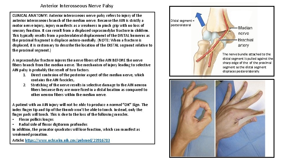
Anterior Interosseous Nerve Palsy CLINICAL ANATOMY: Anterior interosseous nerve palsy refers to injury of the anterior interosseous branch of the median nerve. Because the AIN is strictly a motor nerve injury, injury manifests as a weakness in pinch grip with no loss of sensory function. It can result from a displaced supracondylar fracture in children. This typically results from a posterolateral displacement of the DISTAL humerus as the proximal fragment is displace antero-medially. (NOTE: When a fracture is displaced, it is customary to describe the location of the DISTAL segment relative to the proximal segment. ) A supracondylar fracture injures the nerve fibers of the AIN BEFORE the nerve fibers branch from the median nerve. The mechanism of injury leading to selective AIN palsy is probably the result of two factors. 1. Direct contusion of the posterior aspect of the median nerve, which contains the AIN fascicles, 2. Stretching of the nerve results in selective damage to the AIN neuron fibers because they are more fixed in a distal location as compared to other neuron fibers within the median nerve. A patient with an AIN injury will not be able to produce a normal “OK” Sign. The index finger tip and tip of the thumb won’t be able to touch. Instead, only the finger pads will touch. This is due to the loss of the following muscles. • Flexor pollicis longus • Radial side of flexor digitorum profundus In addition, the pronator quadratus will lose function, which can manifest as weakened pronation. Article: https: //www. ncbi. nlm. nih. gov/pubmed/23916783 Distal segment = posterolateral As distal The nerve bundle attached to the distal segment is pulled against the sharp edge of the proximal segment as the distal segment displaces posterolaterally.
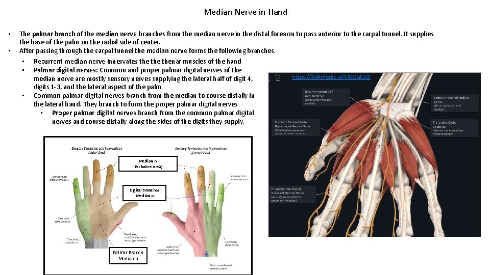
Median Nerve in Hand • • The palmar branch of the median nerve branches from the median nerve in the distal forearm to pass anterior to the carpal tunnel. It supplies the base of the palm on the radial side of center. After passing through the carpal tunnel the median nerve forms the following branches. • Recurrent median nerve innervates the thenar muscles of the hand • Palmar digital nerves: Common and proper palmar digital nerves of the https: //3 d 4 medic. al/VMQaf 9 Ql median nerve are mostly sensory nerves supplying the lateral half of digit 4, digits 1 -3, and the lateral aspect of the palm. • Common palmar digital nerves branch from the median to course distally in the lateral hand. They branch to form the proper palmar digital nerves • Proper palmar digital nerves branch from the common palmar digital nerves and course distally along the sides of the digits they supply. Median n. (Exclusive Area) Digital Branches Median n Palmar Branch Median n
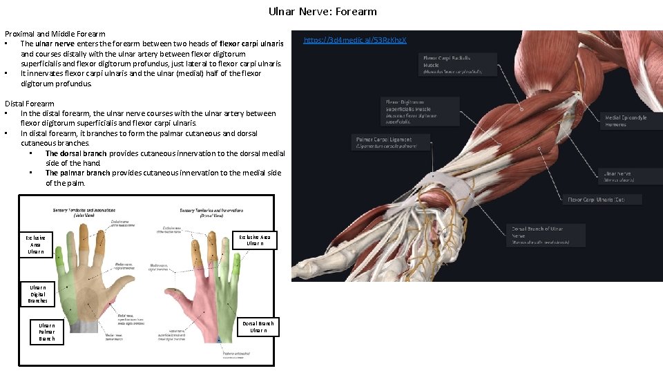
Ulnar Nerve: Forearm Proximal and Middle Forearm • The ulnar nerve enters the forearm between two heads of flexor carpi ulnaris and courses distally with the ulnar artery between flexor digitorum superficialis and flexor digitorum profundus, just lateral to flexor carpi ulnaris. • It innervates flexor carpi ulnaris and the ulnar (medial) half of the flexor digitorum profundus. Distal Forearm • In the distal forearm, the ulnar nerve courses with the ulnar artery between flexor digitorum superficialis and flexor carpi ulnaris. • In distal forearm, it branches to form the palmar cutaneous and dorsal cutaneous branches. • The dorsal branch provides cutaneous innervation to the dorsal medial side of the hand. • The palmar branch provides cutaneous innervation to the medial side of the palm. Exclusive Area Ulnar n Digital Branches Ulnar n Palmar Branch Dorsal Branch Ulnar n https: //3 d 4 medic. al/53 Rz. Khz. X
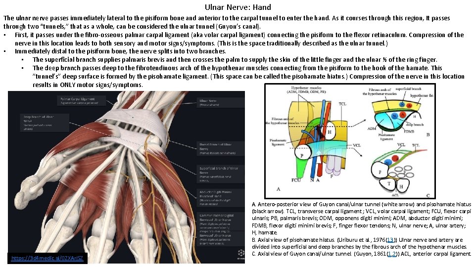
Ulnar Nerve: Hand The ulnar nerve passes immediately lateral to the pisiform bone and anterior to the carpal tunnel to enter the hand. As it courses through this region, It passes through two “tunnels, ” that as a whole, can be considered the ulnar tunnel (Guyon’s canal). • First, it passes under the fibro-osseous palmar carpal ligament (aka volar carpal ligament) connecting the pisiform to the flexor retinaculum. Compression of the nerve in this location leads to both sensory and motor signs/symptoms. (This is the space traditionally described as the ulnar tunnel. ) • Immediately distal to the pisiform bone, the nerve splits into two branches. • The superficial branch supplies palmaris brevis and then crosses the palm to supply the skin of the little finger and the ulnar ½ of the ring finger. • The deep branch passes deep to the fibrotendinous arch of the hypothenar muscles connecting from the pisiform to the hook of the hamate. This ”tunnel’s” deep surface is formed by the pisohamate ligament. (This space can be called the pisohamate hiatus. ) Compression of the nerve in this location results in ONLY motor signs/symptoms. https: //3 d 4 medic. al/0 ZXArj. SZ A. Antero-posterior view of Guyon canal/ulnar tunnel (white arrow) and pisohamate hiatus (black arrow). TCL, transverse carpal ligament ; VCL, volar carpal ligament; FCU, flexor carpi ulnaris; PB, palmaris brevis; ODM, opponens digiti minimi; ADM, abductor digiti minimi; FDMB, flexor digiti minimi brevis; F, finger flexor tendons; N, ulnar nerve; A, ulnar artery; H, hamate. B. Axial view of pisohamate hiatus. (Uriburu et al. , 1976(13)) Ulnar nerve and artery are divided into superficial and deep branches by the fibrous arch of the hypothenar muscles. C. Axial view of Guyon canal/ulnar tunnel. (Guyon, 1861(1, 2)) ACL, anterior carpal ligament.
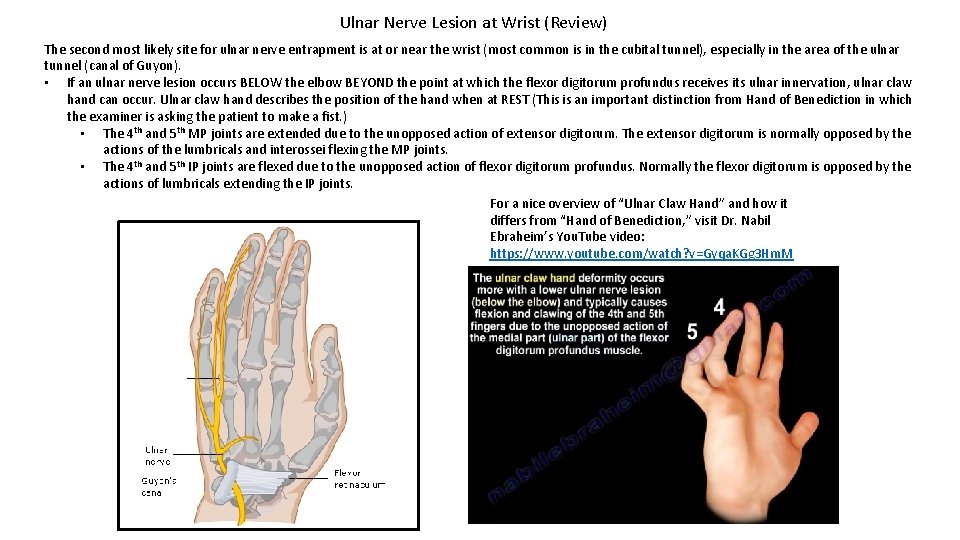
Ulnar Nerve Lesion at Wrist (Review) The second most likely site for ulnar nerve entrapment is at or near the wrist (most common is in the cubital tunnel), especially in the area of the ulnar tunnel (canal of Guyon). • If an ulnar nerve lesion occurs BELOW the elbow BEYOND the point at which the flexor digitorum profundus receives its ulnar innervation, ulnar claw hand can occur. Ulnar claw hand describes the position of the hand when at REST (This is an important distinction from Hand of Benediction in which the examiner is asking the patient to make a fist. ) • The 4 th and 5 th MP joints are extended due to the unopposed action of extensor digitorum. The extensor digitorum is normally opposed by the actions of the lumbricals and interossei flexing the MP joints. • The 4 th and 5 th IP joints are flexed due to the unopposed action of flexor digitorum profundus. Normally the flexor digitorum is opposed by the actions of lumbricals extending the IP joints. For a nice overview of “Ulnar Claw Hand” and how it differs from “Hand of Benediction, ” visit Dr. Nabil Ebraheim’s You. Tube video: https: //www. youtube. com/watch? v=Gyqa. KGg 3 Hm. M
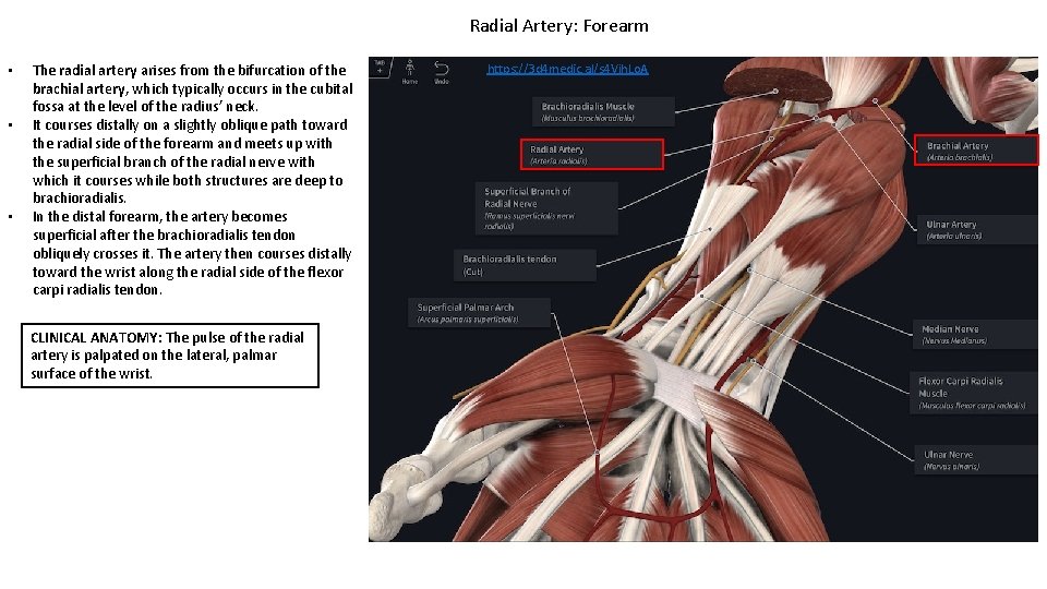
Radial Artery: Forearm • • • The radial artery arises from the bifurcation of the brachial artery, which typically occurs in the cubital fossa at the level of the radius’ neck. It courses distally on a slightly oblique path toward the radial side of the forearm and meets up with the superficial branch of the radial nerve with which it courses while both structures are deep to brachioradialis. In the distal forearm, the artery becomes superficial after the brachioradialis tendon obliquely crosses it. The artery then courses distally toward the wrist along the radial side of the flexor carpi radialis tendon. CLINICAL ANATOMY: The pulse of the radial artery is palpated on the lateral, palmar surface of the wrist. https: //3 d 4 medic. al/s 4 Vjh. Lo. A
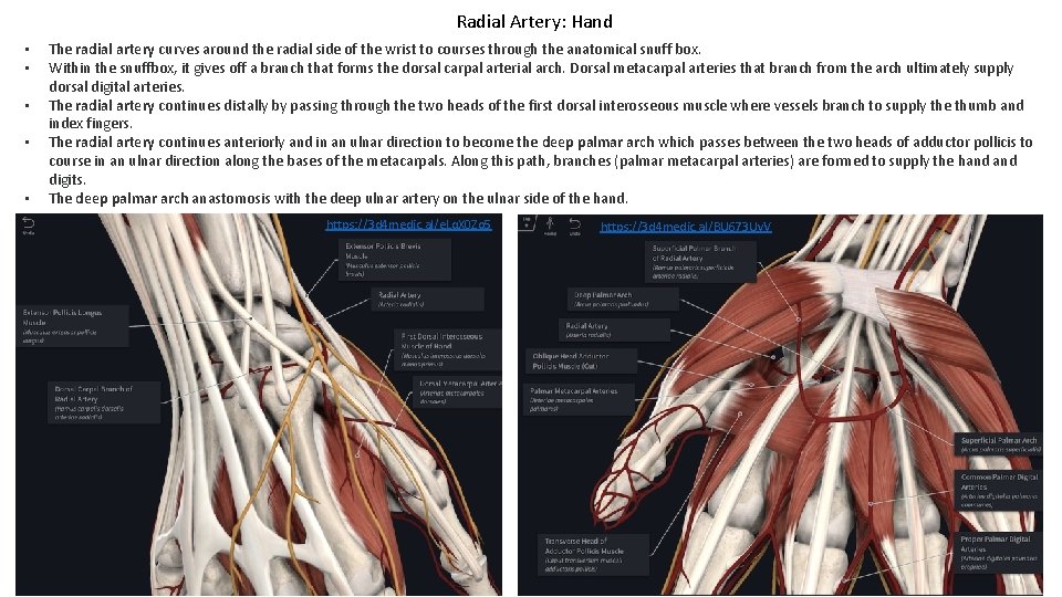
Radial Artery: Hand • • • The radial artery curves around the radial side of the wrist to courses through the anatomical snuff box. Within the snuffbox, it gives off a branch that forms the dorsal carpal arterial arch. Dorsal metacarpal arteries that branch from the arch ultimately supply dorsal digital arteries. The radial artery continues distally by passing through the two heads of the first dorsal interosseous muscle where vessels branch to supply the thumb and index fingers. The radial artery continues anteriorly and in an ulnar direction to become the deep palmar arch which passes between the two heads of adductor pollicis to course in an ulnar direction along the bases of the metacarpals. Along this path, branches (palmar metacarpal arteries) are formed to supply the hand digits. The deep palmar arch anastomosis with the deep ulnar artery on the ulnar side of the hand. https: //3 d 4 medic. al/e. Lq. X 0 Zo 5 https: //3 d 4 medic. al/BU 673 Uv. V
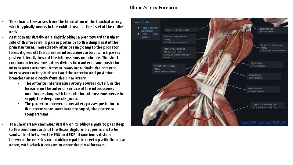
Ulnar Artery Forearm • • • The ulnar artery arises from the bifurcation of the brachial artery, which typically occurs in the cubital fossa at the level of the radius’ neck. As it courses distally on a slightly oblique path toward the ulnar side of the forearm, it passes posterior to the deep head of the pronator teres. Immediately after passing deep to the pronator teres, it gives off the common interosseous artery, which passes posterolaterally toward the interosseous membrane. The short common interosseous artery divides into anterior and posterior interosseous arteries. Note: In many individuals, the common interosseous artery is absent and the anterior and posterior branches arise directly from the ulnar artery. • The anterior interosseous artery courses distally in the forearm on the anterior surface of the interosseous membrane along with the anterior interosseous nerve to supply the deep muscle group. • The posterior interossesous artery passes posterior to the interosseous membrane to supply the posterior compartment. The ulnar artery continues distally on its oblique path to pass deep to the tendinous arch of the flexor digitorum superficialis to be sandwiched between the FDS and FDP. It continues distally between the muscles on an oblique path to meet up with the ulnar nerve, with which it courses to enter the distal forearm. https: //3 d 4 medic. al/Psbi. ODXM
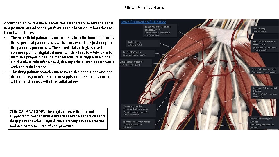
Ulnar Artery: Hand Accompanied by the ulnar nerve, the ulnar artery enters the hand in a position lateral to the pisiform. In this location, it branches to form two arteries. • The superficial palmar branch courses into the hand forms the superficial palmar arch, which curves radially just deep to the palmar aponeurosis. The superficial arch gives rise to common palmar digital arteries, which ultimately bifurcate to form the proper digital palmar arteries that supply the digits. On the ulnar side of the hand, the superficial arch anastomosis with the radial artery. • The deep palmar branch courses with the deep ulnar nerve to the deep region of the palm to supply the deep palmar arch, which anastomosis with the radial artery. CLINICAL ANATOMY: The digits receive their blood supply from proper digital branches of the superficial and deep palmar arches. Digital veins accompany the arteries and are common sites of venipuncture. https: //3 d 4 medic. al/BU 673 Uv. V
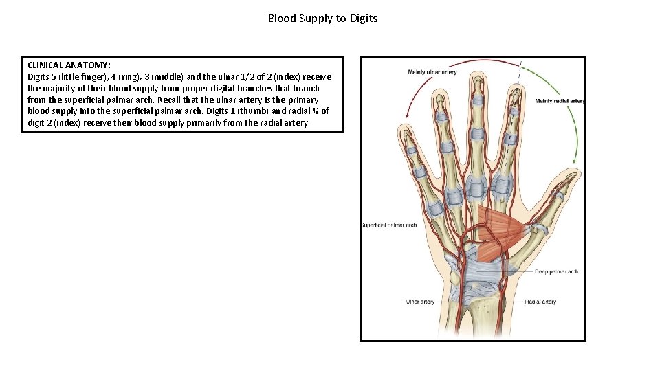
Blood Supply to Digits CLINICAL ANATOMY: Digits 5 (little finger), 4 (ring), 3 (middle) and the ulnar 1/2 of 2 (index) receive the majority of their blood supply from proper digital branches that branch from the superficial palmar arch. Recall that the ulnar artery is the primary blood supply into the superficial palmar arch. Digits 1 (thumb) and radial ½ of digit 2 (index) receive their blood supply primarily from the radial artery.
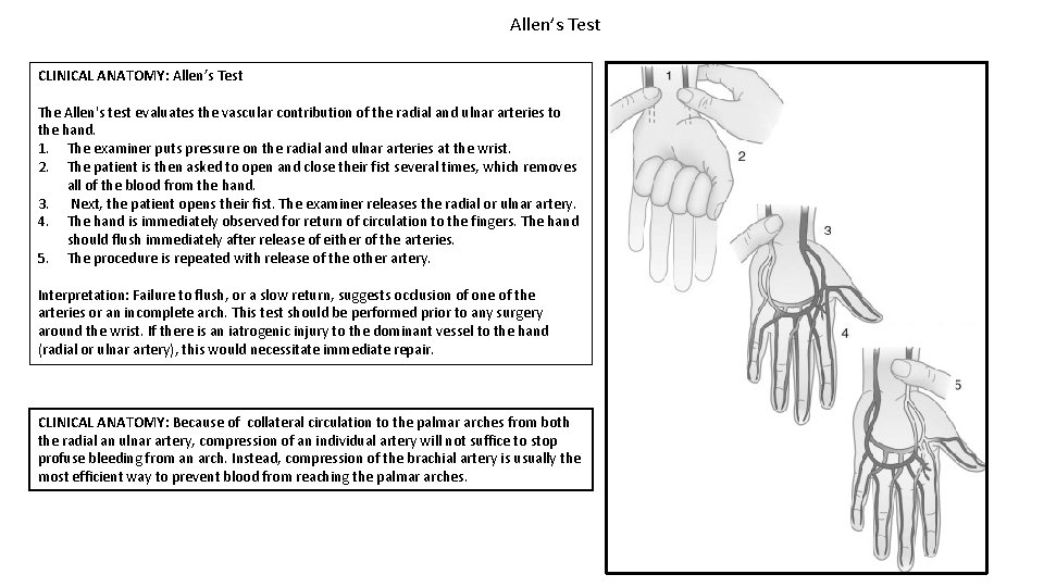
Allen’s Test CLINICAL ANATOMY: Allen’s Test The Allen's test evaluates the vascular contribution of the radial and ulnar arteries to the hand. 1. The examiner puts pressure on the radial and ulnar arteries at the wrist. 2. The patient is then asked to open and close their fist several times, which removes all of the blood from the hand. 3. Next, the patient opens their fist. The examiner releases the radial or ulnar artery. 4. The hand is immediately observed for return of circulation to the fingers. The hand should flush immediately after release of either of the arteries. 5. The procedure is repeated with release of the other artery. Interpretation: Failure to flush, or a slow return, suggests occlusion of one of the arteries or an incomplete arch. This test should be performed prior to any surgery around the wrist. If there is an iatrogenic injury to the dominant vessel to the hand (radial or ulnar artery), this would necessitate immediate repair. CLINICAL ANATOMY: Because of collateral circulation to the palmar arches from both the radial an ulnar artery, compression of an individual artery will not suffice to stop profuse bleeding from an arch. Instead, compression of the brachial artery is usually the most efficient way to prevent blood from reaching the palmar arches.
- Slides: 38