Postural Reflexes What Is Posture Posture is the


 -Involve sustained contraction of muscles B-Phasic reflexes")
 Reflexes §Maintain posture at rest §Include the following reflexes: A- Spinal B-Medullary")
 1 - local static reflexes: Confine")


 +")
 except the §")
")
: q studied")
 : §pressure on side")
 • Studied in intact animal with destroyed labyrinth and")
( center in C. C): q. Maintain posture during")


 Features of decerebrate")
 • Reflexes that")


 Placing Reaction , Hopping Reaction •")

- Slides: 32
Postural Reflexes
What Is Posture? § Posture is the attitude taken by the body in any particular situation like standing posture, sitting posture, etc. even during movement, there is a continuously changing posture § The basis of posture is the ability to keep certain group of muscles in sustained contraction for long periods. Variation in the degree of contraction and tone in different groups of muscle decides the posture of the individual.
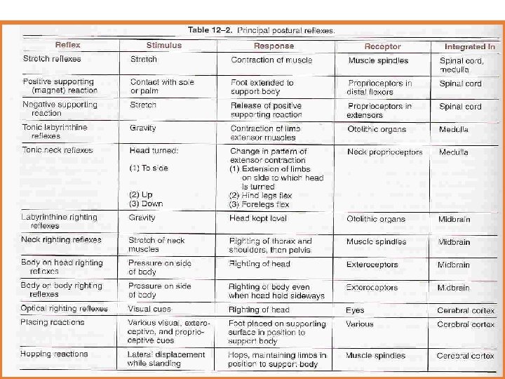
Postural Reflexes q. These reflexes resist displacement of the body caused by gravity or acceleratory forces, and they have the following functions: 1. Maintenance of the upright posture of the body 2. Restoration of the body posture if disturbed 3. Providing a suitable postural background for performance of voluntary movements
Postural Reflexes-2 § Postural reflexes depends on the following receptors: § 1 - Vestibular apparatus receptors as § Maculae (utricle & saccule) & SCC cristae. § 2 - Visual (vision) & auditory(hearing) receptors: Vision can compensate for loss of auditory, vestibular & proprioception § -(Tabes dorsalis +ve Rombergism)
Postural Reflexes-3 § 3 -Proprioceptors of muscles , tendons , ligaments & joints: - a- Neck Proprioceptors: • detect head position in relation to trunk b- Body Proprioceptors proprioceptors of anti-gravity muscles c- Pressure receptors • as in sole of feet initiate positive supporting reaction ( magnet reflex)
Stretch reflexes & postural reflexes can be modified by coordinated activity ; - § § § Spinal cord Medulla Midbrain Cerebral cortex Cerebellum
Postural reflexes are: A-Static reflexes( at rest) -Involve sustained contraction of muscles B-Phasic reflexes ( Statokinetic reflexes) -Involve transient contraction of muscles
A-Static (statotonic) Reflexes §Maintain posture at rest §Include the following reflexes: A- Spinal B-Medullary reflexes C-Righting reflexes ( midbrain )
A-Spinal reflexes: - ( Center in S. C) 1 - local static reflexes: Confine to stimulated limb. i -Stretch reflex This is the most important local static reflex which controls the tone in those extensor muscles which keep the body upright (antigravity muscles) Positive supporting reaction ( magnet reflex) - Deep pressure on the sole lead to contraction of both flexors & extensors to convert the whole lower limb into a rigid column to support body weight ii.
2 - Segmental static reflexes: - mediated by one segment of the spinal cord as : - A. Crossed extensor reflex - B. Negative supporting R (which disappearance of +ve supporting reaction - ( receptors are proprioceptors of extensors of the released limb) N. B spinal R can be studied in spinal animal with cut at neck b/w the S. C& brain stem so all S. C is intact.
B- Medullary Static Reflexes • Center=medulla oblongata • Two types; • Tonic neck reflexes • & tonic labyrinthine reflexes
B- Medullary Static Reflexe-2 • 1 - Neck static reflexes • ( studied in a decerebrated animal cut above medulla + labyrinth destroyed) • -Stimulus is : -changing head position that (+) neck proprioceptors
1 - Ventroflexion of head Flexion of forelimbs + extension of hindlimbs 2 -Dorsiflexion of head Extension of forelimbs + flexion of hindlimbs. 3 - Turning head to one side— Extension of limbs on that side + flexion of other side.
• 2 - Labyrinthine static reflex: § ( in decerebrated animal ) + elimination of neck proprioceptors ( labyrinth intact) - Receptors are otolith organs (maculae) § -Stimulus is gravity 1 -placing the animal in prone position----- 4 limbs flexion 2 - the animal in supine position)-----4 limbs extended.
C- Righting Reflexes § Center is midbrain visual in C. C) except the § Theses reflexes are for correction of disturbed posture § Head correction is first followed by body correction § Studied in a midbrain animal ( cut above midbrain) § Initiated by signal from otlith organs, neck proprioceptors , pressure receptors of the body as well as from visual receptors
A- Midbrain Righting Reflexes 1. Labrinthine RR that correct head position q (cover eyes) & animal held in air from pelvis) q The body is not in the proper position q As in tilting the head (+) otolith organs >>>>-(+) neck muscles to correct the head level, when head is not in proper site. q receptors; otolith organs, q response; contraction of neck muscles lead to righting of head
• 2 - Body RR corrects head position(Body on head RR): q studied in mid brain animal with destroyed labyrinth qstim: pressure on side of body& head is free q receptors; body pressure receptors q. Response/ reflex correction of head. • 3 - Neck righting reflexes corrects body position) : • Correction of the head by previous 2 reflexes lead to twisting of the neck. This initiates reflex righting of the body • stim: stretch of neck muscles • -righting of shoulders & body. • receptors; proprioceptors of neck muscles • response; righting of body.
4 - Body RRcorrects body position (Body on body RR) : §pressure on side of the body and head is fixed) §Receptors/ body pressure receptors §response /reflex correction of body
B- Visual Righting Reflexes( cortical) • Studied in intact animal with destroyed labyrinth and cutting upper 3 cervical nerves - If this animal is thrown in air, visual image can correct position of head & body. § center is c. c § stim: visual stim, § receptors; eye receptors,
B- Phasic reflexes (statokinetic reflexes )( center in C. C): q. Maintain posture during motion q. Integrated in the cerebral cortex a- Hopping reaction: • When animal is pushed laterally }}}}} reflex hopping to keep limbs in position to support body. • The receptors ; muscle spindle. • b- Placing reaction : • Blind folded animal suspended in air & moved towards a supporting surface, the feet will be placed firmly on the supporting surface ( receptors are touch receptors& proprioceptors in soles of feet)
Decerebrate rigidity& Decorticate rigidity
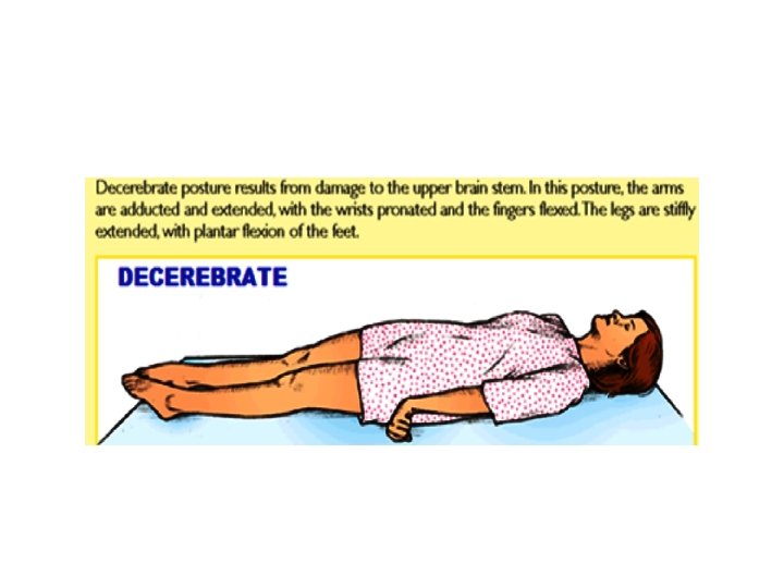
Decerebrate Rigidity üSite of lesion between the superior and inferior colliculi of the midbrain , lesion below red nucleus resulting in üextensive extensor posture of all extremities Rigidity of all 4 limbs ü All limbs extended arms extended by the sides & rotated internally ü (hallmark elbows extended) üHead may be arched to the back ü -In human is rare and may be caused by vascular lesion of brain stem between red N & vestibular nucleus ü
In a decerebrate animal : damage to (level below red nucleus) Features of decerebrate rigidity - hyperextension of all four limbs - dorsiflexion (hyperextension) of tail and head - extreme hyperextension of the spine(opisthotonus) produces concave configuration of the back • - the animal can be made to stand on four limbs but is easily toppled by slight push • •
• Reflexes that are lost/absent • Righting Reflexes ( optical) • Reflexes that are retained /still present ( i. e. , reflexes those which have their centers in SC, medulla or pons ): • Stretch reflex, positive & negative supporting reaction, crossed extensor reflex • Tonic Labyrinthine reflexes • Tonic Neck Reflexes • Mechanism of Decerebrate Rigidity • Diffuse facilitation of stretch reflex due to: 1. increase excitability of motor neuron 2. increase gamma discharge ülesion below red nucleus , resulting in block normal inhibitory signals from brain & red nucleus in midbrain to tonically active pontile reticular formation & Vestibular. N.
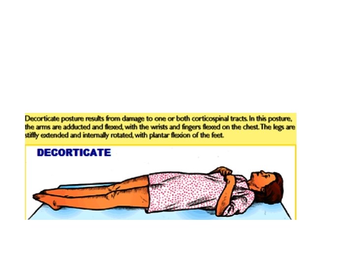
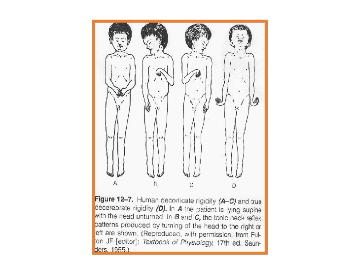
Decorticate Rigidity • In humans , where true decerebrate rigidity is rare , since the damage to the brain centers involved in it are lethal. However decorticate rigidity can be caused by bleeding in the internal capsule which causes UMNL (damage to upper motor neurons). Typical features in decorticated man consist of: § Full extension of the legs § Arm lying across the chest § Semiflexoin at the elbow § Slight pronation of forearm § Flexion of wrist and fingers § Decorticate rigidity is seen at rest § Turning the head to one side initiates tonic neck reflexes e. g turning head to the left >>>>>>>extension of limbs on left side & flexion of right side
§ In decorticate rigidity the lesions is above the red nucleus so rubrospinal are intact together with pontine reticulospinal and the vestibulospinal this leads to the characteristic flexion posturing of the upper extremities and extensor posturing of the lower extremities. § Normally suppressor area 4 strip in the anterior edge of precentral gyrus inhibit red nucleus , if this inhibition is lost by decortication >>>>disinhibition of the red nucleus , so facilitate the rubrospinal tract to flex U. L § Also/ there is loss of inhibitory cortical signals (from suppressor area 4 to gamma motor
• Reflexes that are lost/absent • (1) Placing Reaction , Hopping Reaction • (2) Visual righting reflex • Reflexes that are retained /still present ( i. e. , reflexes the do not depend primarily on cerebral cortex : • (1) Tonic Labyrinthine reflexes • (2) Tonic Neck Reflexes • (3) Other Righting Reflexes
THANK YOU