
Postmortem in Veterolegal cases Purpose Insurance claim Write
- Slides: 20
Postmortem in Vetero-legal cases
Purpose • • • Insurance claim. Write off animal. Veterolegal cases. Record keeping (at farm etc. ). For treatment of healthy animals.
• Now a days there are many cases comes at animal clinics as a vetero legal cases and that should be processed carefully. • The objective of the Post mortem examination of an animal is to ascertain the time since death and the cause of death.
• A Vetero-legal Post mortem examination should be done by written order from the police officer or the executive magistrate. • Before examination carefully read the police report. • Post mortem examination should be thorough and complete. • All the details observed by the veterinary officer should be carefully noted in the Post mortem report on the spot of the Post mortem examination.
• A veterinarian should have a fair knowledge of the normal and pathological appearance of the viscera. • Time & date of arrival of carcass should be noted. • There should be no unnecessary delay in holding Post mortem examination.
External examination: • Identification of the Animal: – Species, sex, colour, tag number, ear marks, branding any permanent mark on the skin (injuries, scars, malformations, muzzle prints etc. ) • Age of the Animal – Body size. Dentition. Carcass bones Horn Rings (Cattle)
• Position of the Animal at Postmortem – Sitting posture. – Fully extended. – Struggled before death etc. • General Condition of the Carcass – Well nourished. – Emaciated. – Stiffening (rigor mortis).
• Evidence of Postmortem Changes – Determine the time of death and observe postmortem changes. • Examination of Natural Orifices – Nose, mouth, ears, urethra, vagina, prepuce, anus. – Colour of mucus membranes. • Discharge from the Orifices – Watery, thick, frothy, blood mixed. – Any smell of insecticides or other chemical.
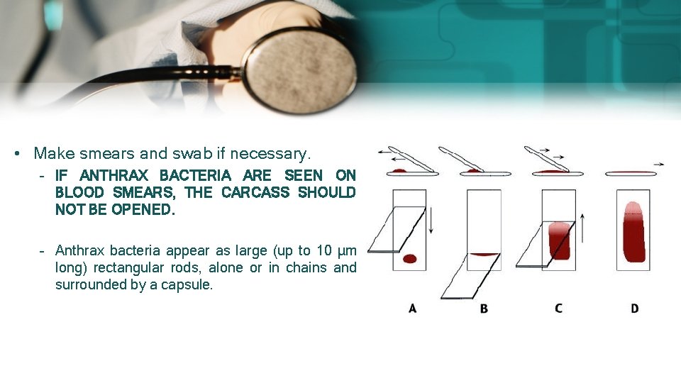
• Make smears and swab if necessary. – IF ANTHRAX BACTERIA ARE SEEN ON BLOOD SMEARS, THE CARCASS SHOULD NOT BE OPENED. – Anthrax bacteria appear as large (up to 10 μm long) rectangular rods, alone or in chains and surrounded by a capsule.
• Examine the Visible Mucus Membranes – Anaemia (pale). – Jaundice/icterus (Yellow) etc. • Eyes – Colour. – Sunken/normal. • Limbs, Bones Feet/Hoofs, Joints – Fractures. Inflammatory lesions. – Rickets, osteomalacia. Dislocation, etc.
Examination of the Body Surface • • • Skin, hair coat colour and hair loss. Toughness. Abscesses. Wounds (antemortem or postmortem), Burns, scald. Parasites or mycotic infections. Emphysema, herniation, umbilical hernia. Signs of diarrhoea (littering of hind quarters). Local lymph nodes (enlargements). Throat oedema (HS).
External Genitalia: • Udder Mastitis, abscesses, growths etc. • Vagina/vulva Discharge, swelling, abscesses, growth etc. • Male organs Abscesses, swelling, paraphymosis (penis), growth etc.
• Histo-pathological examination in many cases is helpful in diagnosing the cause of death. • Tissues for histo-pathological examination should be preserved in as nearly as possible the same state in which they are removed from the animal. • The tissue must be fixed as soon as they are removed from the animal. • The ideal fixing agent should quickly penetrate the tissue and preserve it for longer period.
• The different fixing agents are formaldehyde, alcohol, mercuric chloride, chromates, osmic acid, picric acid, etc. the most common fixative is used 10% formalin. • The specimen taken for examination should be represervative of the lesion and if possible, should include some of the normal tissues. • The specimen should then be cut in size of 0. 5 to 1 cm thick and placed in the fixative (10 times the volume of the tissue). • The labeled bottles are then packed carefully to prevent leakage or breakage.
• Post mortem report: Post mortem no……………. Date: -…. . FIR No……. . . Date………… Police station…… District… • Ruminants, the carcasses should be placed with left sides down (left lateral recumbency). • Horse should be placed right side down (right recumbency]. • Small animals (dog, cat, calves etc. ), dorsum should be down.
POSTMORTEM CONDUCTED UNDER SPECIAL PRECAUTIONS • • • Anthrax [DO NOT CONDUCT] Glanders Rabies (carnivores, ungulates) Scrapie and Bovine Spongiform Encephalopathy (Sheep, cattle) Echinococcus (hydatid Disease) (carnivores) Psittacosis in birds.
Must do after Postmortem
DEEP BURIAL • • Burial along with rendering — most widely used. Largest risks to human health – ground and surface water. Carcasses must be buried within 36 hours. A common practice is to dig a trench and fill over time with carcasses and soil — risky. • Decomposing carcasses can pose a risk to groundwater. • The burial site should consist of clay and silt.
• The burial pit should be at least 100 feet away from production facilities to lessen risk of disease transmission by rodents. • Burial place should be at least 300 ft away from watercourse, sinkhole or from any other source of water used for domestic purposes. • Area with high ground water level should be avoided. • The dimension of the pit should be enough to accommodate the whole carcass. • A pit of 7 feet wide and 9 feet deep can accommodate a mature bovine or equine carcass, 5 mature sheep or goats, 100 mature chickens, or 40 mature turkeys.
• Cover with at least 6 ft of soil. • The covering soil should not be compacted. • A thick layer of lime should be laid under and over the carcass. • Then a layer of mud, a layer of thorny shrubs and again a thick layer of mud. • It is important to burry the bedding and soil contents.