Posterior Pituitary Hormones 1 The posterior pituitary gland

 q HISTOLOGY neural connections between the hypothalamus and the")

 is secreted mainly by: supraoptic nucleus")


 Changes")
 q Initiated by receptors called")


- Slides: 14
Posterior Pituitary Hormones 1
The posterior pituitary gland (neurohypophesis) q HISTOLOGY neural connections between the hypothalamus and the posterior lobe of the pituitary gland. q Posterior pituitary is made up of: q Pituicytes are the fusiform cells derived from glial cells, act as supporting cells and do not secrete any hormone. q Unmyelinated Nerve Fibers come from supraoptic and paraventricular nuclei of the hypothalamus through the pituitary stalk. These fibers stores & releases hormones into the close capillaries.
HORMONES OF POSTERIOR PITUITARY
ANTIDIURETIC HORMONE Source of Secretion: Antidiuretic hormone (ADH) is secreted mainly by: supraoptic nucleus of hypothalamus. It is also secreted by paraventricular nucleus in small quantity (one sixth). Chemistry and Half-life Antidiuretic hormone is a polypeptide containing 9 amino acids. Its half-life is 18 to 20 minutes. Actions Antidiuretic hormone has two actions: 1. Retention of water 2. Vasopressor action.
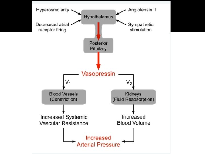
Physiological Functions of ADH Vasopressin V 2 receptor activation. The binding of arginine vasopressin (AVP) to the V 2 vasopressin receptor (V 2 R) stimulates a Gs-coupled protein that activates adenylyl cyclase, in turn causing production of c. AMP to activate protein kinase A (PKA). This pathway increases the exocytosis of aquaporin 2 water channel–containing vesicles (AQMCV) and inhibits endocytosis of the vesicles, both resulting in increases in aquaporin 2 (AQP 2) channel formation and apical membrane insertion. This allows an increase in the permeability of water from the collecting duct (CD). A. C. , adenylyl cyclase; AP 2, aquaporin-2 gene; AQP 2, aquaporin-2; CRE, c. AMP response element; CREB-P, phosphorylated c. AMP response element-binding protein; -P, phosphorylated proteins.
Physiological Functions of ADH 2. Vasopressin V 2 receptor activation The binding of arginine vasopressin (AVP) to its V 1 receptor (V 1 R) stimulates membrane-bound phospholipase (PLCB) via stimulation of a Gcoupled protein (Gq), which in turn results in inositol triphosphate (IP 3) formation and mobilization of intracellular Ca 2+ (ic. Ca 2+). A separate phosphorylation cascade occurs via diacylglycerol (DAG) and protein kinase C (PKC), which has downstream effects, including vascular smooth muscle (VSM) vasoconstriction. Blood vessels Vasoconstriction
Regulation of ADH Secretion 1. The osmolality of the body fluids (Osmotic factor) Changes in the osmolality of body fluids play the most important role in regulating secretion of ADH; changes as minor as 1% are sufficient to alter it significantly. Set point : from 275 to 290 m. Osm/kg H 2 O (average, ≈280 to 285 m. Osm/kg H 2 O) Preganacy the set point Osmolality Stimulates Osmoreceptors Inhibits Osmoreceptors Stimulate inhibit ADH-synthesizing/secreting cells located in the supraoptic and paraventricular nuclei
Regulation of ADH Secretion 2. Blood Volume/Pressure (hemodynamic factor) q Initiated by receptors called Baroreceptors q These receptors are in the lowpressure (left atrium and large pulmonary vessels) and the high-pressure (aortic arch and carotid sinus) sides of the circulatory system. q volume/pressure Stimulates Baroreceptors ADHsynthesizing/ secreting cells located Afferent fibers of the in the vagus and The sensitivity of the supraoptic glossopharyngeal nerves baroreceptor system is less and than that of the Stimulate paraventricul osmoreceptors, and a 5% to ar nuclei 10% decrease in blood volume brainstem (solitary tract or pressure is required before nucleus of the medulla ADH secretion is stimulated. oblongata) 9
OXYTOCIN Source of Secretion: Oxytocin is secreted mainly by paraventricular nucleus of hypothalamus. It is also secreted by supraoptic nucleus in small quantity (one sixth). Chemistry and Half-life Oxytocin is a polypeptide having 9 amino acids. It has a half-life of about 6 minutes.
Oxytocin Some target tissues affected by oxytocin 11
Action of oxytocin on mammary glands milk ejection reflex
Action of Oxytocin on uterus
Slide 34 / Lecture 1 correction of a Typo mistake in the table