Posterior pituitary gland Embryonic origin Infundibulum Base of

Posterior pituitary gland

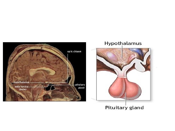
Embryonic origin • Infundibulum – Base of mesencephalon – Downward growth • Composition – Axons of hypothalamic neurons • Magnicellular neurons – Blood vessels • Part of peripheral circulation
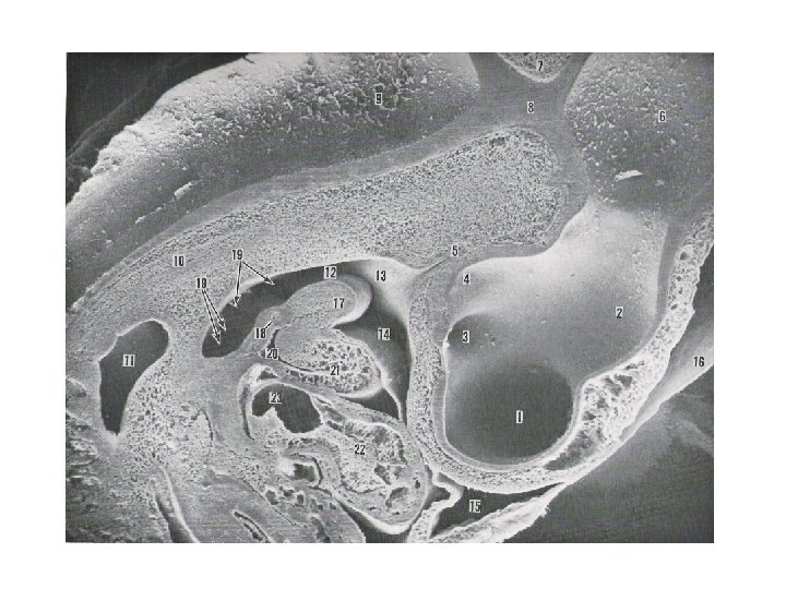


Cellular composition • Pituicytes – Unknown function
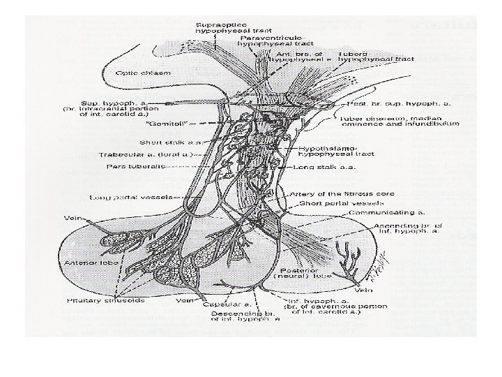

Hormone secretion • Magnicellular neurosecretion – Hypothalamic nuclei • Hormone production • Extend axons to posterior pituitary gland • Specific nuclei produce specific hormone – Oxytocin – Vasopressin/ADH • Specific localization of nuclei within the specific region of hypothalamus – Supraoptic – Paraventricular

Regulation of hormone secretion • Sensory stimuli – Changes in blood pressure – Suckling stimuli – Generation of neural impulses • Interaction between magnicellular neurons and higher brain – Generation of action potential • Travels through axons to the posterior pituitary

• Action potential – Increased Ca permeability and increased Ca influx • Migration and fusion of secretory granules – Release of hormones into perivascular space • Into the capillary subsequently

• Rate of hormone production – Transcription level • Increased transcription after receiving stimuli • Transport of hormones from hypothalamic neural cells – Transport stops when synthesis stops • Some storage in the posterior pituitary gland – Asynchrony between synthesis and release of hormones

• Paracrine/autocrine regulation of hormone production – Intrinsic pattern of firing by neurons • Oxytocin – high amplitude burst followed by long pause (pumping action of smooth muscle within the mammary gland) • VP – strongly and weakly active neurons alternatively fire • Structural plasticity – Contraction of dendrites during firing • Efficient propagation of action potential

Posterior pituitary hormones • Nonapeptide – 9 amino acids • Formation of ring via disulfide bridge • Highly conserved amino acid sequences – Pigs have lysinevasopressin instead of argininevasopressin • Structurally similar – Completely different function

Vasopressin • Synthesis and secretion – Two systems • Osmotic • Pressure-volume • Action – Different receptors • V 1 (blood vessels) • V 2 (renal collecting duct) • V 3 (corticotrophs)

Vasopression • Function – Regulation of osmolarity • Control/conservation of water – Simple relationship • Regulation of Na concentrations in plasma – Pressure-volume – Different system (renin-angiotensin system and aldosterone) – Complicated
 •")
• Regulation of osmolarity – Osmoreceptors in brain (outside of blood-brain barrier) • Hypothalamic neurons • Cells in organum vasculosum of lamina terminalis

• Extracellular fluid osmolarity – Affected greatly by vasopressin • Change in plasma osmolarity • Change in urine volume

• Vasopressin – Acts on the collecting duct of the kidney • Increased water permeability and uptake – Increased number of aquaporin (water channel) of the cell surface (c. AMP) • Result in production of concentrated urine and decreased urine output (antidiuresis)

Thirst • Replacement of water in the body – Urine production – Insensible water loss • Thirst – Defense mechanism – Triggered by changes in osmolarity or volume • Strongly triggered by hypovolemia • Decrease in blood pressure • Generally people ingest excess fluid

Vasopressin and thirst • Water balance – Osmolarity • Changes are usually too small to trigger thirst – 1 to 2 % of basal level – Under normal condition • Regulated by water excretion – Vasopressin

Oxytocin • Physiological regulation of oxytocin secretion – Complicated • Difference among species • Extrapituitary synthesis of oxytocin – Ovaries (corpus luteum) – Uterus in some species – Regulated by suckling stimuli • Classical regulatory mechanism

Function of oxytocin • Lactation – Critical for milk let-down • Oxytocin receptors – Grandular cells in the mammary alveoli – Myoepithelial layers in the mammary ducts • Contraction of myoepithelial layer – Secretion stimulated by suckling • Tactile response • Regulated by the CNS

Function of oxytocin • Contraction of smooth muscle around uterus during parturition – Uterine myometrium • Relaxed during pregnancy – Progesterone and relaxin (hormone from cervix) • Become responsive to oxytocin as parturition approaches – Increased number of receptors – Formation of gap junctions (synchronous contraction) • Works in concert with prostaglandin F 2 a

Function of oxytocin • Contraction of smooth muscle around uterus during parturition – Burst of oxytocin secretion by the posterior pituitary gland during labor • Pulsatile manner • Triggered by Fergusson reflex (dilation of cervix and vagina)

• Postpartum secretion of oxytocin – Primed by changes in steroid hormone concentrations during parturition • Increase in estradiol • Decrease in progesterone • Affects oxytocin responsiveness – Receptors in the mammary gland

• Other functions – Action at the CNS level • Maternal behavior • Sexual arousal – Regulation of reproductive cycle • Ruminants
- Slides: 26