POSTERIOR ANTERIOR TRIANGLE OF NECK Dr H A


 region of neck")
 region of neck trapezius muscle Sternomastoid muscle")
 region of neck clavicle Sternomastoid muscle")
 region of neck q. Occipital triangle qsupraclavicular triangle (greater supraclavicular")
 region of neck q. Occipital triangle qsupraclavicular triangle (greater supraclavicular")


 Arises from chiefly from 4 th cervical nerve;")
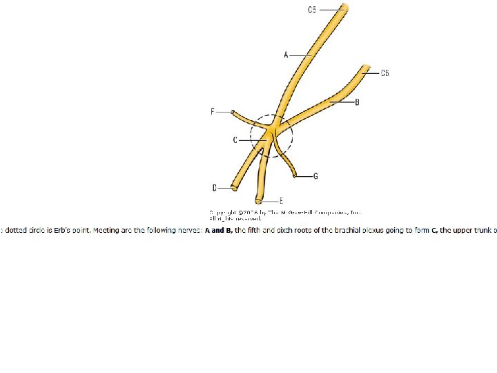
 § arises as a contribution of : § C")


- Slides: 143
POSTERIOR & ANTERIOR TRIANGLE OF NECK Dr. H. A. Jaafar Al-Nahrain Universitycollege of Medicine Dept. Of Anatomy
Objectives: At the end of this lecture we should be able to know : 1. The boundaries, roof, floor & contents of Anterior triangle 2. The boundaries, contents of posterior triangle 3. The role of Sternomastoid muscle in main anatomical division of the neck. 4. What are the careful and careless area of posterior triangle 5. What are the importance of diagastric and omohyoid muscles as a land marks for subdivision of the two triangular regions of neck.
Posterior triangle
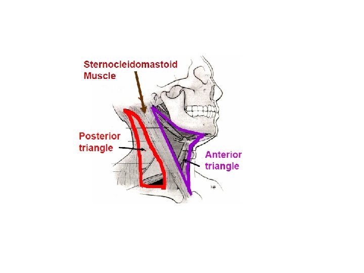
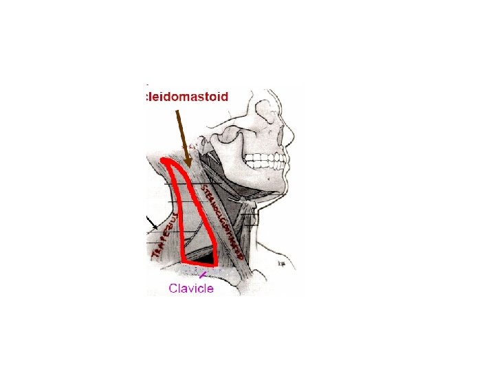
A. Posterior triangle Is bounded by : § § § posterior border of sternocleidomastoid muscle, anterior border of trapezius muscle, superior border of clavicle. roof ………formed by : • • platysma investing (superficial) layer of deep cervical fascia. floor : ……. formed by : § § § splenius capitis levator scapulae muscles anterior, middle, posterior scalene muscles.
Posterior cervical triangle Trapezius Sternocleidomastoid
Is divided into : ……. by omohyoid posterior belly I. occipital triangle. II. subclavian (supraclavicular) triangle. Contains: 1. accessory nerve, 2. cutaneous branches of cervical plexus, 3. external jugular vein, 4. transverse cervical 5. suprascapular vessels, 6. subclavian vein & artery, 7. Inferior belly of omohyoid, 8. roots and trunks of brachial plexus. 9. nerve to subclavius 10. dorsal scapular, 11. suprascapular, 12. long thoracic nerves.
Triangles of posterior (lateral) region of neck
Triangles of posterior (lateral) region of neck trapezius muscle Sternomastoid muscle
Triangles of posterior (lateral) region of neck clavicle Sternomastoid muscle
Triangles of posterior (lateral) region of neck q. Occipital triangle qsupraclavicular triangle (greater supraclavicular fossa)
Triangles of posterior (lateral) region of neck q. Occipital triangle qsupraclavicular triangle (greater supraclavicular fossa)
Sternomastoid muscle
Sternomastoid muscle trapezius muscle
Sternomastoid muscle trapezius muscle clavicle
Omohyoid muscle Sternomastoid muscle trapezius muscle clavicle
Omohyoid muscle Scalenus anterior muscle Sternomastoid muscle trapezius muscle clavicle
Omohyoid muscle Scalenus medius muscle Sternomastoid muscle trapezius muscle clavicle
Omohyoid muscle Sterno thyroid m Sternomastoid muscle trapezius muscle clavicle
Omohyoid muscle thyrohyoid m Sternomastoid muscle trapezius muscle clavicle
Omohyoid muscle Sternohyoid m Sternomastoid muscle trapezius muscle clavicle
apex trapezius muscle Investing layer of deep fascia roof Sternomastoid muscle Middle 1/3 of clavicle – base
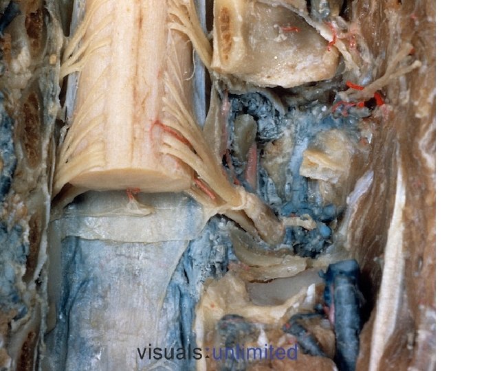
Nerves in posterior triangle 1 -Cervical plexus 1 - four muscular branches 2 - four cutaneous branches 3 - spinal root plexus of accessory nerve (11 th cranial nerve ) 2 -Brachial Roots , trunks and their branches 1 - dorsal scapular nerve –c 5(nerve to rhomboids ) 2 - nerve to subclavius – c 5 &c 6 3 - nerve to serratus anterior –c 5, 6 &7 It is the most important structure in the occipital triangle
Spinal part of accessory nerve
Platysma
Cutaneous nerves and superficial veins Lesser occipital n. External jugular vein Transverse nerve of neck Anterior jugular vein Greet auricular n. Supraclavicular n.
Anterior triangle
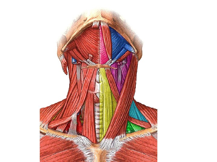
Anterior triangles Suprahyoi dmuscles Infrrahyoid muscles Scalene muscles Prevertebral muscles
B. Anterior triangle Is bounded by: § anterior border of sternocleidomastoid, § anterior midline of neck, § inferior border of mandible. roof : ……formed by: • • platysma investing layer of deep cervical fascia. Is further divided by : § omohyoid superior belly § digastric anterior and posterior bellies into : § § digastric (submandibular), submental (suprahyoid), carotid, muscular (inferior carotid) triangles.
Omohyoid muscle Sternomastoid muscle
Omohyoid muscle Sternomastoid muscle
Omohyoid muscle Sternomastoid muscle
Omohyoid muscle Sternomastoid muscle
apex trapezius muscle Investing layer of deep fascia roof Sternomastoid muscle Middle 1/3 of clavicle – base
Anterior triangles Suprahyoi dmuscles Infrrahyoid muscles Prevertebral muscles Scalene muscles
Posterior belly of digastric Stylohyoid Anterior belly of digastric muscle Sternohyoid muscle
Digastric triangle Above ……………. lower border of mandible Below & infront …. . anterior belly of Digastric muscle Below & behind …. . posterior belly of Digastric & stylohyoid muscles. Floor : Anteriorly : ……. . mylohyoid muscle Posteriorly ………part of hyoglossus muscle
Contents of Digastric triangle § submandibular salivary gland § submandibular lymph nodes § lie on the surface of the gland § facial artery § deep to posterior end of submandibular salivary gland § facial vein § lies superficial to submandibular salivary gland § hypoglossal nerve § nerve to mylohyoid muscle
Anterior belly of digastric • d Posterior belly of digastric Mylohyoid muscle forms the floor of the mouth Digastric triangle
Hyoid bone
C. Hyoid bone……. . Is a U-shaped bone consisting of : § a median body, § lesser horns (cornua) laterally, …………. paired § greater horns (cornua) posteriorly……… paired 1. Body Provides for attachments for : § § Geniohyoid , mylohyoid, Omohyoid , sternohyoid. muscle 2. Greater horn Provides attachments for : § § § middle constrictor, hyoglossus, digastric (anterior and posterior) bellies, stylohyoid, thyrohyoid muscle. 3. Lesser horn Provides attachment for : • stylohyoid ligament, ligament which runs from styloid process to lesser horn
Styloid process
D. Styloid process § Is a slender projection of variable length § extends downward & forward from temporal bone. Gives origin to : three muscles: § § § stylohyoid, styloglossus, Stylopharyngeus two ligaments : • • stylohyoid stylomandibular.
Carotid triangle boundary : • Behind : …………. . sternomastoid muscle • Infront & above : posterior belly of digastric muscle • Infront&below : superior belly of omohyoid muscle Floor : infont : hyoglossus muscle ( above ) and the thyrohyoid muscle (below) • Behind: the middle constrictor muscle of the pharynx (above ) and the inferior constrictor muscle of the pharynx (below )
• d Posterior belly of digastric Carotid triangle
apex trapezius muscle Investing layer of deep fascia roof Sternomastoid muscle Middle 1/3 of clavicle – base
Internal carotid artery External carotid artery Common carotid artery
Contents of the Carotid triangle • 1 - The carotid sheath and its contents : • - common carotid artery : in the lower part of the triangle. • - internal carotid artery : in the upper part of the triangle. • Internal jugular vein : lateral • Vagus nerve : between the artery and the vein but in a more posterior plane.
• 2 - the external carotid artery : gives most of its branches in the carotid triangle ( superior thyroid artery , lingual artery , facial artery , ascending pharyngeal artery and occipital artery ) • 3 - hypoglossal nerve • 4 - Descendes cervicalis (C 2, 3) anterior to carotid sheath. • 5 - sympathetic trunk adherent to the posterior wall of carotid sheath
Contents of the Muscular triangle • It contains the infrahyoid muscle
Contents of the Submental triangle • 1 - submental lymph nodes • 2 - beginning of the anterior jugular vein
The infrahyoid muscles • They are strap like muscles occupy each side of the midline of the neck , from the hyoid bone to the manubrium sterni. • They consist of 4 muscles arranged into 2 layers : • 1 - superficial layer : a- sternohyoid • B- omohyoid • 2 - deep layer: a- sternothyroid • B- thyrohyoid
Suprahyoid muscles • • • 1 - digastric muscle 2 - mylohyoid muscle 3 - hyoglossus muscle 4 - geniohyoid muscle 5 - stylohyoid muscle
Geniohyoid muscle Hyoglossus
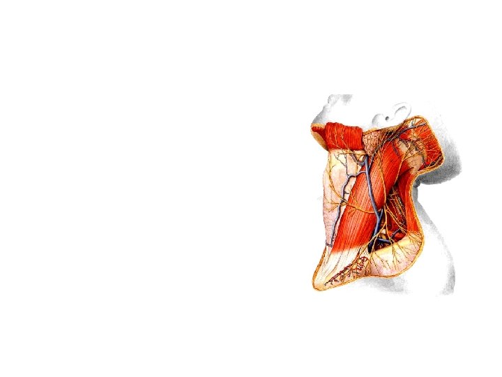
Submendibular gland Digastric Accessory n. Hypoglossal n. Superior thyroid a. Ansa cervicalis Sternothyroid Sternohyoid Vagus n. Cervical plexus Phrenic n. Omohyoid
III. Nerves
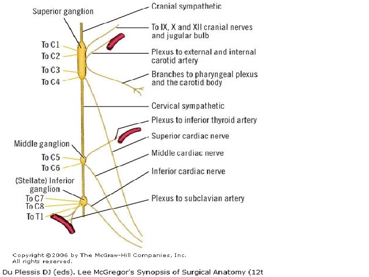
A. Accessory nerve Is formed by union of : • cranial & • spinal roots. Has both spinal and cranial portions, traverse jugular foramen, foramen they interchange fibers. cranial roots : • arise from medulla oblongata below roots of vagus spinal roots : § arise from lateral aspect of cervical segment of spinal cord § between C 1 – C 5 § unites to form a trunk § ascends between dorsal & ventral roots of spinal nerves in vertebral canal § passes through foramen magnum
cranial portion § contains motor fibers. . join vagus nerve § innervate soft palate, palate pharyngeal constrictors, constrictors & larynx spinal portion § innervates sternocleidomastoid & trapezius muscles. § Lies on levator scapulae in posterior cervical triangle § passes deep to trapezius
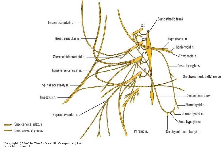
B. Cervical plexus Is formed by ventral primary rami of C 1 to C 4. 1. Cutaneous branches a- Lesser occipital nerve (C 2) • Ascends along posterior border of sternocleidomastoid to scalp • behind auricle. b- Great auricular nerve (C 2 -C 3) • Ascends on sternocleidomastoid • innervate skin behind auricle & on parotid gland. C- Transverse cervical nerve (C 2 -C 3) • Turns around posterior border of sternocleidomastoid • innervates skin of anterior cervical triangle. D-Supraclavicular nerve (C 3 -C 4) Emerges as a common trunk from under sternocleidomastoid divides into anterior, middle, & lateral branches to skin over clavicle &shoulder.
2. Motor branches Ansa cervicalis………………Is a nerve loop formed by union of : § superior root (C 1) § inferior root (C 2 --C 3; descendens hypoglossi descendens cervicalis . Lies superficial to carotid sheath in anterior cervical triangle. Innervates infrahyoid (strap) muscles: § omohyoid, § sternothyroid muscles, with exception of thyrohyoid muscle, muscle innervated by C 1 via hypoglossal nerve.
Phrenic nerve (C 3 -C 5) Arises from chiefly from 4 th cervical nerve; contains : § motor, § sensory, § sympathetic nerve fibers; Provides: § motor supply to diaphragm § sensation to its central part. • Descends on anterior surface of anterior scalene muscle • under cover of sternocleidomastoid muscle. • Passes between subclavian artery & vein at root of neck • enters thorax by crossing in front of origin of internal thoracic artery, artery • it joins pericardiacophrenic branch of this artery. • Passes anterior to root of lung • • between mediastinal pleura & fibrous pericardium supply sensory fibers to these structures. longus capitis and cervicis or colli, sternocleidomastoid, trapezius, levator scapulae, scalene muscles.
Accessory phrenic nerve (C 5) § arises as a contribution of : § C 5 to phrenic nerve or § a branch of nerve to subclavius (C 5), § descends lateral to phrenic nerve, § enters thorax by passing posterior to subclavian vein, § joins phrenic nerve below first rib to supply diaphragm.
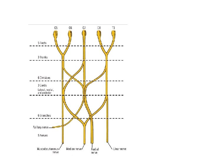
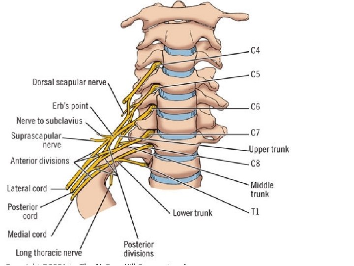
C. Brachial plexus § Is formed by union of ventral primary rami of C 5 to T 1 § passes between anterior scalene & middle scalene muscles. 1. roots give rise to : Dorsal scapular nerve (C 5) o Emerges from behind anterior scalene muscle o runs downward and backward through middle scalene muscle o deep to trapezius. o Passes deep to levator scapulae o descends along with dorsal scapular artery on deep surface of rhomboid muscles along medial border of scapula, Innervating: § levator scapulae § rhomboid muscle. Long thoracic nerve (C 5 --C 7) § Pierces middle scalene muscle, § descends behind brachial plexus, § enters axilla innervate ……. . serratus anterior
C. Brachial plexus 2. Iupper trunk gives rise to : Suprascapular nerve (C 5 -C 6) § Passes deep to trapezius § joins suprascapular artery in a course toward shoulder. § Passes through scapular notch under transverse scapular ligament Supplies : • supraspinatus • infraspinatus muscles Nerve to subclavius muscle (C 5) § Descends in front of plexus and behind clavicle § innervate subclavius. § Communicates with phrenic nerve as accessory phrenic nerve
Omohyoid muscle Sternomastoid muscle
apex trapezius muscle Investing layer of deep fascia roof Sternomastoid muscle Middle 1/3 of clavicle – base
Posterior belly of digastric Stylohyoid Anterior belly of digastric muscle Sternohyoid muscle