Posterior Abdominal Wall ABDOMINAL AORTA The abdominal aorta

Posterior Abdominal Wall

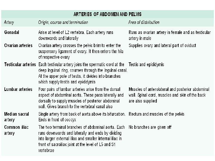
ABDOMINAL AORTA The abdominal aorta begins in the midline at the aortic opening of the diaphragm, opposite the lower border of vertebra T 12 and ends in front of the lower part of the body of vertebra L 4, about 1. 25 cm to the left of the median plane, by dividing into the right and left common iliac arteries. Branches The branches of the abdominal aorta are classified.

The abdominal aorta, inferior vena cava and associated lymph

• Ventral branches, which develop from ventral splanchnic or vitelline arteries and supply the gut. These are as follows. a. Coeliac trunk gives left gastric, common hepatic and splenic branches. b. Superior mesenteric artery gives inferior pancreaticoduodenal, middle colic, right colic, ileocolic and 12– 15 jejunal and ileal branches. c. Inferior mesenteric artery gives left colic, sigmoid arteries and continues as superior rectal.

• Lateral branches, which develop from the lateral splanchnic or mesonephric arteries and supply the viscera derived from the intermediate mesoderm. These are right and left: a. Inferior phrenic arteries. b. Middle suprarenal arteries. c. Renal arteries. d. Testicular or ovarian arteries. • Dorsal branches represent the somatic intersegmental arteries and are distributed to the body wall. These are: a. Lumbar arteries—four pairs. b. Median sacral artery—unpaired.

Contd…

Contd…

Contd…

Contd…

Inferior Vena Cava The inferior vena cava is formed by the union of the right and left common iliac veins on the right side of the body of vertebra L 5. It pierces the central tendon of the diaphragm at the level of vertebra T 8, and opens into the lower and posterior part of the right atrium. Tributaries 1. 2. 3. 4. 5. The common iliac veins The third and fourth lumbar veins The right testicular or ovarian vein The renal veins The right suprarenal vein

Lymph Nodes of Posterior Abdominal Wall The external iliac nodes 8 to 10 lie along the external iliac vessels, being lateral, medial and anterior to them. They receive afferents from: 1. Inguinal lymph nodes 2. Deeper layers of the infraumbilical part of the anterior abdominal wall 3. Adductor region of the thigh 4. Glans penis or clitoris 5. Membranous urethra 6. Prostate 7. Fundus of the urinary bladder 8. Cervix uteri 9. Part of the vagina.

Their efferents pass to common iliac nodes. • The common iliac nodes, 4 to 6 in number lie along the common iliac artery. They receive afferents from the external and internal iliac nodes, and send their efferents to the lateral aortic nodes. • The lumbar or aortic nodes are divided into preaortic and lateral aortic groups. The preaortic nodes are divisible into coeliac, superior mesenteric and inferior mesenteric groups. They receive afferents from gastrointestinal tract, the liver, the pancreas and the spleen. Their efferents form the intestinal trunks which enter the cisterna chyli. The lateral aortic nodes lie on each side of the abdominal aorta.

Cisterna Chyli This is an elongated lymphatic sac, about 5 to 7 cm long. It is situated in front of the first and second lumbar vertebrae, immediately to the right of the abdominal aorta. It is overlapped by the right crus of the diaphragm. Its upper end is continuous with the thoracic duct. It is joined by the right and left lumbar and intestinal lymph trunks. Muscles of the Posterior Abdominal Wall

Contd…
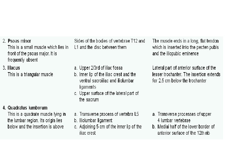

Muscles of the posterior abdominal wall

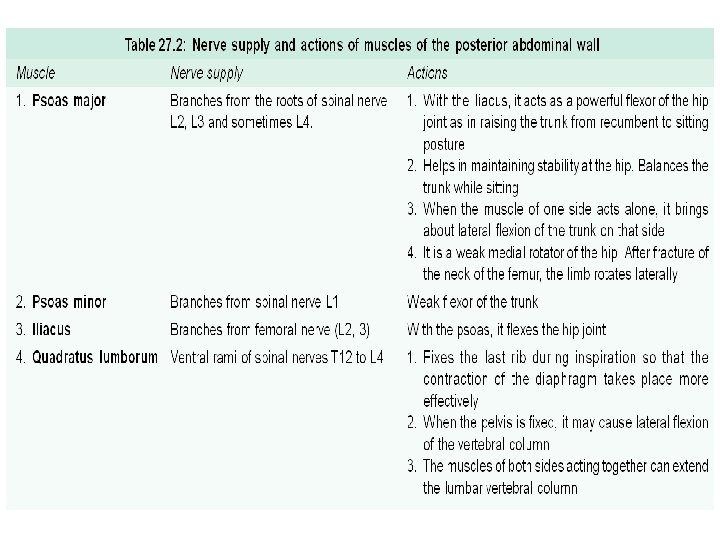
CLINICAL ANATOMY Psoas abscess: Pus from tubercular infection of the thoracic and lumbar vertebrae may track down through the sheath into the thigh, producing a soft swelling in the femoral triangle. Nerves of the Posterior Abdominal Wall Lumbar Plexus The lumbar plexus formed by the ventral rami of the upper four lumbar nerves. The branches of the lumbar plexus are summarised below.
 Ilioinguinal Nerve (L 1) Genitofemoral Nerve Lateral")
• • Iliohypogastric Nerve (L 1) Ilioinguinal Nerve (L 1) Genitofemoral Nerve Lateral Cutaneous Nerve of the Thigh Femoral Nerve Obturator Nerve Lumbosacral Trunk Abdominal Part of the Autonomic Nervous System The abdomen is supplied by both sympathetic and parasympathetic nerves. The sympathetic nerves are derived from two sources. 1. The lumbar sympathetic trunk supplies somatic branches to the lower abdominal wall and the lower limb; and visceral branches for the pelvic organs.

2. The coeliac plexus, formed by splanchnic nerves from the thorax, supplies all the abdominal organs, including the gonads. The parasympathetic nerves are also derived from two sources. a. The vagus joins the coeliac plexus. b. The pelvic splanchnic nerves join the inferior hypogastric plexus. Coeliac Ganglia and Coeliac Plexus The coeliac ganglion is the largest ganglion in the body, situated one on each side of the coeliac trunk. It receives greater splanchnic nerve, and lesser splanchnic nerve.

The coeliac plexus or solar plexus is closely related to the coeliac ganglion. The fibres making up the plexus are as follows: a. Preganglionic sympathetic fibres reach it through the greater and lesser splanchnic nerves. b. Postganglionic sympathetic fibres arising in the coeliac ganglion. c. Preganglionic vagal fibres are derived from the posterior vagal trunk containing fibres from both the right and left vagal nerves. The fibres from the right vagus predominate. d. Sensory fibres from the diaphragm reach the coeliac plexus along the inferior phrenic arteries.

Branches The coeliac plexus forms a number of secondary plexuses which surround branches of the aorta. 1. Phrenic plexus 2. Hepatic plexus 3. Left gastric plexus 4. Splenic plexus 5. Suprarenal plexus 6. Renal plexus 7. Testicular plexus 8. Ovarian plexus 9. Superior mesenteric plexus 10. Abdominal aortic plexus or

Formation of plexuses

Layers of the Abdomen The following 6 layers in the abdomen are shown in figures. Anterior abdominal wall rectus sheath

Liver, stomach and greater omentum

Most of small intestine, appendix, caecum and colon

Duodenum with pancreas

Kidneys with large blood vessels

Muscles of posterior abdominal wall

FACTS TO REMEMBER • Branches of abdominal aorta are: – 3 ventral unpaired visceral: Coeliac axis, superior mesenteric and inferior mesenteric arteries – 4 lateral paired visceral: Inferior phrenic, middle suprarenal, renal and gonadal arteries – 4 dorsal paired: 4 pairs of lumbar arteries – 3 terminal: One median sacral and 2 common iliac arteries • Inferior vena cava is the largest and widest vein. • Coeliac plexus is chiefly sympathetic. It also contains fibres of parasympathetic system through vagus nerve.

• Pelvic splanchnic nerves arise from S 2, S 3 and S 4 segments and supply parasympathetic fibres to the distal parts of digestive tube, urinary bladder, urethra, prostate including the genital organs. • Cisterna chyli is the largest lymphatic sac.
- Slides: 32