POST TOF REPAIR Dr PRASANTH S TOF post


 Regurgitation orifice area (ROA) (2) RV")


 related outcome")

 ICR- Early mortality – Risk")


 � RV systolic dysfunction")


 ü Usually transient, requires pacing")


-60% to 90% of repaired Fallot pts have")


 PR duration: from the onset in early diastole to the")


 analysis of flow through the")

 30, 356– 361 PR volume and PR fraction")

")


 and after (B) PVR")


- Slides: 77
POST TOF REPAIR Dr. PRASANTH S
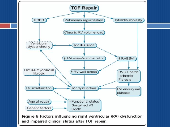
TOF- post repair. Pathophysiology of repaired TOF. Post repair survival. Early outcome determinants. Long term outcome determinants and evaluation. Impact of pulmonary regurgitation and requirement of PVR. Arrhythmias and sudden death.
Pathophysiology of repaired TOF
Pulmonary Regurgitation After TOF Repair Relief of RVOT obstruction in TOF often involves disruption of pulmonary valve integrity, which leads to PR in the majority patients. Inevitable consequence of TAP and/or pulmonary valvotomy.
Determinants of the degree of pulmonary regurgitation (1) Regurgitation orifice area (ROA) (2) RV compliance (3) Diastolic pressure difference between the main pulmonary artery (MPA) and the RV (4) Capacitance of the pulmonary arteries (5) Duration of diastole (6) Pulmonary vascular resistance (7) LV function
Pulmonary regurgitation Immediate postop TOF: despite a relatively large ROA, hypertrophic RV and low RV compliance, PA are hypoplatic or their diameters low-normal(low capacitance of PA), relatively high HR (relative short duration of diastole) => minimized the impact of PR.
RV mechanics after TOF repair Myocardium: a relatively thin compact layer + a prominent layer of trabeculations. The orientation of the myofibers in the RV -more horizontal and contraction is predominantly from base-to-apex (longitudinal) with a lower degree of angular motion (twist). Supplied by a single coronary artery with ~50% of the flow occurring during diastole as oppose to ~90% in the LV. Conduction system in the RV comprises a single fascicle with a long course and a long delay in activation between the base and the distal infundibular free wall, resulting in peristalsis like motion Although RV function impacts LV function, the reverse is much more pronounced with 63% of RV pressure rise accounted for by LV contraction. Geva Journal of Cardiovascular Magnetic Resonance 2011, 13: 9
RV-LV Interaction After TOF Repair The alterations in the size and function of the RV lead to LV dysfunction, a phenomenon termed ‘reversed Bernheim effect’. They share myofibers, septum, coronary blood flow and pericardial space. Abnormal coronary artery, prolonged periods of deep cyanosis, LV volume overload after palliative shunts – causes LV hypoxic/ischemic damage. 3 independent predictors of LVEF 24 yrs post repair : (Davlouros et al) � 1. RVEF � 2. Duration of palliative prerepair � 3. Aortic regurgitation
Survival after TOF repair
Post TOF survival Actuarial survival of 105 patients after repair of TOF Annals of Surgery. 204(4): 490, October 1986
Murphy JG et al. Long-term outcome in patients undergoing surgical repair of tetralogy of Fallot. N Engl J Med 1993
Post surgery outcome – Age and procedure (TAP) related outcome
Early outcome of TOF Repair
Early outcome of TOF Repair ( pre op. factors) ICR- Early mortality – Risk factors Depends on era of Sx. v Age – unfavorable < 3 months and > 20 yrs v Associated Lesions- Pulmonary atresia, abnormal PA anatomy, multiple VSD, abnormal coronaries v Prior AP shunts (due to PAH and PA distortion) v Hypoplastic RVOT and PA. v High hematocrit (reflecting prolonged hypoxia) v
JACC Vol. 30, No. 5 November 1, 1997: 1374– 83
Residual or unrecognized additional VSD q. Even small 3 -4 mm residual VSDs -poorly tolerated because of ass. PR, non compliant RV and unprepared LV for volume overload. q. Results in high filling pressures. q. PA saturation (>80%) Residual RVOT Obstruction Ø Inadequate relief of subpulmonary obstruction or an obstructed or restrictive pulmonary vascular bed (small PAs). Ø Well tolerated immediate post op Ø Present with murmur and raised RV pressure Ø In long term is associated with RV dysfunction, arrhythmia and need for re operation.
Ventricular dysfunction- restrictive physiology Low cardiac output (C. O. ) � RV systolic dysfunction due to post op stunning (esp. if ventriculotomy is done). � severe PR. Manifests as elevated CVP, Hepatomegaly, edema and pleural effusions. Usually recovers in 3 - 5 DAYS. Treatment§ § Drugs- diuretics, digoxin, ionotropes and ionodilators. Extended Ventilation
RV diastolic dysfunction Antegrade late diastolic flow in the PA → atrial contraction is transmitted to the PA → the stiff RV acts as a passive conduit with little or no true RV filling during the late diastole. -Low cardiac output syndrome - Related to the degree of myocardial damage during repair - Inversely related to age at operation - Independent of type of outflow tract repair.
Doppler examination of pulmonary arterial flow in a patient with restrictive right ventricular physiology.
ECG-GATED CINE PHASE CONTRAST MR restrictive RV physiology
Electrophysiological abnormalities Brady arrhythmias q Complete heart block (CHB) ü Usually transient, requires pacing if hemodynamically unstable. Bifascicular block- 8 -12% q RBBB- almost all cases of ventriculotomy q Tachy arrhythmias � JETü AV dissociation with JR of 200 -300/min. If hemodynamically unstable, requires treatment (Amiodarone, overdrive pacing, cooling, correct acidosis, electrolytes) � Rarely VT
Complete heart block The incidence of surgically acquired CHB occurred in 10% in pts operated between 1954 -55. (Lillehei et al) Incidence came down to 0. 6 -1. 3% according to recent trials. Knowledge about the course of conduction tissue and its relationship to VSD is crucial in reducing this complication. Transient CHB persisting beyond 3 rd POD- strongly correlated with sudden death. Hokanson JS, Moller JH. Significance of early transient complete heart block as a predictor of sudden death late after operative correction of
Late Outcomes after TOF repair
Late Outcomes after TOF repair Residual RVOT obstruction at several levels VSD: Swiss cheese/ multiple RV diastolic dysfunction Sequelae RV/LV dysfunction Pulmonary regurgitation Tricuspid regurgitation Arrhythmias Atrial: Atrial Fl/F Ventricular: VT
JACC Vol. 30, No. 5 November 1, 1997: 1374– 83
Echocardiography in adults with TOF Assessment of physiologic and hemodynamic parameters that influence outcome � 1. RV and LV size and function � 2. Pulmonary regurgitation and / or stenosis � 3. Tricuspid regurgitation Assessment of anatomic criteria of unknown significance on outcomes : RVOT aneurysm, aortic dilatation and aortic regurgitation
Role of CMR Quantitative assessment of LV and RV volumes, mass, SV and EF. Evaluation of regional wall motion abnormalities. Imaging the anatomy of the RVOT, pulmonary arteries, aorta and aorto-pulmonary collaterals. Quantification of PR, tricuspid regurgitation, cardiac output and pulmonary-to-systemic flow ratio. Assessment of myocardial viability with particular attention to scar tissue in the ventricular myocardium.
Pulmonary regurgitation Shinebourne and Anderson(Paediatric Cardiology, 2002)-60% to 90% of repaired Fallot pts have some degree of pulmonary regurgitation. Worse late outcome� Late age of repair � had undergone a large right ventriculotomy, excision of extensive muscular trabeculae and a large TAP.
Pulmonary regurgitation Significant PR � Right ventricular dilatation � Impair right ventricular performance � Tricuspid regurgitation � Predispose to atrial flutter/fibrillation, ventricular arrhythmias and sudden cardiac death. Restrictive RV diastolic physiology – � delay/inhibit progressive right ventricular dilatation and dysfunction � by reducing the amount of pulmonary regurgitation.
Evaluation of PR: PR grade by 2 D Echo J Am Soc Echocardiogr 2003; 16: 777 -802
Echocardiographic Assessment of PR. The ratio of jet width / RV outflow diameter (measured at valve level): mild ≤ 1/3 � moderate 1/3 -2/3 � severe ≥ 2/3 � Ratio of duration of PR/ duration of diastole < 0. 77 correlates with PR regurgitant fraction > 24. 5% by CMR Pressure half time <100 ms correlates with hemodynamically significant PR Presence of diastolic flow reversal in branch
Color flow and CW Doppler
PR severity (CW Doppler) PR duration: from the onset in early diastole to the end of the PR Doppler signal Total diastolic time: measured from the end of forward pul flow (coinciding with the onset of the retrograde PR flow) to the beginning of the next forward pulmonary flow curve The ratio btw duration of PR and total diastolic time = PR index (Pri) � Mild : through diastole � Moderate: late diastole � Severe: mid-diastole or earlier <0. 77 : significant PR Am Heart J 2004; 147: 165– 172
Pressure half-time / PR J Am Soc Echocardiogr 2003; 16: 1057– 1062 PHT < 100 ms : significant PR
Quantifying Pulmonary Regurgitation in repaired TOF ØPW in MPA ØThe ratio of diastolic / systolic time velocity integral (DSTVI)- >0. 72 =RF>40% Circ Cardiovasc Imaging 2012; 5; 637 -643
CMR-Quantification of PR: two distinct method Phase contrast (PC) analysis of flow through the MPA & retrograde flow � Indexed PR volume (m. L/m 2) and PR fraction. Ventricular stroke volume (SV) differential measurements derived from steady-state freeprecession(SSFP)cine imaging � Indexed PR volume (RVSV –LVSV) (m. L/m 2) � PR fraction (RVSV –LVSV / RVSV x 100 %) European Heart Journal (2009) 30, 356–
ECG-GATED CINE PHASE CONTRAST MR
MRI-PR assessment European Heart Journal (2009) 30, 356– 361 PR volume and PR fraction are not interchangable. PR volume may be a more accurate reflection of RV preload and may better represent physiologically significant PR as compared with PR fraction.
Timing and indications of PVR Most of the symptomatic c/c severe PR pts referred for PVR- have markedly dilated RV with RV/LV dysfunction. Most of the pts with RV EDV<150 ml/m 2, RV ESV <82 ml/m 2, RVEF>48%- RV size returned to normal 1 yr post PVR. The timing and indications for PVR- must balance the benefits of elimination of RV volume load before irreversible dysfunction occurs and the disadvantages of a premature surgical or transcatheter procedure.
Indications for Pulmonary Valve Replacement Moderate or severe pulmonary regurgitation (regurgitation fraction ≥ 25%) Asymptomatic patient with two or more of the following criteria � RV end-diastolic volume index >150 ml/m 2 or Z-score >4. (In patients whose body surface area falls outside published normal data: RV/LV EDV ratio >2 ) RV ESV index >80 ml/m 2 � RV EF <47% � LV EF <55% � Large RVOT aneurysm � QRS duration >140 ms � Sustained tachyarrhythmia related to right heart volume load � Other hemodynamically significant abnormalities: RVOTO, severe branch PS, moderate TR, residual L->R shunt (Qp/Qs ≥ 1. 5), severe AR, Severe aortic dilatation (diameter ≥ 5 cm) �
Indications for Pulmonary Valve Replacement Symptomatic patients: � Symptoms and signs attributable to severe RV volume load documented by CMR or alternative imaging modality, fulfilling ≥ 1 of the quantitative criteria detailed above. 1. 2. 3. Exercise intolerance not explained by extracardiac causes with documentation by exercise testing (≤ 70% predicted peak VO 2 for age and gender). Signs and symptoms of heart failure. Syncope attributable arrhythmia. Geva Journal ofto Cardiovascular Magnetic Resonance 2011, 13: 9
Indications for Pulmonary Valve Replacement Special considerations: a. Due to higher risk of adverse clinical outcomes in patients who underwent TOF repair at age ≥ 3 years , PVR may be considered if fulfill ≥ 1 of the quantitative criteria. b. Women with severe PR and RV dilatation and/or dysfunction may be at risk for pregnancy-related complications. Geva Journal of Cardiovascular Magnetic Resonance 2011, 13: 9
PVR after TOF repair
Survival after Pulmonary valve replacement The actuarial survival: 95% ± 3% at 10 years 87% ± 8% at 15 yrs Yemets et al; Ann Thoracic Surg 1997; 64: 526 -530
Benefits of PVR Highly effective in eliminating or significantly reducing PR. Significant improvement in NYHA functional class. RV EDV and ESV reduce by 30 -40% as compared with preop values. Degree of TR tends to improve. Therrien et al reported incidence of VT lower after PVR (9% vs 23%). Data regarding effects of PVR on QRSd, objective exercise parameters- inconsistent.
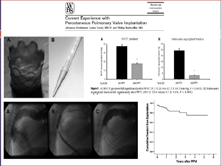
Transcatheter PVI First reported by Bonhoeffer et al, Lancet. 2000; 356: 1403– 1405 � Valved segment of bovine jugular vein sewn within a balloonexpandable stent Melody valve A bovine jugular vein valve sutured within a platinum iridium stent. � One size valve (18 mm) that is crimped to 6 mm and reexpanded from 18 mm to 22 mm. � Thin, compliant leaflets open fully and close readily with a minimum of pressure. � Balloon-in-balloon catheter delivery system with a retractable PTFE sheath covering. � Nylon inner and outer balloons available in three sizes: 18 mm, 20 mm and 22 mm. � At inflation, the inner balloon is half the diameter of the outer balloon. �
Transcatheter PVI Provide new non-surgical option for the treatment of failed bioprosthetic pulmonary valve. Currently limited to mostly patients with RV-topulmonary artery conduits. (size and geometry of RVOT). Further development of this technology may reduce the need for reoperation after pulmonary valve implantation.
Indications for Transcatheter PV placement � Severe PR with RV dilation / dysfunction in FC I � moderate PR + FC II and above � RVOT gradient > 40 mm Hg for FC I � RVOT gradient > 35 mm Hg + FC II and above
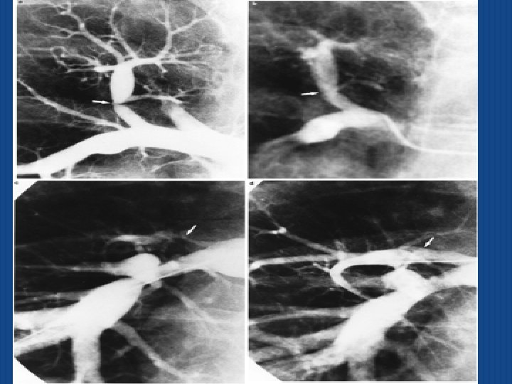
Lateral still-frame PA angiograms showing PT and RVOT before (A) and after (B) PVR Percutaneous implantation of a stented valve within the previous valved conduit.
US Melody valve trial
Branch Pulmonary Artery Angioplasty: Indication Dilation may be considered when RV pressure is more than 50% of the systemic level or at lower pressure when there is RV dysfunction. It may also be considered when there is unbalanced pulmonary blood flow (eg: 75% and 25%), or unexplained dyspnea with severe vascular stenosis and evidence of segmental hypo perfusion by radionuclide studies
Post TOF repair complications : Sudden Death and Arrhythmias Risk of sudden death – 0. 5% to 6% Predictors of Sudden death: q. Severe PR q. Younger age at the time of repair q. R V dilatation, outflow tract aneurysms q. History of sustained VT q. QRS duration >180 ms q. Moderate or severe LV dysfunction
Mechanoelectrical interactions Chronic PR- direct mechanical effects+ electrical deterioration of heart. (both are pathophysiologically linked). QRSd ≥ 180 msec- sensitive & specific predictor for later symptomatic VT and/or sudden death. Rate of QRSd progression(>5 msec/yr over 5 yrs)predicts sudden cardiac death. QRSd ≥ 180 ms was 35% sensitive and 97% specific for induced sustained monomorphic ventricular tachycardia. 2. Balaji S. QRS prolongation is associated with inducible ventricular tachycardia after 1. Gatzoulis MA et al; risk factors for arrhythmia and sudden cardiac death late after repair of tetralogy of Fallot. Am J Cardiol 1997 TOF , multicentre study; Lancet 2000.
Nonsustained ventricular arrhythmia on ambulatory electrocardiographic recordings did not predict sudden death. Cullen et al; J Am Coll Cardiol 1994 Vigorous pharmacotherapy of VT not associated with reduced risk of sudden death. saul et al Right atrial approach to repair of tetralogy of Fallot significantly reduced the risk of life threatening arrhythmias without increasing the risk of supraventricular arrhythmias Dietl CA et al. Life-threatening arrhythmias and RV dysfunction after surgical repair of tetralogy of Fallot. Comparison between transventricular and transatrial approaches. Circulation 1994
Post TOF repair complications : Sudden Death and Arrhythmias Pulmonary Regurgitation: main underlying hemodynamic lesion for patients with sustained VT and SCD Hemodynamic substrate in patients with sustained tachyarrhythmia and SCD late after repair of. Gatzoulis TOF et al, Lancet 2000; 356: 975 -981
Impact of PVR on Arrhythmia Propensity Late After Repair of Tetralogy of Fallot Change in incidence of clinical arrhythmia after PVR. Dashed area represents de novo arrhythmia after PVR Circulation. 2001; 103: 2489 -2494.
Aortic root dilatation Infants with TOF with severe RVOTO are born with dilated ascending aorta( absolute diameter increased as a result of volume overload on developing aorta) Prevalence of > mild AR- 6. 6% of pts 15 yrs post repair. Aortic dissection- rare complication. 88 children post repair(median age 7 months)serial aortic root measurements taken. Annulus and sinotubular z scores returned to normal within 7 yrs. et al. The fate of aortic root after early repair of TOF. Eur J Francois Cardiothorac Surg 2010.
Atrial flutter and fibrillation Associated with older age at repair, a higher frequency of hemodynamic abnormalities and increased morbidity. Among 242 pts with repaired TOF, 29 pts (12%) sustained episodes of serious atrial arrhythmias. The development of atrial arrhythmias was associated with substantial morbidity including congestive heart failure, reoperation, ventricular tachycardia, stroke and death (combined events, 20 of 29 pts; 69%). Patients with atrial arrhythmias were older at surgical repair (25 ± 16 vs. 10 ± 9 yrs, P = 0. 001) and at most recent assessment were aged 48 ± 12 vs. 32 ± 10 yrs (P = 0. 001), as compared with arrhythmia free pts. Harrison DA et al. Sustained atrial arrhythmias in adults late after repair of tetralogy of Fallot. Am J Cardiol 2001; 87: 584– 8.
Infective endocarditis Well known complication post TOF repair. Frequency increases after PVR. Affect aortic valve, tricuspid valve, pulmonary artery, residual VSD, AML in its area contiguous with AV. Morris et al; 30 yr incidence of endocarditis in repaired TOF 1. 3%. Require life long infective endocarditis prophylaxis. Importance of maintaining good oral hygeine.
Contraception and pregnancy Issues reg. genetics, recurrence risk and fetal screening to be discussed. Caution with OCP in women with significant ventricular dysfunction, atrial arrhythmias b/c associated risk of thromboembolism. Pregnancy usually well tolerated. The risks of pregnancy depends on severity of residual lesions, degree of ventricular dysfunction and likelihood of developing arrhythmia.
Procedures for Rerepair of Tetralogy of Fallot in Adults Pulmonary valve replacement Residual subvalvular obstruction or pulmonary artery stenosis Residual VSD closure Sx for Aneurysm of RVOT MAPCAs coiling Atrial and ventricular arrhythmia- RFA or surgery. Tricuspid valve repair for significant tricuspid regurgitation AVR for aortic regurgitation Closure of residual PFO or ASD, if any. Replacement of ascending aorta for dilatation(very rare).
MCQs
First TOF repair by an open heart procedure done by A. B. C. D. Kirklin and wallace, 1946 Lillehei and Varco, 1954 Murphy and Mair, 1956 Parry and Mc. Elhinney, 1958
According to ACC/AHA 2008 guidelines, echocardiographic assessment should be done in TOF repaired pts at intervals of A. B. C. D. 6 months One yr. Two yrs Three yrs
All are indications for Pulmonary Valve Replacement in aymptomatic post TOF repair pts, except A. B. C. D. RV end-diastolic volume index >150 ml/m 2 RV ESV index >80 ml/m 2 regurgitation fraction ≥ 40% RV EF <47%
Commonest arrhythmia in early post op period A. B. C. D. Atrial fibrillation Junctional ectopic tachycardia. Ventricular tachycardia CHB
Commonest cause of mortality in late post TOF repair A. B. C. D. VT CCF SCD SVT
Pri- PR index A. B. C. D. The ratio of diastolic / systolic time velocity integral The ratio duration of PR / total diastolic time Regurgitant jet width / RVOT diameter None of these
Significant PR by echocardiography indicated by all except A. B. C. D. The ratio of jet width / RV outflow diameter ≥ 2/3 PHT >100 ms. PR index <0. 77. Presence of diastolic flow reversal in branch pulmonary arteries.
All are determinants of degree of PR, except A. B. C. D. E. RV compliance Diastolic time duration Degree of Tricuspid regurgitation Capacitance of the pulmonary arteries LV function
Which of the following statement is incorrect A. B. C. D. Transient CHB beyond 72 hrs postop among TOF repaired pts, strongly correlated with SCD. NSVT in Holter monitoring predict sudden death in post TOF repair pts. LV systolic dysfunction and QRSd >180 ms predicts sudden death in TOF repaired pts. Transatrial approach for TOF repair reduces the risk of late life threatening arrhythmias.
THANK YOU
3 D Echo in TOF The mean RV EF were 42 ± 8% on 3 D ultrasound and 44 ± 7% on MRI (r = 0. 89, P <. 0001). The mean EDV were 249 ± 66 and 274 ± 82 m. L and the mean ESV 147 ± 50 and 159 ± 60 m. L on 3 D ultrasound and MRI, respectively. JASE 2010 Feb; 23(2): 12733. Three-dimensional echocardiography underestimated ESV and EDV (P < 0. 001) but agreement between 3 DE and MRI was excellent (ICC = 0. 88 and 0. 87, respectively). Ejection fraction was 47. 7 +/- 7. 8 with 3 DE and 47. 9 +/- 6. 7 with MRI, agreement between both methods was good (ICC = 0. 72). Eur J Echocardiogr. 2009 Aug; 10(6): 784 -92 However, the accuracy of 3 DE echo diminishes with larger RV volumes, in part due to current difficulty to include the entire RV in the imaged sector.