Positioning difficulties encountered in periapical radiography Placing the


- Slides: 21
Positioning difficulties encountered in periapical radiography
Placing the film packet in the positions is not always possible, so many difficulties you may face it during taking radiograph, the main difficulties encountered involve.
Problems of the edentulous ridge: Patients who are missing some or all of their teeth may present some difficulties for the radiographer. Decreased alveolar bone height and missing or migrated teeth can make receptor placement and retention less than ideal. Possible solution In partially dentate patients, the parallel technique can be used. If the edentulous area causes the film holder to be displaced; the deficiency can be built up by using cotton woll rolls.
In edentulous patients , the lack of height in the palate or loss of lingual sulcus depth contraindications the parallel technique and all periapical radiographs should be taken using a modified bisecting angle technique. The long axis of the film and the alveolar ridge are assessed and the x-ray tube head is perpendicular to the bisector.
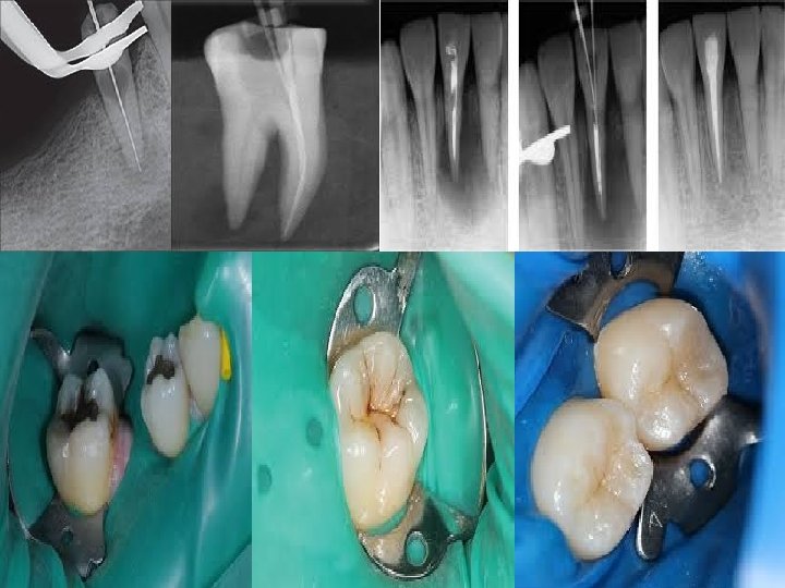
Problems encountered during endodontics The main difficulties involve: 1 - Film packet placement and stabilization when endodontic instruments, rubber dam and rubber dam clamps are in position 2 - Identification and separation of root canals. 3 - Assessing root canal lenghts from foreshortened or elongated radiograph.
Possible soluations 1 - Using a simple film holder which is positioned in the mouth and then held in place by the patient. 2 - The problem of identifying and separating the root canals lenghts can be solved by: -Taking an accurate paralleling technique and measuring the lenghts of the root directly from the radiograph. -- Calculating mathematically the actual length of root canal from a distorted bisecting angle technique , by this formula: Radiographic tooth length x Actual instrument length -Actual tooth length =……………………………………………. Radiographic instrument length
Patient with neuromuscular problem: Patient unable to remain immobile during intraoral film placement Possible soluations 1 - Use parallel technique 2 - Decrease exposure time , by increase k. Vp, increase m. A, and decrease tube –object distance and use of fast film. 3 - Give sedation 4 - Use extra oral film , the film of choice is oblique lateral radiograph , while panoramic radiograph is not recommended in this condition, because it need the patient to remain stabile for 15 sec.
Problems of gagging: The gag reflex is particularly strong in some patients. This makes the placement of the film packet in the desired position particularly difficult, especially in the upper and lower molar regions. Possible solutions 1 - Swabbing the soft palate with topical anesthesia 2 - Placing the film packet flat in the mouth , so it dose not touch the palate 3 - Avoid hot weather and direct the fan toward the patient face 4 - Asking the patient to concentrate on breathing deeply while the film packet is in the mouth.
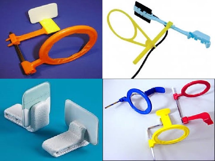
Problems posed by mandibular third molars The main difficulty is placement of the film packet sufficiently posteriorly to record the entire third mandibular molar (particularly when it is horizontally impacted) Possible solutions 1 - Using specially holders to hold and position the film in position, as follows: a- The holder is clipped on the top edge of the film packet.
b- With mouth open , the film packet is positioned gently in the lingual sulcus as far posteriorly as possible c- The patient is asked to close the mouth ( to relaxing the tissue of the floor of the mouth ) and at the same time the film packet is eased further back into the mouth , if required d- The patient is asked to bite on the holder and to to support it in position. e- The x-ray tubehead is positioned at right angle to the third molar and the film packet , and centered 1 cm up from the lower border of the mandible , on a vertical line dropped from the outer corner of the eye
2 - Taking 2 radiographs of the third molar using 2 different horizontal tubehead angulations, as a- The film is positioned as posteriorly as possible using the holder b- The x-ray tube head is positioned with the ideal horizontal angulation, the x-ray beam passes between the second and the third molars ( when the third molar horizontally impacted, the apex may not be recorded c- The second film packet is placed in the same position as before , but the x-ray tube head is positioned further posteriorly aiming forwards to project the apex of the third molar on the film
The vertical angulation of the x-ray is the same for both projections Radiographic surveys Survey : this terminology is used to describe a collection of periapical radiogrphs showing the full dentition , not every tooth is radiographed individually , but enough films are taken to include all the teeth
Survey of adult: Minimum of 14 periapical film size 1. 2 for all teeth (2 posteriorly for each quadrant and 6 anteriorly , 3 for upper and 3 for lower) Maximum of 17 periapical film size 1. 1 for anterior and size 1. 2 for posterior (5 films for upper anterior and 4 films for lower anterior + 2 films for each quadrant posteriorly) If you can use posterior bitewing films : minimum 2 posterior bitewing, one for each side Maximum using 4 posterior bitewing , two for each side
Survey of children: A minimum of 10 periapical films , size 1. 1 (anteriorly 6 films , 3 films for upper and 3 films for lower – posteriorly , one film for each quadrant) -From age 10 -12 year survey is basically identical to that of adults Survey of edentulous: -14 periapical films size 1. 2 (anteriorly 6 films for anterior region , posteriorly 2 films for each quadrant)
THANK YOU