Porphyrias Introduction The porphyrias are caused by deficiencies

Porphyrias

Introduction -The porphyrias are caused by deficiencies of enzymes involved in heme biosynthesis which lead to blockade of the porphyrin pathway and subsequent accumulation of porphyrins and their precursors. - Either genetic (autosomal dominant, autosomal recessive and X-linked) or acquired. - Heterozygotes are asymptomatic in between acute attacks. - Classified depending on site of overproduction and accumulation of porphyrin, overlapping features common Hepatic Erythropoietic ↓ ↓ - Neurologic, mental disturbances - Cutaneous photosensitivity - Abdominal pain - Extremity pain, paresthesias - Motor neuropathy (long wave UV) - light excites porphyrins in skins causing: 1 - Cell damage 2 - Hemolytic anemia

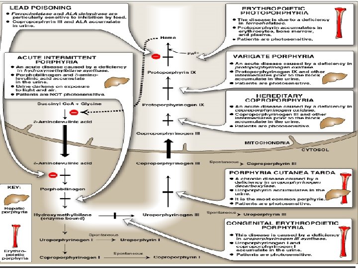
Heme Synthesis Pathway

Classification of the Porphyrias - Multiple ways to categorize porphyrias: - Hepatic vs. Erythropoietic: organ in which accumulation of porphyrins and their precursors appears - Cutaneous vs. Non- cutaneous - Acute and chronic forms - Acute: - ALA dehydratase deficiency porphyria (ALAD) - Acute intermittent porphyria (AIP) - Hereditary coproporphyria (HCP) - Variegate porphyria (VP) - Chronic: - Porphyria cutanea tarda (PCT) - Erythropoietic protoporphyria (EPP) - Congenital erythropoietic porphyria (CEP) - Hepatoerythropoietic porphyria (HEP)

Porphyria categories A- Bone Marrow - Erythropoietic protoporphyria - Congenital erythropoietic porphyria B- Liver - Porphyria cutanea tarda - Acute intermittent porphyria - Variegate porphyria - Hereditary coproporphyria - Hepatoerythropoietic porphyria

Overview of the four acute porphyrias - Four acute porphyrias cause acute, self-limiting attacks that lead to chronic and progressive deficits - Symptoms of acute attacks increase the potential for misdiagnosis. - Acute porphyrias are clinically indistinguishable during acute attacks, except the neurocutaneous porphyrias (variegate porphyria and hereditary coproporphyria) can cause dermatologic changes - Acute attacks lead to an increase in PBG and ALA which can be detected in urine - Diagnosis is difficult because of variable clinic course, lack of understanding about diagnostic process, and lack of a universal standard for test result interpretation

- Cutaneous features are not seen in acute intermittent porphyria or the very rare ALA dehydratase deficient porphyria. - Erythropoietic protoporphyria and congenital erythropoietic porphyria are characterized by porphyrins produced mainly in the bone marrow. - The reminder are primarily hepatic porhyrias. - Excessive concentrations of porphyrins exposed to day-light generate free radicals, leading to cell membrane damage and cell death. - The type of cellular damage depends on the solubility and tissue distribution of the porphyrins. - Two main patterns of skin damage are seen in the porphyrias: 1 - accumulation of water soluble uro - and coproporphyrins leads to blistering. 2 - accumulation of the lipophilic protoporphyrins leads to burning sensations in the exposed skin.

Category Type Clinical presentation Inheritance Hepatic ALA dehydratase deficiency Acute attacks Autosomal recessive Acute intermittent porphyria Acute attacks Autosomal dominant Porphyria cutanea tarda Skin disease Usually acquired; a minority are inherited (autosomal dominant) Skin disease, acute attacks Autosomal dominant Variegate porphyria Skin disease, acute attacks Autosomal dominant Congenital erythropoietic porphyria Skin disease Autosomal recessive Erythropoietic protoporphyria Skin disease: specific presentation with immediate photosensitivity Autosomal dominant: severe forms have complex inheritance Hereditary coproporphyria Erythropoietic

Diagnosis - Overlapping, may be difficult to determine exactly - Check plasma, urine, stool porphyrin excretion Porphyria Symptoms Diagnostic findings U= Urine, F=Feces, E=Erythrocytes ALA dehydratase deficiency Neurovisceral ↑ ALA (U) Acute intermittent porphyria Neurovisceral ↑ ALA and PBG (U) Congenital erythropoietic porphyria Photocutaneous ↑ uroporphyrin I and coproporphyrin I (U & E) Porphyria cutanea tarda Photocutaneous ↑ 7 - carboxylate porphyrin (U) and isocoproporphyrin (F) Hereditary coproporphyria Photocutaneous ↑ ALA, PBG and coproporphyrin (U) and neurovisceral and coproporphyrin (F) Variegate porphyria Photocutaneous ↑ ALA, PBG (U) and protoporphyrin and neurovisceral (F) Erythropoietic protoporphyria Photocutaneous ↑ protoporphyrin (F & E) and in plasma

Acute intermittent porphyria - Prevalence of 5 -10 per 100, 000 and thought to be higher in psychiatric populations - More frequent in women than men. - Heterozygotes are asymptomatic between acute attacks. - Risk factors for exacerbation include medications, diet, weight loss, surgery, infection, menstrual hormones, smoking - Common symptoms include: - Abdominal pain. - Tachycardia, arrhythmia. - Orthostatic hypotension. - Psychiatric symptoms including anxiety, depression, hallucinations and paranoia - Peripheral neuropathy Diagnosis: Caused by a deficiency of PBG deaminase resulting in an accumulation of PBG and ALA Treatment: - Discontinue all unnecessary or potentially harmful drugs as Sulfa drugs, barbiturates, ACEI, Antiepileptics and Antifungals - Treat any infection. - Pain control with Morphine - Treat sympathetic hyperactivity with propranolol. - 300 -400 grams of carbohydrates per day. - IV heme at 3 -5 mg/kg/day.

Porphyria cutanea tarda - Most common porphyria which causes skin manifestations - Deficiency of hepatic urodecarboxylase - Cutaneous photosensitivity → fluid filled vesicles on sun exposed areas, friable skin, wounds heal slowly and hyperpigmentation on face - No neurologic manifestations - Higher incidence of hepatocellular carcinoma - Precipitants frequently include alcohol, estrogen and iron Treatment: - Avoid sunlight, use sunscreen - Chloroquine or hydroxychloroquine to form complexes with porphyrins to enhance excretion - Superactivated charcoal - β- carotene may increase tolerance of sunlight through Vitamin A.

Erythropoietic protoporphyria - It is the most common childhood porphyria. - It is usually evident by 2 years of age. - Protoporphyrin levels are elevated because of deficient activity of ferrochelatase enzyme. Congenital erythropoietic porphyria (Gunther's disease) - It is a very rare autosomal recessive disorder. - Patients usually present during infancy and rarely present in adult life with milder forms. - It is caused by elevation of both water-soluble and lipid-soluble porphyrin levels due to deficiency of uroporphyrinogen III synthase enzyme. Clinical features -Very severe photosensitivity with phototoxic burning and blistering leading to burning sensation in the light exposed parts. - Hypersplenism. - Hemolytic anemia. - Thrombocytopenia Treatment - Superactivated charcoal - Hypertransfusion - Splenectomy - Bone marrow transplantation

Pseudoporphyria - In certain settings patient develop blistering and skin fragility identical to PCT with the histological features but with normal urine and serum porphyrins. -This condition called → pseudoporphyria. - Most commonly due to medications especially NSAIDs and tetracycline. - Some patients on hemodyalisis develop a similar PCT-like picture. Neurotoxicity mechanisms - Most current thinking focuses on accumulations of toxic metabolites. - ALA and PBG are neurotoxins. - ALA may be a false transmitter for GABA, it also blocks one of ATPases (perhaps a sodium pump). - Another hypothesis: unsaturation of hepatic tryptophan pyrrolase secondary to liver heme deficiency leads to altered tryptophan delivery to CNS → ↑ tryptophan excretion.
- Slides: 14