POLYTRAUMA Objectives Approach to Multiple Injurepolytrauma Patients Learn

POLYTRAUMA
 Patients • Learn to diagnose, start initial management and")
Objectives: Approach to Multiple Injure(polytrauma) Patients • Learn to diagnose, start initial management and know when to refer a patient with a condition that requires urgent specialist management • Implement Management as per Advanced trauma life support(ATLS) protocol • Learn about in-line immobilization of cervical spine, in the context of managing the airway • Understand the function of spinal board as a transfer tool only • Review emergent orthopedic conditions that are critical, and their initial management; e. g. open book pelvis fracture, bilateral femur fractures, mangled extremity • Promote the Importance of interpersonal communication skills
 Multisystem trauma Terminology: • Injury = the result of a harmful event that")
(Polytrauma) Multisystem trauma Terminology: • Injury = the result of a harmful event that arises from the release of specific forms of energy • “polytrauma” = Multisystem trauma = injury of two or more systems, or system with dereanged vital signs

INTRODUCTION UK - > 18, 000 deaths annually. > 60, 000 hospital admission. > Costing 2. 2 billion pounds. USA - > 120, 000 deaths annually. > 100 billion dollars.

MECHANISMS OF INJURY Types of injury • Penetrating • blunt • Blast • Thermal • Chemical • Others - crush & barotrauma.
 0 to 1 hr Early death (30%)")
TRIMODAL DISTRIBUTION OF DEATH Immediate death (50%) 0 to 1 hr Early death (30%) 1 to 3 hrs Late death ( 20%) 1 to 6 wks Golden Hour

Trauma deaths First peak • Within minutes of injury • Due to major neurological or vascular injury • Medical treatment can rarely improve outcome (die on scene) Second peak • Occurs during the 'golden hour' • Due to intracranial haematoma, major thoracic or abdominal injury • Primary focus of intervention for the Advanced Trauma Life Support (ATLS) methodology Third peak • Occurs after days or weeks • Due to sepsis and multiple organ failure
 PHILOSOPHY Treat lethal injuries first Reassess Treat again/Transfer")
ADVANCED TRAUMA LIFE SUPPORT (ATLS) PHILOSOPHY Treat lethal injuries first Reassess Treat again/Transfer

PREHOSPITAL RETRIEVAL & MANAGEMENT AIMS Gain access to the patient Smooth transfer APPROACHES ‘’Scoop & Run policy’’ ‘’Stay & Play policy’’

ATLS – COMPONENT STEPS Primary survey Identify what is killing the patient. Resuscitation Treat what is killing the patient. Secondary survey Proceed to identify other injuries. Definitive care Develop a definitive management plan.

ORGANISATION OF TRAUMA CENTRES LEVEL 1 – REGIONAL TRAUMA CENTRES LEVEL 2 – COMMUNITY TRAUMA CENTRES LEVEL 3 – RURAL TRAUMA CENTRES

MANAGEMENT IN HOSPITAL THE TRAUMA TEAM comprised as per hospital policy for eg initially of 4 Doctors At least 1 Anaesthetist 1 Orthopaedician 1 General surgeon 5 Nurses 1 Radiographer

LEADER OF THE TRAUMA TEAM Most experienced Preferably a general surgeon Takes all TRIAGE decisions Should be familiar with each members’ skills Prioritize procedures Communicate with consultants & family members

Multiple casualties • Several causalities at the same time. 1. Alert ER services 2. Assess the scene - without putting your safety at risk 3. Triage 'do the most for the most'
 • Ability to walk • Airway • Respiratory rate • Pulse")
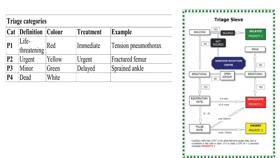
Triage(START triage algorithm) • Ability to walk • Airway • Respiratory rate • Pulse rate or capillary return

1. Can the patient walk? Yes No delayed check for breathing 2. Is the patient breathing? No How to triage? open the airway Are they breathing now? Yes IMMEDIATE No DEAD Yes count the rate <10 & > 30 / min – IMMEDIATE 10 – 30 /min – check circulation 3. Check the circulation Capillary refill> 2 sec- IMMEDIATE Capillary refill < 2 secs - urgent
 – to separate dead & the walking")
TRIAGE SEIVE/SORT algorithm TRIAGE SIEVE(on the field) – to separate dead & the walking from the injured TRIAGE SORT(2 nd step) – to categorize the casualties according to local protocols. Cat 1 : critical & cannot wait. Cat 2 : urgent – can wait for 30 mins at most Cat 3 : less serious injuries. Cat 4 : expectant – survival not likely.

1. Make the area safe • protect yourself, the casualty and other road users • • Park your car safely, turn lights on, set hazard lights flashing Do not across a busy motorway to reach other side Set others to warn other coming drivers Set up warning triangles or lights 200 metres in each direction • Switch off ignition of any damaged vehicle • Is anyone smoking?

2. Check all casualties • quick assess • not moving • apply life-saving treatment

How to move unconscious casualty • do not move the casualty unless it is absolutely necessary • assume neck injury until proved otherwise • support head and neck with your hands, so he can breathe freely Apply a collar, if possible • There should be only 1 axis (head, neck, thorax) no moving to sides, no flexion, no extension. • Move with help of 3 -4 other people 1 support head (he is directing others), other one shoulders and chest, other one hips and abdomen, last one - legs.

Skill Video Demonstations • Applying cervical collar • log rolling and spinal board • Inline Immobilization • spinal clearance
 • Penetrating injuries • Two or more proximal bone")
TRAUMA TEAM CALL-OUT CRITERION(in hospital) • Penetrating injuries • Two or more proximal bone fractures • Flail chest & pulmonary contusion • Evidence of high energy trauma - fall from > 6 ft -changes in velocity of 32 kmph - 35 cm displacement of side wall of car - ejection of the patient - roll-over - death of another person in same car - blast injuries

-Assessment approach to trauma patient in hospital setting. ATLS algorithm Primary survey & resuscitation follows ABCDE sequence Only imaging permitted during this phase is - AP supine chest X-ray - AP plain pelvic film - FAST(focused assessment by ultrasonography) - cross table lateral C- spine X-ray(outdated)

Assessment of the injured patient • Primary survey and resuscitation • • • A = Airway and securing cervical spine B = Breathing C = Circulation and haemorrhage control D = Dysfunction of the central nervous system E = Exposure • Adjunct to primary survey: Xrays , U. S • Secondary survey • Definitive treatment • Consider Early Transfer

Airway and cervical spine • Always assume that patient has cervical spine injury • If patient can talk then he is able to maintain own airway • If airway compromised initially attempt a jaw thrust and clear airway of foreign bodies, suction, adjuncts to open airways. • Remember to avoid causing harm NP tube, nasopharyngeal airway in base skull fracture • Give 100% Oxygen (face mask, bag valve) • Assist airway & breathing including ’’definitive airways’’ (endotracheal tube/cricothyroidotomy)
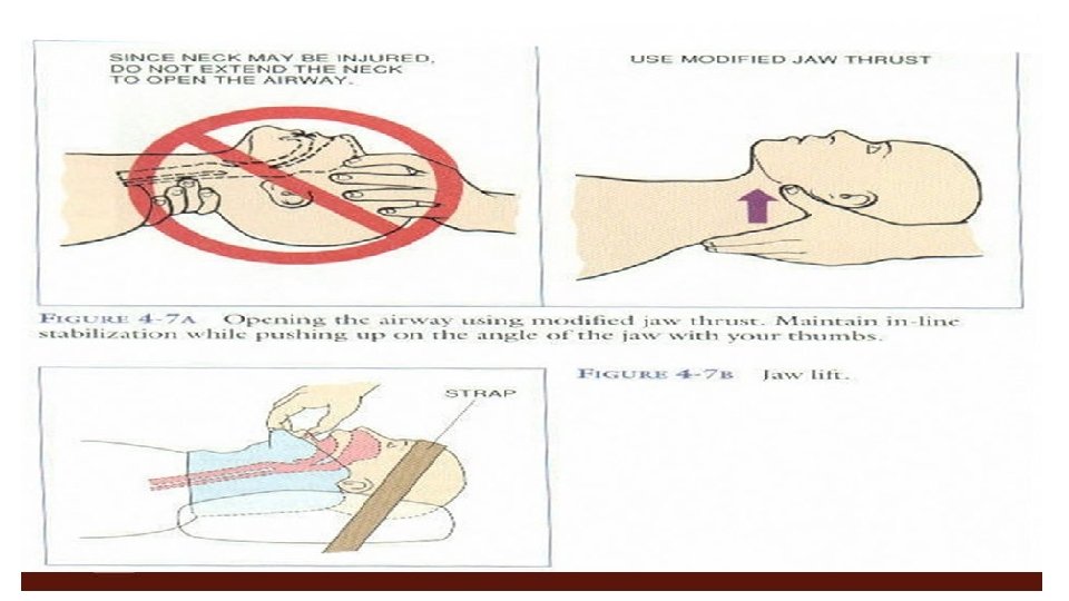

ATLS- PRIMARY SURVEY A – Airway maintenance & Control of C. Spine If conscious- Ask the pt’s name If unconscious-Look for added sounds (stridor, cyanosis etc) If the pt does not respond to any questions- resuscitate.
 Jaw")
ATLS- PRIMARY SURVEY A-AIRWAY Sequence of events: chin lift(if no c spine concern) Jaw thrust suction Oropharyngeal/ orotrachial tube endotracheal intubation Cricothyroidotomy Tracheastomy

ATLS- Primary Survey B- Breathing & ventilation • Exposure • Inspection • Palpation • Movement • Auscultation The aim is to hunt out & treat the life threatening thoracic conditions which include:

ATLS- Primary Survey B- Breathing & ventilation Tension pneumothorax C/F Respiratory distress Tracheal deviation Diminished breath sounds Distended neck veins needle decompression video Immediate needle thoracocentesis thro’ 2 nd intercostal space in mid clavicular line reqd.

ATLS- Primary Survey B- Breathing & ventilation Five life threatening thoracic conditions: 1. Tension Pneumothorax 2. Massive Pneumothorax/heamthorax 3. Open pneumothorax 4. Flail segment 5. Cardiac tamponade

Breathing • If open chest wound seal with occlusive dressing • Definitive treatment for hemopneumothorax will include chest tube placement • https: //chest Tube insertion

ATLS- Primary Survey B- Breathing & ventilation Suction pneumothorax: Sealing of the wound and Tube thoracostomy Flail segment: Endotrachial intubation Mechanical ventilation

ATLS- Primary Survey B- Breathing & ventilation Cardiac tamponade (almost always seen with a penetrating wound) Beck’s triad: Hypotension distended neck veins Muffled heart sounds Treatment: needle pericardiocentes Thoracotomy & repair as def managemnt

ATLS- Primary Survey C- Circulation and hge control Adults-consider up to 2 lit of fluids if patient hypotensive, cardiac arrest(until blood available) Children- 20 mg/kg of body wt Response to initial fluid challenge: • Immediate & sustained return of vital signs. • Transient response with later deterioration • No improvement.

Circulation and haemorrhage control • Assess pulse, capillary return and state of neck veins • Identify exsanguinating haemorrhage and apply direct pressure • Place two large calibre intravenous cannulas Give intravenous fluids (crystalloid or colloid) • Attach patient to ECG monitor

ATLS- Primary Survey C- Circulation and hge control Tachycardia in a cold patient indicates shock Causes of shock following injury: 1. Hypovolemic 2. Obstructive 3. Cardiogenic 4. Neurogenic 5. Septic

ATLS- Primary Survey C- Circulation and hge control Assessment of blood loss External or obvious Internal or covert chest abdomen pelvis limbs Resuscitation Arrest bleeding Obtain vascular access

ATLS- Primary Survey C- Circulation and hge control Immediate responders-<20% blood loss Bleeding ceases spontaneously Transient respondersbleeding within body cavities Surgical intervention reqd. Non responders- >40% of blood vol lost require immediate surgery Continued IV fluids detrimental

Classification of Hypovolaemic Shock and Physiologic Changes Class III Class IV Up to 0. 75 -1. 5 -2. 0 >2 15% 30% 40% >40% Pulse rate < 100 >120 >140 Blood pressure Normal Decreased Pulse pressure Normal or inc Decreased 14 -20 20 -30 30 -40 >35 Urine output > 30 ml/hr 20 -30 5 -15 Negligible Mental status Slightly anxious Mildly anxious Anxious/confused Confused/lethargic Crystalloid and blood Crystalloid and Blood loss (liter) % TBV Respiratory rate Fluid Replacement What is your fluid replacement regimen?

ATLS- Primary Survey C- Circulation and hge control Estimation of blood loss

VIDEO: TESTING PELVIC STABILITY

Fluid resuscitation - DEBATE Sho ck due hae to p m orrh rima Firs age ry O t. H it ngo resu ing b l scit Sec atio eeding ond n re 2 O g Hit? ime n Voluminous crystalloid ● dilutes coagulation factors ● causes hyperchloremic and lactate acidosis ● supplies inadequate O 2 to under-perfused tissue Hypothermia Coagulopathy Lethal Triad of Death Acidosis

Current concepts Permissive hypotension Maintain systolic B. P. at 85 - 95 mm of Hg Turn off the tap and do not infuse much of fluid and blood products too

Paradigm Shift in Resuscitation To DCR in 1990 s HD Triage: Stable, Borderline, Unstable and In Extremis C A B C D E DAMAGE CONTROL RESUSCITATION Permissive hypotension and haemorrhage control Haemostatic resuscitation in

Balanced Resuscitation 1. Fluid Replacement in Balanced Resuscitation ● Initial fluid replacement with up to 2 L crystalloid Permissive hypotension to achieve SBP to 80 -90 mm. Hg (radial pulse) until definitive control of bleeding is obtained 2. Haemostatic Resuscitation ● Early blood versus HBOC transfusion decreases MODS ● Packed RBC, FFP and Platelets in 1: 1: 1 ratio ● Cryoprecipitate, Tranexamic acid, Recombinant factor-VIIa ● Storage blood of < 2 weeks to minimize TRALI, MODS

Dysfunction Assess level of consciousness using AVPU method A = alert V = responding to voice P = responding to pain U = unresponsive GCS(glascow coma scale) Assess pupil size, equality and responsiveness

GLASGOW COMA SCALE Eye opening • Spontaneous • To voice • To pain • None Verbal response • Oriented • Confused • Inapp words • Incomp sounds • None 4 3 2 1 5 4 3 2 1 Motor response • Obeys commands 6 • Localises pain 5 • Withdraws 4 • Flexion(pain) 3 • Extension (pain) 2 • None 1 Total 3 -15

Exposure Fully undress patients Avoid hypothermia Hypothermia Prevention and Treatment Strategies ● Limit casualties’ exposure ● Warm IV fluids and blood products before transfusion ● Use forced air warming devices before and after surgery ● Use carbon polymer heating mattress
 • Comprises of head to toe examn of the stable pt")
Secondary survey (ATLS) • Comprises of head to toe examn of the stable pt • Requires Detailed history Thorough examination KEEP MONITORING the vital signs monitoring devices -pulse oximeter -rectal thermometer • Detailed radiographic procedures -C. T. , USG, M. R. I.
 HEAD, ENT • Glasgow coma scale • Reaction and size of")
Secondary survey (ATLS) HEAD, ENT • Glasgow coma scale • Reaction and size of pupils • Plantar response • Signs of rhinorrhoea, otorrhoea(base of skull#) • Nose fracture, septal hematoma
 NECK • Subcut emphysema • Cervical spine fractures (specially C 1,")
Secondary survey (ATLS) NECK • Subcut emphysema • Cervical spine fractures (specially C 1, C 2, C 7) • Penetrating neck injuries
 THORAX Search for potentially life threatening injuries • Pulmonary complication •")
Secondary survey (ATLS) THORAX Search for potentially life threatening injuries • Pulmonary complication • Myocardial contusion • Aortic tear • Diaphragmatic tear • Oesophageal tear • Tracheobronchial tear • Early thoracotomy if initial haemorrhage > 1500 ml
 ABDOMEN • Fingers and tubes in every orifice • Nasogastric and")
Secondary survey (ATLS) ABDOMEN • Fingers and tubes in every orifice • Nasogastric and Urinary catheter for diagnosis and treatment • Rectal exam • Wounds coverage • Eviscerated bowels packed by warm wet mops
 ABDOMEN For rigid and distended abdomen • Ultrasound • Four quadrant")
Secondary survey (ATLS) ABDOMEN For rigid and distended abdomen • Ultrasound • Four quadrant tap • Diagnostic peritoneal lavage • Laparoscopic examination Any deterioration Consider rapid surgical exploration
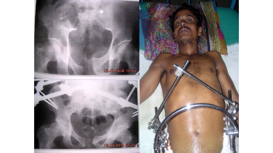
 PELVIS Clinical assessment X-ray stabilize pelvis with fixator/clamps If urethral injury")
Secondary survey (ATLS) PELVIS Clinical assessment X-ray stabilize pelvis with fixator/clamps If urethral injury is suspected—high up prostate in PR blood in meatus Trial catheter perineal haematoma With gentle manipulation t oascending n Fine catheter If urethrogram Lots of lubricants In OT suprapubic cystotomy
 Spinal injury Thorough sensory and motor examination • Prevent further damage")
Secondary survey (ATLS) Spinal injury Thorough sensory and motor examination • Prevent further damage in unstable fractures • Log rolling for full neurological examination-5 people required • Use a long spine board for transportation
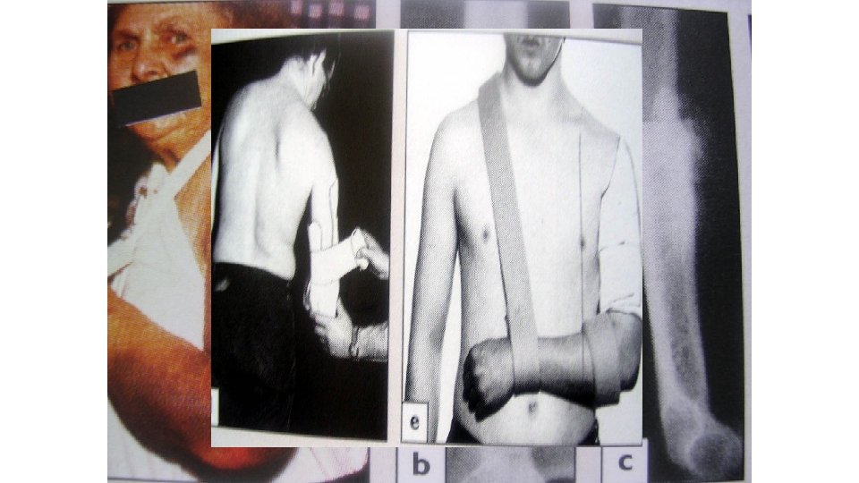
 EXTREMITIES • Full assessment of limbs for assessment of injury •")
Secondary survey (ATLS) EXTREMITIES • Full assessment of limbs for assessment of injury • Always look for distal pulse & neuro-status • Carefully look for skin & soft tissue viability • Look out for impending Compartment syndrome

ATLS-secondary survey F- Fracture management 1. Minor 2. Moderate open # of digits undisplaced long bone or pelvis # 3. Serious 4. Severe closed long bone #s multiple hand/foot #s life threatening open long bone # pelvis # with displacement dislocation of major joints multiple amputations of digits amputation of limbs multiple closed long bone #s

Medication; DON’T FORGET • Tetanus prophylaxis • Anti D immunoglobulin in possible preg female • vasopressor drugs(selective) • Antiobiotics(selective) • Calcium gluconate(selective) • tranexamic acid(TXA)
 Multi-speciality approach ( Inter-disciplinary management ) The most appropriate person in-charge")
Definitive care plan(ATLS) Multi-speciality approach ( Inter-disciplinary management ) The most appropriate person in-charge is the General/trauma surgeon.

Complications • Tetanus • A. R. D. S. • Fat embolism • D. I. C. • Compartment syndrome • Multisystem organ failure (M. S. O. F. )

Complications A. R. D. S. • Tachypnoea • dyspnea • Bilateral infiltrates in CXR Treated with mechanical ‘’low tidal’’ ventilation with PEEP

Complications Fat embolism • Around 72 hours • Tachycardia • Tachypnoea • Dyspnoea • Chest pain • Petechial haemorrhage Treated with ----- mechanical ventilation ------fixation of fractures

Complications Disseminated intravascular coagulation • Follows severe blood loss and sepsis • Restlessness , confusion, neurological dysfunction, skin infarction , oliguria • Excessive bleeding • Prolonged PT, PTT, hypofibrinogenemia Treatment– prevention and early correction and shock, warming fluids, giving less crystalloids

Complications Compartment syndrome/crush syndrome • When a limb remains compressed for many hours/multiple fractures • Increased Compartment pressure and further ischaemia(of limb) • Cardiac arrest due to metabolic changes in blood • Renal failure Treatment • Prevention of renal failure-ensure high urine flow during using IV Crystalloids • Fasciotomy and excision of devitalized muscles • Amputation

Complications M. S. O. F. Progressive and sequential dysfunction of physiological systems Hypermetabolic state It is invariably preceded by a condition known as Systemic Inflammatory Response Syndrome (SIRS) Characterised by two or more of the following • Temperature >38º C or < 36ºC • Tachycardia >90 /min • Respiratory rate >20/min • WBC count >12, 000/cmm or <4, 000/cmm

Complications M. S. O. F. Treatment : Key word is PREVENTION • Prompt stabilisation of fracture • Treatment of shock • Prevention of hypoxia • Excision of all dirty and dead tissue • Early diagnosis and treatment of infection • Nutritional support

Conclusion • Diagnose, prioritize management as per ATLS PROTOCOL • Recognize when to immediately refer a patient that requires urgent specialist management. • Remember A includes in-line immobilization of cervical spine while managing the airway. • Function of spinal board as a transfer tool only • Proper priority to orthopedic conditions affect the patient life/limbs (open book pelvis fracture, bilateral femur fractures, mangled extremity). • Importance of interpersonal and intrapersonal communication skills
- Slides: 72