POLYHYDROMNIOS POLYHYDROMNIOS DEFINITION Anatomically polyhydramnios is defined as


 are associated with polyhydramnios")


 Diabetes (30%) —It is more common in hydramnios. �")
")
 Mild : DVP > 8– 11 cm (b)")


 Early detection and control of diabetes. � (2)")
: � It is commonly found in midtrimester")
: � Principles: � (1) To relieve the")

: � Slow decompression is done at the rate of about")

 II. Malpresentation and persistence")

 The patient looks")
 Internal examination reveals: taking up of the cervix or even")


- Slides: 28
POLYHYDROMNIOS
POLYHYDROMNIOS � DEFINITION: Anatomically, polyhydramnios is defined as a state where liquor amnii exceeds 2, 000 m. L. � Clinical definition : The excessive accumulation of liquor amnii causing discomfort to the patient when amniotic fluid index (AFI) is more than 24 cm (more than 95 th centile for gestational age) and a deepest vertical pocket (DVP) is more than 8 cm. � Incidence: 1 -2%
POLYHYDROMNIOS � It may be the result of deficient absorption as well as excessive production of liquor amnii, which may be temporary or permanent. � The � I. III. IV. V. composition of the liquor amnii, however, remains normal. ETIOLOGY: Idiopathic (50– 60% ) Fetal anomalies Placenta Multiple pregnancy Maternal conditions
ETIOLOGY � FETAL ANOMALIES: Congenital fetal malformations (structural and chromosomal) are associated with polyhydramnios in about 20% cases. � The causes of excessive production of liquor amnii may be due to: (a) transudation from the exposed meninges, (b) absence of fetal swallowing reflex (c) possible suppression of fetal antidiuretic hormone leading to excessive urination.
2. FETAL ANOMALIES: I. Open spina bifida : increased transudation from the meninges. � II. Esophageal or duodenal atresia : preventing swallowing of the liquor. III. �Facial clefts and neck masses : interfering normal swallowing. � IV. Hydrops fetalis : due�to Rhesus isoimmunization, nonimmune hydrops, cardiothoracic anomalies, fetal cirrhosis and fetal infections with TORCH and parvovirus B 19 infection V. Aneuploidy and genetic syndromes
POLYHYDROMNIOS
ETIOLOGY 3. PLACENTA: Chorioangioma of the placenta: Tumor growing from a single villus consisting of hyperplasia of blood vessels and connective tissue results in increased transudation 4. MULTIPLE PREGNANCY: � Hydramnios is more common in monozygotic twins, usually affecting the second sac. � In TTTS the recipient twin develops polydramnios.
ETIOLOGY � 5. MATERNAL: (i) Diabetes (30%) —It is more common in hydramnios. � Rised maternal blood sugar raised fetal blood sugar fetal diuresis hydramnios. (ii) Cardiac or renal disease: may lead to edema of the placenta leading to increase in transudation.
POLYHYDROMNIOS � CLINICAL TYPES: Depending on the rapidity of onset, hydramnios may be: (a) Chronic (most common) : onset is insidious taking few weeks. (b) Acute (extremely rare) : onset is sudden, within few days or may appear acutely on pre-existing chronic variety. The chronic variety is 10 times more common than the acute one.
v Polyhydramnios may be : (a) Mild : DVP > 8– 11 cm (b) Moderate : DVP: 12– 15 cm (c) Severe : DVP ≥ 16 cm
CHRONIC POLYHYDROMNIOS � SYMPTOMS: 1. 2. 3. 4. 5. mainly from mechanical causes. Respiratory : shortness of breath Palpitations Edema of the legs Varicosities in the legs or vulva Hemorrhoids. SIGNS: The patient may be in a dyspneic state in the lying down position. Ø Evidence of preeclampsia (edema, hypertension and proteinuria) may be present. Ø
CHRONIC POLYHYDROMNIOS � ABDOMINAL EXAMINATION: � Inspection: Abdomen is markedly enlarged looks globular with fullness at the flanks skin is tense, shiny with large striae � Palpation: � Height of the uterus is more than the period of amenorrhea. � Girth of the abdomen is more than normal (round the umbilicus) � Fluid thrill can be elicited in all directions over the uterus. � Fetal parts, presentation , position cannot be well-defined � External ballottement can be elicited more easily.
CHRONIC POLYHYDROMNIOS � Auscultation: Fetal heart sound is not heard distinctly, although its presence can be picked up by Doppler ultrasound � Internal Examination: � The cervix is pulled up may be partially taken up � May be dilated, to admit a fingertip through which tense bulged membranes can be felt.
INVESTIGATIONS 1. ABO and Rh grouping : Rhesus isoimmunization may cause hydrops fetalis and fetal ascites. 2. Random blood sugar and if necessary glucose tolerance test 3. Amniotic fluid: Estimation of alpha fetoprotein which is markedly elevated in the presence of a fetus with an open neural tube defect
INVESTIGATIONS 4. Sonography : to detect abnormally large echo-free space between the fetus and the uterine wall a) b) c) d) (largest vertical pocket >8 cm). Amniotic fluid index (AFI) > 25 cm To exclude multiple fetuses To note the lie and presentation of the fetus To diagnose any fetal congenital malformation. (Especially the central nervous system, gastrointestinal system and musculoskeletal system)
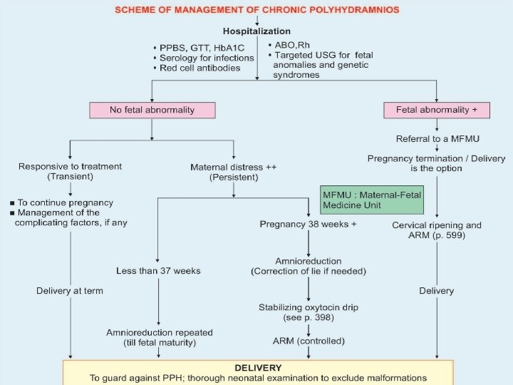
CHRONIC POLYHYDROMNIOS � MANAGEMENT: � (1) Early detection and control of diabetes. � (2) Rhesus isoimmunization is now preventable. � � (3) Genetic counseling in early months and detection of fetal congenital abnormalities with ultrasound and their termination � Treatment cause. of polyhydramnios is usually according to the underlying
� MILD POLYHYDRAMNIOS (DVP: 8– 11 cm): � It is commonly found in midtrimester � usually requires no treatment � The excess liquor is expected to be diminished as pregnancy advances (transient)
� SEVERE POLYHYDRAMNIOS (DVP: ≥ 16 cm): � Principles: � (1) To relieve the symptoms � (2) To find out the cause � (3) To avoid and to deal with the complication.
CHRONIC POLYHYDROMNIOS � Supportive therapy: Bed rest 2. Treatment of the associated conditions like preeclampsia or diabetes on the usual line. 3. The use of diuretic is of little value. 4. Sulindac (COX-2 inhibitor), 200 mg every 12 hours, (under supervision) has been found to be most effective in unexplained cases. � It has been found to decrease amniotic fluid as it reduces fetal urine output. 1.
AMNIOCENTESIS Amniocentesis (amnio reduction): � Slow decompression is done at the rate of about 500 ml/hr and the amount of fluid to be removed should be sufficient enough to relieve the mechanical distress. � Normally amniodrainage is stopped when the AFI is less than 25 cm. � Because of slow decompression, chance of accidental hemorrhage is less but liquor amnii may again accumulate, for which the procedure may have to be repeated. � Amniotic fluid can be tested for fetal lung maturity. �
AMNIOCENTESIS
COMPLICATIONS MATERNAL FETAL 1. During pregnancy : I. Preeclampsia (25%) II. Malpresentation and persistence of floating head III. Premature rupture of the membranes IV. Preterm labor either spontaneous or induced V. Accidental hemorrhage 2. During labor: I. Early rupture of the membranes II. Cord prolapse III. Uterine inertia IV. Increased operative delivery due to malpresentation V. Retained placenta, postpartum hemorrhage (uterine atony) , shock. 3. Puerperium: I. Subinvolution II. Increased puerperal morbidity due to infection resulting from increased operative interference and blood loss 1. prematurity 2. congenital abnormality (40%). 3. Hydrops fetalis 4. Effects of increased operative delivery and accidental hemorrhage
ACUTE POLYHYDROMNIOS � Acute � The � It hydramnios is extremely rare. onset is acute and the fluid accumulates within a few days. usually occurs before 20 weeks of pregnancy. � � It is usually associated with monozygotic twins with TTTS or chorioangioma of the placenta.
ACUTE POLYHYDROMNIOS � SYMPTOMS: Abdominal pain Nausea and vomiting. SIGNS: (i) The patient looks ill (ii) Absence of features of shock (iii) Edema of the legs or presence of other associated features of preeclampsia (iv) Abdomen is hugely enlarged more than the period of amenorrhea; the wall is tense with shiny skin (v) Fluid thrill is present (vi) Fetal parts cannot be felt nor is the fetal heart sound audible
ACUTE POLYHYDROMNIOS � (vii) Internal examination reveals: taking up of the cervix or even dilatation of the os through which the bulged membranes are felt � (viii) Sonography shows multiple fetuses or at times fetal abnormalities
ACUTE POLYHYDROMNIOS �TREATMENT: Most often, spontaneous abortion occurs. � In case with severe TTTS, repetitive amnioreduction until the AFI is normal, may improve the perinatal outcome. � Laser ablation may cure the cause of TTTS whereas amnioreduction only treats the symptoms �
THANKYOU