Pneumothorax Hala Alaween Done by Rawan saif Manar

Pneumothorax Hala Alaween Done by: Rawan saif Manar Dwaghreh

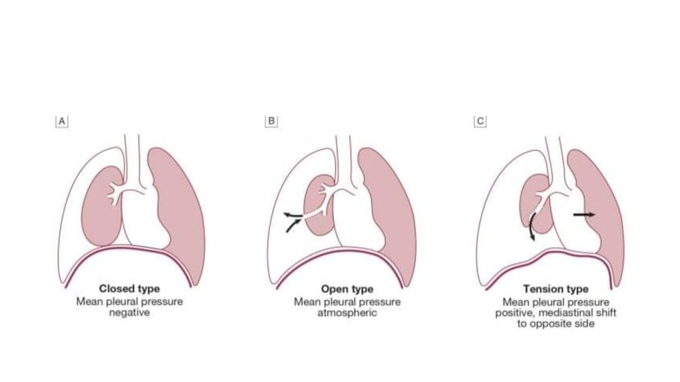
Pneumothorax: Is abnormal collection of air in the pleural space between lung and chest wall ( air in the normally airless pleural space) Pleural space also known pleural cavity , is a thin fluid- filled space between two plumonary pleurae( known as visceral and parietal ) of each lung

• There are two major categories: spontaneous and traumatic pneumothorax result from blunt trauma or pentreating trauma that disrupt the parietal or visceral pleural. Spontaneous: 1. primary : the cause of primary spontaneous pneumothorax is unknown , mean in “healthy” caused by spontaneous rupture of sub pleural blebs( air filled sacs on the lung)
 pneumothorax • Occurs as complication of underlying disease most commonly")
Secondary spontaneous ( complicated) pneumothorax • Occurs as complication of underlying disease most commonly COPD , other undrelying condition include asthma , interstitial lung disease, TB, neoplasms. • Is more life threatening because of lack of plumonary reserve.

• Tension pneumothorax Accumulation of air within the pleural space such that tissue surrounding the opening into the pleural cavity act as valve allowing air to enter but not to escape This cause shift the mediastinum away from the side of pneumothorax.

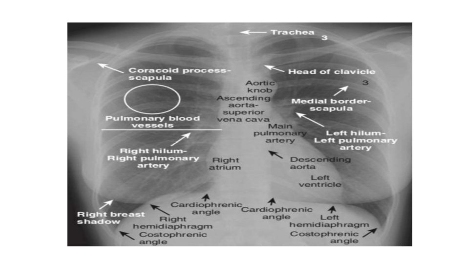
Typical chest x ray Centering patient position: medial end of clavicle equidist from T 4 -T 5 spinous process. Degree of inspiration: Anterior 6 th rib or posterior end of 10 th Rib above the diaphragm Exposure( penetration) : vertebral bodies and disc space behind the heart must be barely visible and vascular marking.

Clinical features • Symptoms 1. Ipsilateral chest pain , sudden onset 2. Dyspnea 3. Cough • Physical signs 1. Decreased breath sound over the affected side 2. Hyper resonance over chest 3. Decreased or absent tactile fremitus on affected side 4. Mediastinal shift

Recognizing apneumothorax Signs to look for : *Visualization of visceral pleural line must for diagnosis * absence of lung marking distal to visceral pleural line ( most time) *periphral space is radio Lucent Compared to adjacent lung * deep sulcus sign * mediastinum shouldn’t shift away from pneumothorax unless tension pneumothorax is present
 with air")
Visceral pleural become Visible as thin white line ( visceral pleural line) with air out lining it on either side Notice how the contour of pneumothorax parallel the curvature of adjacent chest wall

Pneumothorax with pleural adhesion

Absence of lung marking mistaken for a pneumothorax

Deep sulcus sign

Pneumothorax has also be classified based On the presence or absence shift of mobile mediastinal structure such as the heart and trachea • simple : there is usually no shift of mediastinal structure • Tension there is frequently a shift of the mediastinal structures away from side of pneumothorax associated with cardio plumonary compromise

Simple pneumothorax with no shift Large left- sided tension pneumothorax
")
Presence of an air fluid interface in the pleural space ( hydropneumothorax)

Mistaken a skin fold for pneumothorax

Mistaking medical border of the scapula for a pneumothorax

Over penetrated frontal chest radiograp

How large is pneumothorax?

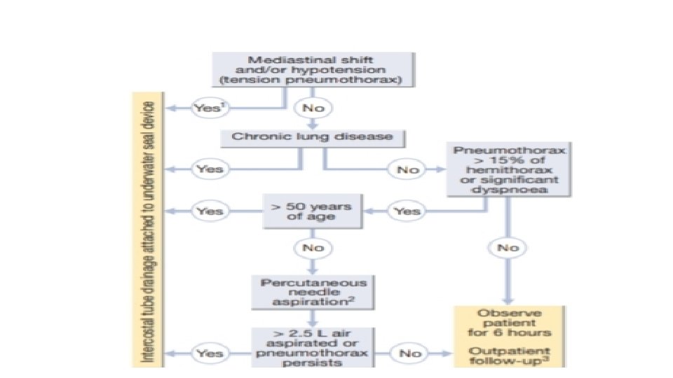
Treatment *Primary spontaneous pneumothorax 1. If small and patient is asymptomatic: . observation should resolve spontaneously in approximately 10 day. small chest tube ( with one way valve) my be benefit some patient. 2 If pneumothorax is large and or patient is Symptomatic: . adminstration of supplemental oxygen , which help with resorption of pleural air. needle aspiration or chest tube insertion to allow air to be released and lung to respond

Treatment tension pneumothorax • Must be treated as medical emergency –if the tension in the pleural space is not relieved the patient is likely to die of hemodynamic compromise(inadequate cardiac output or hypoxemia) Immediately perform chest decompression with a large bore needle (in the second or third intercostal space in the midclavicular line )followed immediately by chest tube placement.

In cases where a pneumothorax is not clearly present on standard frontal chest radiography number of technique can be employed • Lateral decubitus radiography. • Expiratory chest radiography: lung become smaller and denser pneumothorax remain the same side and is thus more conspicuous although some author suggest that there is no difference in detection rate • Ct scan

A 32 year old in a fight with a friend was stabbed in chest and became short of breath what is the diagnosis?

Sudden onset dyspnea Age 40 year Gender Male Left sided pneumothorax with inter coastal drainage tube

Initial chest x ray : left sided pneumothorax Chest xray after recurrent chest pain 20 year Female Recurrent (non trrumatic) Pneumothorax in young people is often du to apical bleb that rupture spontaneousle Chest x ray following bleb excision

Acute sever shortness of no trauma 40 year male Large non traumatic bilateral pneumothorax are un common no mediastinal shift is evident as the bilateral pneumothorax equilibrate any shift The chest cannt be totally coverd on the image but the supine projection indicate bilateral deep sulci a typical feature in this projection
- Slides: 29