Pneumoconiosis Dr Ayman S ElKhateeb Minia University Introduction

Pneumoconiosis Dr / Ayman S. El-Khateeb Minia University

Introduction • Recent decades have seen a marked increase in concern about the adverse health effects of hazardous exposures in the workplace and elsewhere in the environment • The lung – with its extensive surface area, high blood flow and thin alveolar epithelium– – is an important site of contact with these substances in the environment

• Occupational lung diseases are a broad group of diagnoses caused by the inhalation of dusts, chemicals, or proteins • “Pneumoconiosis” is the term used for the diseases associated with inhaling mineral dusts and lung reaction • The severity of the disease is related to the material inhaled and the intensity and duration of the exposure • The incidence of the disease increased dramatically with the development of modern industry.
 • Organic")
Industrial dust • Inorganic dust (consists of particles of minerals and metals) • Organic dust (contains particles of plant and animal origin, and also microorganisms that are on them, and their waste products) • Mixed dust

Classification 1 - Fibrotic pneumoconiosis A- Major pneumoconiosis. B- Minor pneumoconiosis. 2 - Non- fibrotic pneumoconiosis (Benign).

Pathogenesis The effects of an inhaled agent depend on many factors its physical and chemical properties the susceptibility of the exposed person the site of deposition within the bronchial tree

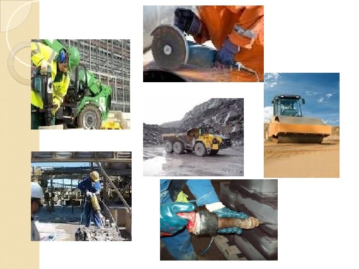
Silica Dust Exposure – Risk Factors Any work that exposes silica dust: ◦ ◦ ◦ ◦ mining stone cutting quarrying road and building construction work with abrasives glass manufacturing sand blasting also, some hobbies can involve exposure to silica (sculptor, glass blower) Silicosis - Sandblasting

Silicosis Ø Silica is silicon dioxide, the oxide of silicon, chemical formula Si. O 2 Ø Si. O 2 is the most abundant mineral on earth. Ø Silicosis is lung disease caused by inhalation of free silica dust, the dust causes inflammation and then scarring of the lungs. Ø There is no effective treatment for any pneumoconiosis, including silicosis
")
Three ‘types’ of silicosis Simple chronic silicosis From long-term exposure (10 - 20 years) to low amounts of silica dust. Nodules of chronic inflammation and scarring, provoked by the silica dust, form in the lungs and chest lymph nodes. Patients often asymptomatic, seen for other reasons. Accelerated silicosis (= PMF, progressive massive fibrosis) Occurs after exposure to larger amounts of silica over a shorter period of time (5 -10 years). Inflammation, scarring, and symptoms progress faster in accelerated silicosis than in simple silicosis. Patients have symptoms, especially shortness of breath. Acute silicosis From short-term exposure to very large amounts of silica dust. The lungs become very inflamed, causing severe shortness of breath and low blood oxygen level.
 Occupational and environmental history – single most helpful tool in the diagnostic")
Diagnosis (1) Occupational and environmental history – single most helpful tool in the diagnostic workup 1. Employment details Job title Type of industry and specific work Name of employer Years employed 2. Exposure information General description of job process and overall hygiene Materials used by worker and others Specific workplace exposures Ventilation / exhaust system Use of respiratory protection Industrial hygiene informations provided by the employer to the employee

3. Environmental non-occupational factors Smoking Diet Hobbies 4. Details about past employments in chronological order 5. Other details Does the patient think symptoms / problem is related to anything at work? Are other workers affected? Work absenteeism Prior pulmonary problems and medications used

Symptoms shortness of breath while exercising fever occasional bluish skin at ear lobes or lips fatigue loss of appetite
Physical examinations Generally un-revealing about specific cause It is most helpful in periodic ex.")
(3)Physical examinations Generally un-revealing about specific cause It is most helpful in periodic ex. nonoccupational causes of respiratory symptoms or diseases (cardiac problems or connective tissue disorders)
 Investigations: A - Chest radiography - is the most important diagnostic test for")
(4) Investigations: A - Chest radiography - is the most important diagnostic test for occupational lung diseases

Note: The chest radiographic findings can be nonspecific. � �Silicosis is a radiological diagnosis. � �personal variations

ILO – International Classification of radiographs of pneumoconiosis, 1971, 2002 Small opacities (less than 1 cm. ) - Regular (round): diameter p : <1. 5 mm. q : 1. 5 – 3 mm. r : 3 - 10 mm. - Irregular : width p : <1. 5 mm. q : 1. 5 – 3 mm. r : 3 - 10 mm.

2. Profusion: Category 0: small rounded opacities may absent, normal bronco-vascular marking. Category 1: small rounded opacities definitely present but few in number, normal bronco-vascular marking. Category 2: small rounded opacities numerous. Bronco-vascular marking are hesitated but still visible. Category 3: small or large opacities very numerous. Bronco-vascular marking are partially or totally obscured

Complications: Increases risk of tuberculosis. Respiratory failure. Heart failure. Bronchogenic carcinoma.

Thanks for attention!
- Slides: 20