PLEURAL EFFUSION Presented by Raheeq dmour Rawan zedaneen

PLEURAL EFFUSION Presented by: Raheeq dmour Rawan zedaneen Supervised by : DR. samah shehata

INTRODUCTI ON Pleura is a serous membrane which folds back onto itself to form a two layered membranous pleural sac • The outer pleura (parital pleura): attached to chest wall • The inner pleura (visteral pleura): cover the lung and adjoining structures • The pleural space : a potential space because the two pleura are adhere to each other (through a thin film of serous fluid of only few milliliters of pleural fluid around 0. 13 ml/kg)

Definition Is the accumulation of fluid in the pleural cavity The nature of fluid : • • Serous fluid (hydrothorax) Pus (empyema) Blood(haemothorax) Chyle(chylothorax)

CALCIFICATI ON : By involvement By mechanism unilateral transudative bilateral exudative

CAUSES: Tansudative: Exudative: Is caused by systemic factors Is caused by inflammation leading to a form of increased microvascular pressure due to disease of pleura or injury in the adjacent lung (e. g. pneumonia or malignancy) causing • Increase in hydrostatic pressure (e. g. chf) • Decrease oncotic pressure (e. g. nephrotic syndrome or cirrhosis ) Because these diseases are increased capillary

hydrostatic pressure / oncotic pressure Unable to remain the fluid with-in intravascular space Fluid shift interstitial space Effusion

Invasion of microbes Initiation of inflammatory reaction Vasodilation increase capillary permeability leak of plasma protein decrease oncotic pressure fluid shift into interstitial space

A transudative effusion needs no further evaluation. It resolves by adequate treatment of the disease An exudative effusion will cause unilateral effusions. This type of effusion need further investigation. In some cases it can be a combination of both It can also be caused by lymphatic obstruction(lymphatic effusion)
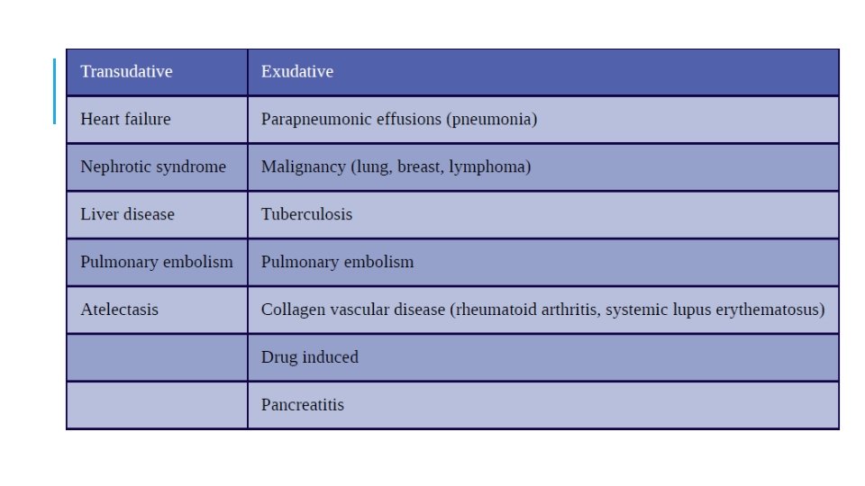

CRITERIA trancudativ Exudative e LDH effusion <200 IU/ML >200 IU/ML LDH effusion/serum ratio <0. 6 >0. 6 Protein efusion/serum ratio <0. 5 >0. 5 Specific gravity < 1. 016 >1. 016 If at least 1 criterion is not met, then this is an exudative effusion

HOW DO WE MAKE THE DISTINCTION BETWEEN THESE TWO? Thoracentesis should be performed for new and unexplained pleural effusion when sufficient fluid is present One of the few conditions that can cause a transudate or exudate is pulmonary embolism (PE). if a patient has a transudative effusion but no apparent cause, consider PE

PARAPNEUMONIC EFFUSION is caused by bacterial pneumonia , lung abscess , bronchiectasis Divided into three types: • Uncomplicated parapneumonic • Complicated parapneumonic • Empyema if complicated not treated by antibiotics

Uncomplicated parapneumonic effusions. The fluid may be cloudy or clear, and it doesn’t contain bacteria. PPE will get better when you take antibiotics to treat pneumonia. Complicated parapneumonic effusions. Bacteria have traveled from the lungs into the pleural space, causing a buildup of fluid and white blood cells. The fluid is cloudy. It will need to be drained. Empyema thoracis. Thick, whitish-yellow pus builds up in the pleural space. This can happen if pneumonia isn’t treated quickly enough.

CLINICAL PRESENTATION Notes : If the patient has minimal effusion with minimal lung compression he might not have any symptoms When the effusion is modret to large size or caused by inflammation symptoms are present It may also lead to respiratory failure in some cases Note that symptoms are related to the underlying cause of effusion

SIGNS AND SYMPTOMS: 1. Dyspnea 2. Caught 3. Peripheral edema 4. Pleuritic chest pain: Results from pleural irritation and raises the likelihood of an exudative etiology. Analysis of pain: • Pain may be mild or severe. • Described as sharp or stabbing. • Exacerbated with deep inspiration. • Pain may be localized to the chest wall or referred to the ipsilateral shoulder or upper abdomen.

PHYSICAL EXAMINATION Physical examination should focus on finding the cause and assessing the degree of respiratory compromise. Physical findings accrue at effusion >300 ml

• Inspection : Tachypnoea Absent or diminished movements off affected side Fullness of chest with bulging of intercostal spaces • Palpation: Asymmetrical chest expansion, with diminished or delayed expansion on the side of the effusion. decreased tactile fremitus Mediastinal shift away from the effusion (effusion of >1000 ml)

• Percussion : Stony dullness to percussion. • 1. 2. 3. Auscultation: Absent of breathing sounds over the effusion Vocal resonance absent There may be signs of pneumonia ( bronchial breathing , crackles)

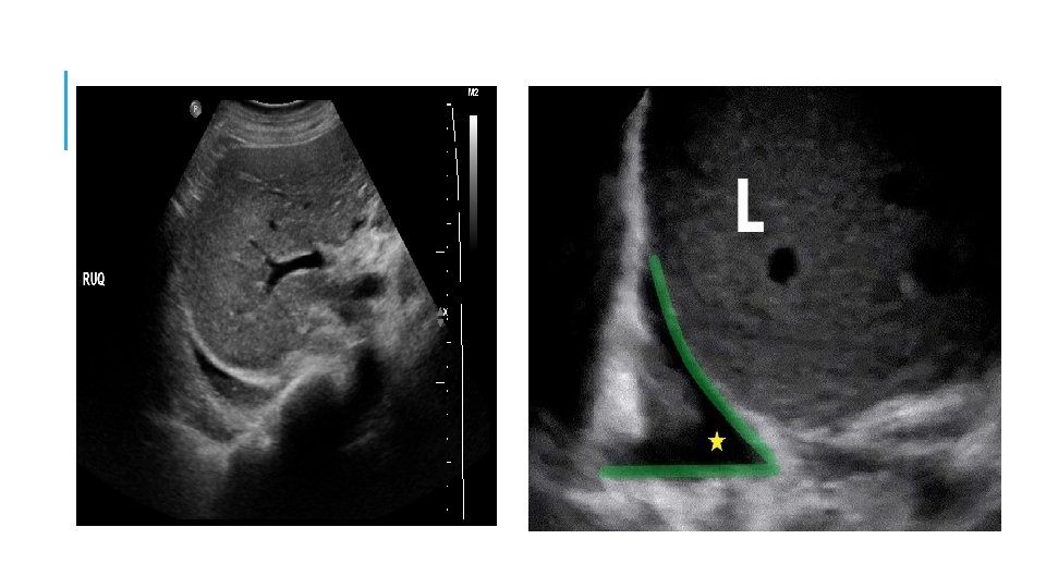
INVESTIGATI ONS • • • 1. 2. 3. • Total and differential leucocyte count CRP AND ESR Radiological examination CXR CT SCAN Ultrasonogram thoracocentesis

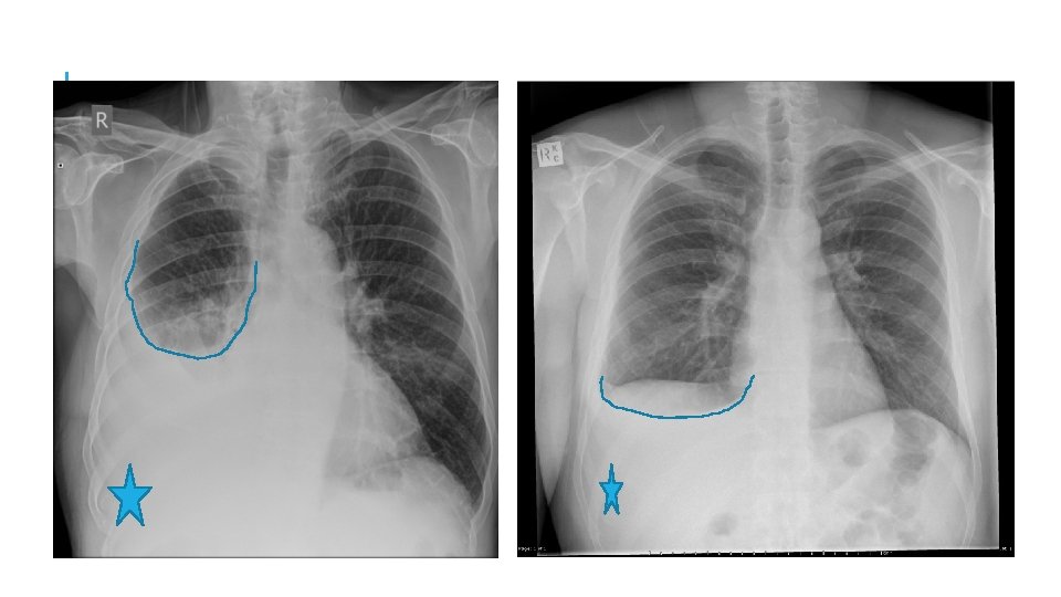
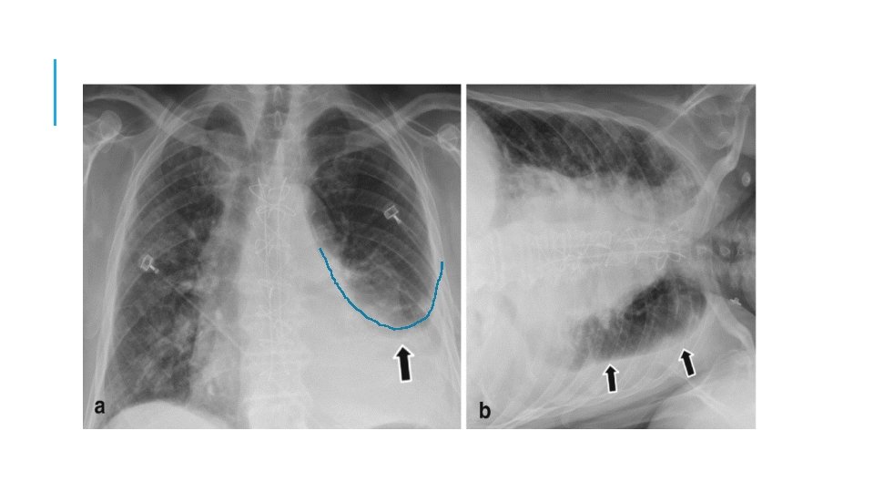
CXR In PA VIEW a total of 300 ml is needed to diagnose pleural effusion In lateral decubitus even 50 ml can be detected, it can also determined whether fluid is free flowing or loculated Findings : • Obliteration costophrenic angles • Tracheal and mediastinal shifts are seen in massive effusion • A curved shadow at lung base that ascend on the chest wall toward the axilla , fluid appear to track up the lateral chest wall (meniscus sign)

CT SCAN More reliable than CXR for detecting effusion , it can displays pleural abnormalities, and determine the volume of the pleural fluid

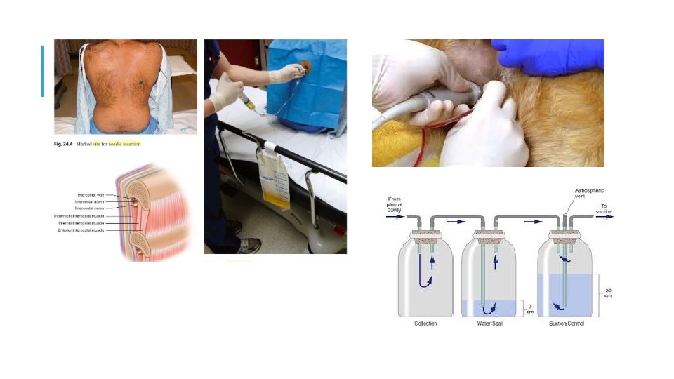
THORACOCENT ESIS Diagnostic: Helps to differentiate between exudates and transudates Therapeutic: 1. Massive collection or rapid collection of pleural fluid 2. Severe respiratory distress 3. Suspected empyema 4. Massive mediastinal shift

is a procedure that drains fluid or air from the space between the lungs and the wall of the chest (the pleural space). It is done using a hollow needle or a plastic tube inserted through the chest wall. Normally only a small amount of fluid is in the pleural space and is done in the most dependent part.

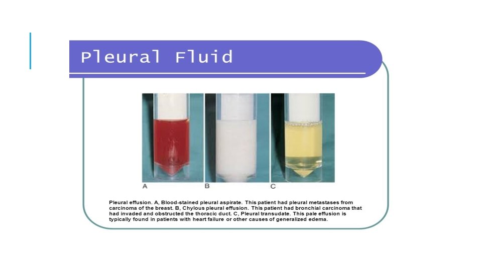
• transudates are clear, and without odor • Frankly purulent fluid indicates an empyema • A milky fluid suggests a chylothorax : lymph in the pleural space. • Grossly bloody fluid may result from trauma, malignancy, post pericardiotomy syndrome, or asbestos-related effusion • Elevated pleural fluid amylase : esophageal rupture , pancreatitis , malignancy. • Exudative effusion that are primary lymphcytic : TB • PH<7. 3 : parapneumonic effusion or empyema

A thoracocentesis is mandatory also in this setting to rule out a complicated parapneumonic effusion (because of the possibility of progression to an empyema). An empyema (or complicated effusion) needs chest-tube drainage to resolve, while an uncomplicated parapneumonic effusion responds to antibiotics alone

MANAGEME NT • Treat the cause Pneumonia : antibiotics CHF: diuretics malignancy • Pleural fluid aspiration 1. Wide bore needle 2. Intercostal drainage under water seal • Thoracic surgery : needs VATS

- Slides: 32