Pleural Effusion ABDULLAH ALHARBI MD FCCP A 67

Pleural Effusion ABDULLAH ALHARBI , MD, FCCP

A 67 -year-old man presents to the emergency department with a 5 -day history of fever and cough that produces green sputum. He has a history of tobacco use and ischemic cardiomyopathy with a left ventricular ejection fraction of 25%. He was admitted with a presumptive diagnosis of pneumonia and is started on antibiotics. A chest radiograph is obtained and shows a left-sided consolidation and moderatesize effusion. Which of the following studies can be used to determine? if the patient’s effusion is due to his CHF (a transudate) or is a Para pneumonic effusion (an exudate)? (A) Pleural fluid p. H (B) Pleural fluid glucose (C) Pleural fluid cell count (D) Lactate dehydrogenase (LDH

A 59 -year-old male presents with a community acquired pneumonia complicated by pleural effusion. A thoracentesis is performed, , but the results are not currently available. Which characteristic of the pleural fluid is most suggestive complicated parapnemonic pleural effusion? A. Presence of more than 30% polymorphonucleocytes (PMNs) B. Glucose less than 150 mg/d. L C. Presence of more than 1000 white blood cells D. p. H less than 7. 20 E. Lactate dehydrogenase (LDH) more than two-thirds of the normal upper limit for serum

A 55 -year-old man presents with progressive shortness of breath. Other than a history of heavy smoker, the patient has no significant past medical history. Breath sounds are absent two-thirds of the way up on the left side of the chest. Percussion of the left chest reveals stony dullness, the trachea appears to be deviated toward the right. Which of the following diagnoses is most likely? (A) Bacterial pneumonia (B) Viral pneumonia (C) Bronchial obstruction (D) Pleural effusion (E) Pneumothorax

Objectives: Describe the pathophysiology of a pleural effusion Describe the main causes of a pleural effusion Differentiate among the manifestations of fluid collections Describe the signs and symptoms of a pleural effusion Explain diagnostic methods Describe the various treatment options
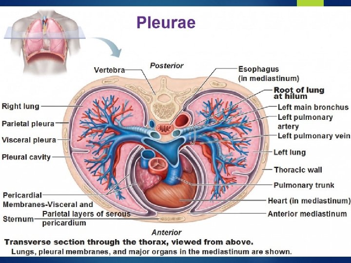
 and visceral pleura")
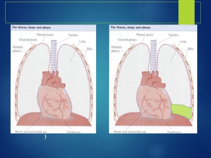
Pleura Serous fluid that allows for the parietal pleura (outer lining) and visceral pleura (inner lining) to glide over each other without separation contains about 5 -15 ml of fluid at one time Pleural fluid is produced by the parietal pleura and absorbed by the visceral pleura as a continuous process. The visceral pleura absorbs fluid, which then drains into the lymphatic system and returns to the blood about 100 -200 ml of fluid circulates though the pleural space within a 24 -hour period

Clinical Presentation History Dyspnea Pleuritic chest pain Cough Other symptoms related to underlying cause Physical exam (Findings usually present for effusions > 300 m. L ) Dullness to percussion, decreased tactile fremitus Asymmetric chest expansion Decreased breath sounds Tracheal shift away from the affected side
 capillary permeability (Pneumonia) intrapleural pressure (atelectasis)")
Development of Pleural Effusion pulmonary capillary pressure (CHF) capillary permeability (Pneumonia) intrapleural pressure (atelectasis) plasma oncotic pressure (hypoalbuminemia) pleural membrane permeability (malignancy) lymphatic obstruction (malignancy) diaphragmatic defect (hepatic hydrothorax) thoracic duct rupture (chylothorax)

Imaging Studies-Chest Radiographs PA - usually around 250 -500 m. L needed before visible Lateral Decubitus – very sensitive, can detect effusions as small as 50 m. L

Imaging Studies CT Scan Better characterization of underlying lung parenchyma and certain processes that may be obscured on radiographs by large pleural effusions Ultrasound Cheap and available at bedside Can help identify free vs. loculated effusions Thoracentesis is facilitated by ultrasound guidance

CT Scan

Thoracocentesis

Indications for thoracentesis Pleural effusion of unknown etiology, with >10 mm depth on lateral decubitus CXR or Ultrasound Therapeutically for symptomatic relief Concern for empyema Air fluid level in pleural space

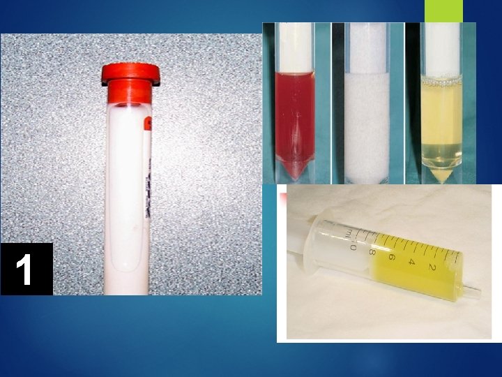
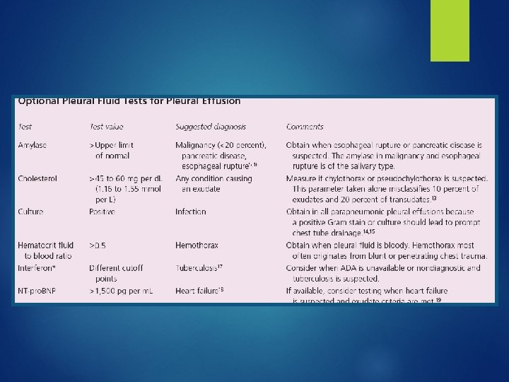
Pleural Fluid Evaluation 5 Cs Color Cytology Culture Cell count Chemistry

Lights Criteria Pleural effusion is exudative if one or more of the following: Ratio of pleural fluid protein level to serum protein level > 0. 5 Ratio of pleural fluid LDH level to serum LDH level > 0. 6 Pleural fluid LDH level > 2/3 the upper limit of normal for serum LDH level. 98% sensitive and 83% specific for exudative effusion using Lights criteria. Absence of all 3 criteria = transudative

Transudative vs Exudative Transudative �Exudative CHF Nephrotic syndrome Hypoalbuminemia Hepatic hydrothorax Atelectasis Hypothyrodism Pneumonia Malignancy PE Inflammatory (pancreatitis, ARDS, uremic pleurisy etc Connective tissue disease

 CA (30 -35%) TB (15 -20%) Trauma Sarcoidosis PTX")
Cell count Lymphocytic (> 50%) CA (30 -35%) TB (15 -20%) Trauma Sarcoidosis PTX CA Asbestos, parasites Pneumonia PMNs Empyema Parapneumonic Rheumatoid Pulmonary infarction Eosinophilic (> 10%) PMN or Lymphocytic RBC > 100, 000/mm CA Conn tissue disease Trauma Post-cardiac injury Pulmonary infarction PE

Treatment Thoracentesis – then treat underlying disease Uncomplicated pneumonia – antibiotics Hemithorax involved/empyema – tube thoracostomy +/ - VATS Malignant effusion- chest tube +/- pleurodesis (sclerosants) VATS

Summary Pleural effusions are commonly encountered on wards Thoracentesis is not immediately indicated if there is a obvious explanation for pleural effusion without atypical features Pleural effusions are classified as transudative vs exudative. CHF, pneumonia, malignancy and PE comprise the vast majority of causes for pleural effusions.
- Slides: 25