Plasma Proteins Blood plasma has 100 different proteins

Plasma Proteins

Blood plasma has 100 different proteins § Transport proteins § Fibrinogen and other clotting factors § Acute-phase reaction proteins § Complement components § Enzyme inhibitors (proteinase inhibitors) § Precursors of substances such as angiotensin and bradykinin § Hormones § Enzymes

• Plasma proteins are synthesized at the liver at ribosomes that bound to the membrane • All plasma proteins are in glycoprotein structure • Contains N and O glycoside-linked oligosaccharide residues • Only albumin does not contain any sugar residue

• Plasma proteins showing polymorphism • α 1 antitrypsin • haptoglobin • transferrin • ceruloplasmin • and the immunoglobulins

• Each plasma protein has a specific half-life in circulation. • Half-life of albumin 20 days • Haptoglobin half-life 5 days • In acute inflammation and tissue injury plasma protein levels increase

CRP stimulates the path of inflammation, α 1 antitrypsin neutralizes some proteases during inflammation Changes in plasma protein levels are induced by cytokines released from the site of injury

• Functions of plasma proteins • They provide osmotic and oncotic pressure of blood • Transport of many substances in the cell • Keeping the plasma water content within the vessel • Effects on blood viscosity • Contribution to maintaining acid-base balance • Maintain protein need of the tissues • Protection of an Organism against infections and harmful substances

• Increases and decreases in total protein concentration are called dysproteinemia • Dysproteinemia are two types, hyperproteinemia (increased serum protein concentration) and hypoproteinemia (decrease in serum protein concentration). • The presence of proteins that are not normally present in the blood and without special functions is called paraproteinemia.
 Relative hyperproteinemia (relative hyperproteinemia) may occur in cases of hemoconcentration")
Causes of hyperproteinemia 1) Relative hyperproteinemia (relative hyperproteinemia) may occur in cases of hemoconcentration where plasma water content decreases. 2) Depending on the emergence of paraprotein, hyperproteinemia may occur. 3) In some chronic diseases, hyperproteinemia occurs due to globulin increase.

• Hemoconcentration states 1. Loss of water from the digestive tract with bad and vomiting 2. loss of water through the skin in hot conditions and fever 3. renal failurenephritis 4. diabetes mellitus 5. diuretic treatment states 6. oliuria in the kidneys through water loss 7. restriction of water intake

• The emergence of paraproteins: • lenforeticular malignancies • Some autoimmune diseases • chronic infections • cirrhosis of the liver

Some chronic diseases with -globulin increase: • chronic inflammatory events such as severe chronic polyarthritis, endocarditis etc. • tropical diseases • cirrhosis of the liver
 Relative hypoproteinemia occurs in hemodilution cases where plasma water content")
Causes of Hypoproteinemia 1) Relative hypoproteinemia occurs in hemodilution cases where plasma water content increases. 2) Excessive protein loss 3) When there is a decrease in protein synthesis 4) It may be essential hypoproteinemia due to protein metabolism disorder.
")
Hemodilution conditons: • Excessive hydration due to over-intake and excess fluid intake (water poisoning) • heart failure condition • taking the sample from the point (extremity) of fluid replacement

Conditions for excessive protein loss: • Nephrotic syndrome, chronic glomerulonephritis, . . . • Sore, irrigated wound and skin lesions, psoriasis. . . • Protein losing enteropathy, ulcerative gastritis, • Surgical and traumatic shocks. • Depletion of excess fluid from body cavities. • Hypertiroidizm. diabetes mellitus. • Pregnancy toxemia

Conditions for Decrease in protein synthesis • Kwashiorkor • Severe Malabsorption Conditions • protein-poor nutrition • severe liver diseases

Albumin: • MA: 66 k. D, the small globular protein • 50% of total plasma protein • Alb is found in 40% pl and 60% in extracellular compartments. • 2 -3 g Alb is produced per day in the liver in average size dog

• The most abundant protein in most extravascular body fluids such as CSF, interstitial fluid, urine, and amniotic fluid • Alb synthesis is reduced by inflammatory cytokines, release is secreased in hypokalemia • Catabolism occurs by pinocytosis in all tissues

Function • Regulates colloid osmotic pressure in vascular and extracellular spaces • Carries ligands • Stores • Sources for endogenous aacids • Maintaining of oncotic pressure

Examples • Free Fatty Acids • Phospholipids • Metallic Ions • Hormones • Bilirubin Alb is effective in the metabolism and detoxification of many of these substances above.

• Bound drugs • Sulfanamides • barbiturates • Albumin is an important component of the antioxidant activity of pl • Alb (-) is an acute phase reactant

Clinical significance • Alb rises in acute dehydration, clinically insignificant • Alb reduction ------ reduction of synthesis • Depends on catabolism or both reasons

• In analbuminemia; other globulins increase when decrease in synthesis due to a genetic disorder. (Alb levels are 0. 5 g / L) • Bisalbuminemia, the molecular structure is out of the ordinary • Double band is seen instead of single band in electrophoresis • Bisalbuminemia occurs during treatment with some drugsinflammation • Acute, chronic inflammation, hemodilution, excessive catabolsim by cells, decreased synthesis are the causes of hypoalbuminemia

• Globulins • Hyperglobulinemia • a. Globulin concentration tends to increase with – infection and inflammation – near term during pregnancy. – In birds, globulins increase just prior to egg laying. • b. Acute-phase proteins cause mild increases in globulin concentration – Most acute-phase proteins are α-globulins, but some are β-globulins – Hepatic synthesis of positive acute-phase proteins begins shortly after (24 hours) after acute tissue injury (e. g. , inflammation, necrosis, surgery, infection, tumors, immunemediated processes, etc. ), and also may be produced in response to estradiol, physical stress etc.

• Acute-phase proteins play a role in – the immune response, provide protection against oxidative stress generated in the course of an inflammatory response. • Detection of acute-phase proteins may be useful – in the early diagnosis of tissue injury – in monitoring the response to treatment and resolution of tissue trauma or inflammation. • They are often more sensitive than the WBC count in identifying an inflammatory response. • High values often correlate with severity of disease but do not necessarily predict outcome. • Sustained high values of rapid-responding acute phase proteins, despite treatment, may indicate poor prognosis.
 can cause marked increase in the globulin fractions")
• Immunoglobulins (chronic phase proteins) can cause marked increase in the globulin fractions on the serum electrophoretogram. • Immunoglobulins are observed in the β and γ regions of the serum electrophoretogram

• Selective increased concentrations of βglobulins seldom occur; they usually are associated with increased concentration of other globulins. • Bridging of β and α regions in the serum electrophoretogram highly suggests chronic and active hepatitis.

• Polyclonal gammopathies are characterized by an increased globulin fraction with a wide electrophoretic peak comprised of a heterogeneous mixture of immunoglobulins – Polyclonal gammopathy is associated with chronic antigenic stimulation occurs in inflammatory diseases, immune-mediated disorders and liver diseases
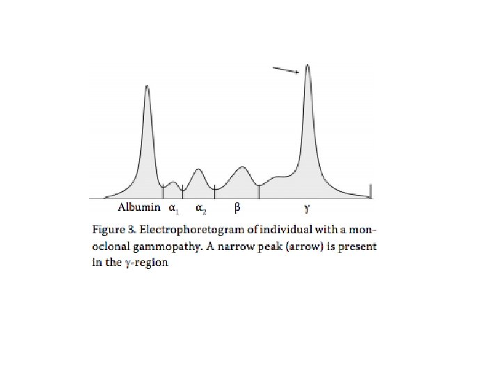
 is characterized by an increased globulin fraction with a")
• Monoclonal gammopathy (paraproteinemia) is characterized by an increased globulin fraction with a narrow-based electrophoretic peak that is no wider than the albumin peak – The monoclonal peak or spike is caused by a homogeneous immunoglobulin molecule that is produced by a single clone of B lymphocytes or plasma cells. – Monoclonal peaks can either be observed in γ, β-, or α-globulin regions.

• Monoclonal gammopathy occurs most commonly with lymphoid neoplasias • Infrequently, monoclonal gammopathy may be observed with non-neoplastic disorders such as canine amyloidosis, canine ehrlichiosis, canine etc. • Monoclonal gammopathy may be accompanied by overproduction of immunoglobulin light chains. Light chains (Bence-Jones proteins) are rapidly filtered from plasma into urine by the kidneys. Bence-Jones proteins are best detected by electrophoresis of concentrated urine specimens.
- Slides: 31