Plasma membrane and Cellular Transport Plasma membranes Membranes

Plasma membrane and Cellular Transport Plasma membranes : Membranes cover the surface of every cell and also surround most organelles within cells. They have a number of functions, such as: • keeping all cellular components inside the cell • Allowing selected molecules to move in and out of the cell • Isolating organelles from the rest of the cytoplasm, • allowing cellular processes to occur separately. • A site for biochemical reactions , allowing a cell to change shape Membrane models 1925 Gorter and Grendel propose lipid bilayer structure for cell membranes; surface area covered by lipids extracted from red blood cells on water surface is twice as large as original surface of red blood cells. polar nonpolar

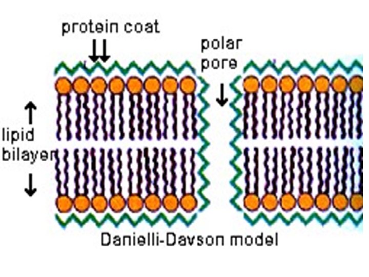
the polar hydrophilic groups of the molecules being situated on the outside and hydrophobic ends standing at right angles to the surface are oriented inside. 1935 Danielli and Davson (sandwich Model) membrane model of globular proteins on surface of lipid bilayer; was basically a "sandwich" of lipids covered on both sides with proteins

Unit membrane Model: This model was proposed by Robertson in 1959. unit-membrane model The description of all the membranes of a cell (i. e. the cell membrane and membranes enclosing organelles) as having a common structure, revealed by electron microscopy as two dark bands, each about 20 A° thick, separated by a lighter band about 35 A° thick. The plasma membranes of prokaryotes and eukaryotes are unit membranes. Again the membranes of endoplasmic reticulum, Golgi bodies, Mitochondria, lysosomes, plastids and nucleus are unit membranes.

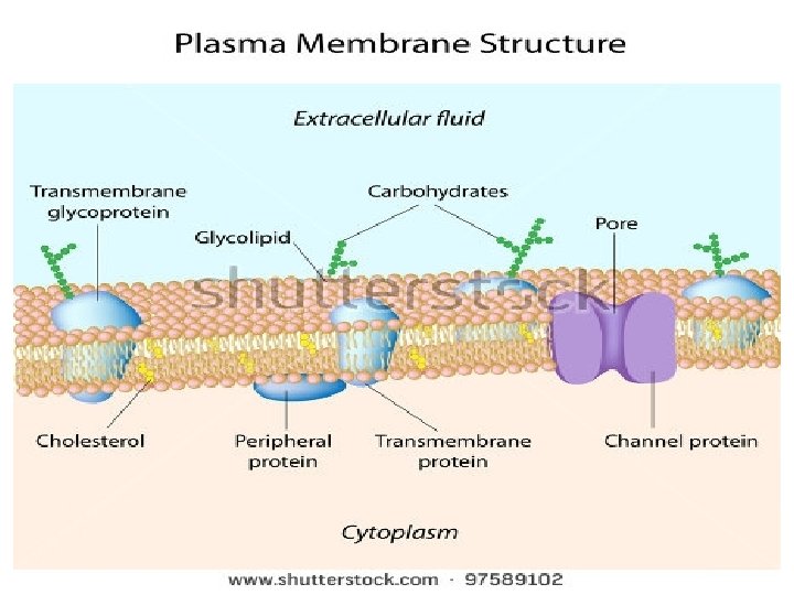
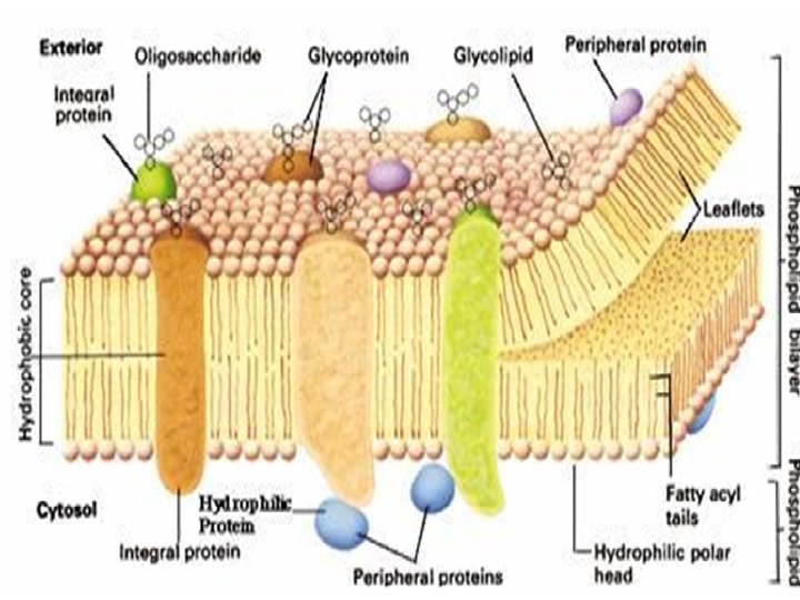
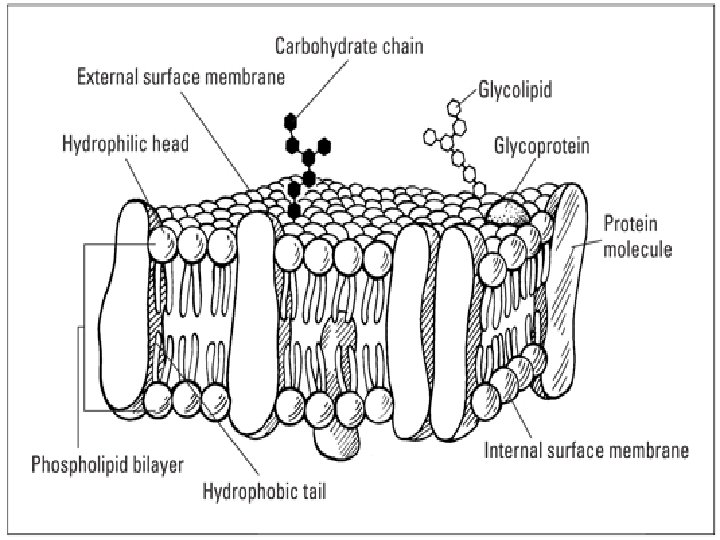
Fluid Mosaic Model: In the year 1972 Singer and Nicolson proposed a model for explaining the membrane structure, According to this model, cell membrane consists of a highly viscous fluid matrix of a bilayer of phospholipids having globular proteins associated with them. This model came to be known as fluid mosaic model. The phospholipid molecules in the cell membrane have their polar, hydrophilic heads towards outer surface and the nonpolar, hydrophobic tails towards inner surface. The proteins in cell membrane are of two kinds. Some of the proteins are found at the periphery, partly projecting out of the lipid layer. These are the peripheral proteins. These proteins can be easily extracted. Some of the protein molecules are found totally embedded in the phospholipid matrix. Those are the integral proteins, which represent nearly 70% of the membrane proteins. These proteins cannot be extracted. Some of the integral proteins have very large molecules that extend throughout the phospholipid matrix, projecting out on both surfaces.

These proteins are called tunnel proteins or transmembrane proteins. They are believed to have channels for the passage of water-soluble substances. Some proteins have a polysaccharide attached to them. These are called Glycoproteins and are important in cell recognition and essential to the immune response. Saccharides are also attached to some lipids, forming glycolipids. The phospholipid bi-layer and associated proteins is called the fluid mosaic model of the membrane's structure. 'Fluid' because the positions of the constituents are always changing and 'mosaic' because the membrane is made from different types of molecules.

Types of Cellular Transport The membrane is selectively permeable and able to regulate what enters and exits the cell. The movement of substances across the membrane can be either passive transport or active transport • Passive Transport (cell doesn’t use energy) 1 -Simple diffusion 2 - Facilitated diffusion 3 - Osmosis
 1 -Protein Pumps 2 - Endocytosis")
• Active Transport (cell does use energy) 1 -Protein Pumps 2 - Endocytosis 3 - Exocytosis 1 -Passive Transport - cell uses no energy - molecules move randomly - Molecules spread out from an area of high concentration to an area of low concentration. - (High concentration Low concentration)
 from")
1. Simple diffusion • simple diffusion is the random movement of particles (molecules) from a region of high concentration to a region of low concentration. • NO ENERGY required • this process will continue until a dynamic equilibrium reached. example – Oxygen diffuses from the blood cells in the blood stream into muscles. Simple diffusion through plasma membrane
 ü Facilitated diffusion")
2 -Facilitated diffusion (diffusion with the help of transport proteins ) ü Facilitated diffusion is the passive movement of molecules or ions down a concentration gradient (high concentration to an area of low concentration) ü It is utilized by molecules that are unable to freely cross the phospholipid bilayer (e. g. large, polar molecules and ions) ü This process is mediated by two types of transport proteins – channel proteins and carrier proteins in the plasma membrane ü This process don’t required energy example- the absorption of glucose and amino acid from the villi into the blood capillaries Facilitated diffusion through a pore protein in the plasma membrane

Facilitated diffusion through a carrier protein in the plasma membrane
 Define osmosis : The diffusion of water across a")
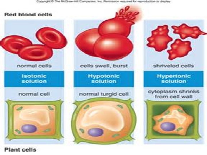
3. Osmosis (diffusion of water) Define osmosis : The diffusion of water across a selectively permeable membrane ü The random movement of water molecules ü from a region of high concentration of water molecules to a region of low concentration of water molecules ü through a partially permeable membrane. ü Define selectively permeable membrane : a membrane that allows only certain materials to cross it ü Materials pass through pores in the membrane • Example – the reabsorption of water molecules from the nephrons into the blood capillaries. How Does Osmosis Affect Cells? Osmosis allows cells to regulate the balance of water inside the cells. The concentration of solutes outside the cells determines whether a cell is isotonic, hypotonic or hypertonic.

water will move rapidly into or out of the cell by osmosis, Solutions with concentrations equal to the concentration of the cytoplasm are described as isotonic. -A solution that is less concentrated than the intracellular fluid is described as hypotonic. a cell placed in a hypotonic solution draws water in, swells and may burst. -If a cell is placed in a hypertonic solution, which is high concentrated than the cellular fluid, it loses water to the surrounding fluids and shrinks, A plant cell in a hypertonic solution undergoes plasmolysis (shrinking of the cytoplasm) and the plant often wilts.
 1 -Protein Pumps 2 - Endocytosis 3")
§ Active Transport (cell does use energy) 1 -Protein Pumps 2 - Endocytosis 3 - Exocytosis • Process that moves materials across the plasma membrane • Requires energy from the cell in the form of ATP • Materials move against the concentration gradient: low concentration high concentration 1 -Active Transport Pumps 1 -An ATP molecule breaks down into ADP, releasing a phosphate group and a whole lot of energy. 2 -The phosphate group attaches to a protein pump, causing it to change its shape so that it can move a small molecule or ion across the plasma membrane. 3 -The protein changes shape again so that the molecule can be released on the other side. • There are many types of carrier proteins and they only carry specific molecules across the plasma membrane.

Example of active transport: sodium-potassium pump Sodium ions are kept at low concentrations inside the cell and potassium ions are at higher concentrations Outside the cell, For each molecule of ATP used, 2 K+ are pumped into the cell and 3 Na+ are pumped out of the cell. 1. 3 Na+ and 1 ATP bind to the protein “pump. ” 2. ADP is released, causing a change in the pump’s shape. 3. 3 Na+ are released as 2 K+ bind to the pump. 4. Pi is released, causing the pump’s shape to change and releasing 2 K+

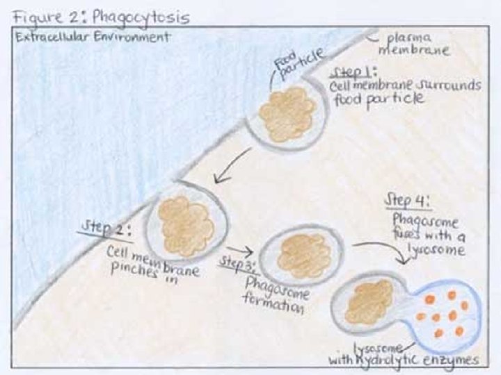
Endocytosis and Exocytosis The movement of macromolecules such as proteins or polysaccharides into or out of the cell is called bulk transport. There are two types of bulk transport, exocytosis and endocytosis and both require of energy (ATP). 2 -Endocytosis, is the process by which materials move into the cell. There are three types of endocytosis: phagocytosis, pinocytosis, and receptormediated endocytosis. In phagocytosis or “cellular eating, ” the cell’s plasma membrane surrounds a macromolecule or even an entire cell from the extracellular environment and buds off to form a food vacuole or phagosome (vacuole in the cytoplasm of a cell, containing a phagocytosed particle enclosed within a part of the cell membrane), The newly-formed phagosome then fuses with a lysosome whose hydrolytic enzymes digest the “food” inside.
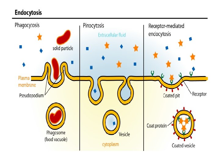


pinocytosis or “cellular drinking, ” the cell engulfs drops of fluid by pinching in and forming vesicles.

receptor-mediated endocytosis is a process by which cells absorb metabolites, hormones, other proteins - and in some cases viruses - (endocytosis) by the inward budding of plasma membrane vesicles containing proteins with receptor sites specific to the molecules being absorbed.

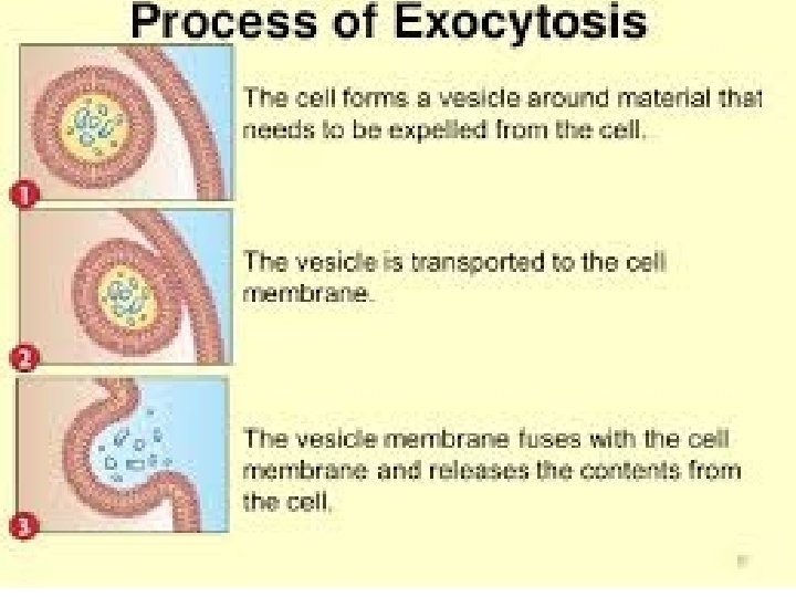
3 -Exocytosis In exocytosis, materials are exported out of the cell via secretory vesicles. In this process, the Golgi complex packages macromolecules into transport vesicles that travel to and fuse with the plasma membrane. This fusion causes the vesicle to spill its contents out of the cell. Exocytosis is important in expulsion of waste materials out of the cell and in the secretion of cellular products such as digestive enzymes or hormones.

- Slides: 28