
PLACENTA PREVIA Dr M Moshfeghi OBSGYN fellowship of


PLACENTA PREVIA Dr. M. Moshfeghi OBS&GYN fellowship of perinatology RUYAN INSTITUTE
prevalence higher among older women . Nulliparous women ≥ 40 years of age have a 10 fold increased risk
risk factors Previous placenta previa Previous cesarean delivery Multiple gestation Multiparity Advanced maternal age Infertility treatment Previous abortion Previous intrauterine surgical procedure Maternal smoking Maternal cocaine use Male fetus Non-white race
PREVALENCE • 3. 5 to 4. 6 per 1000 births
Placenta previa severe bleeding preterm birth, need for cesarean delivery.
beyond 20 weeks painless vaginal bleeding. sonographic before EXAM
PATHOGENESIS unknown presence of areas of suboptimal endometrium in the upper uterine cavity due to previous surgery or pregnancies promotes implantation of trophoblast in, or unidirectional growth of trophoblast toward, the lower uterine large placental in multiple gestation response to reduced uteroplacental perfusion,
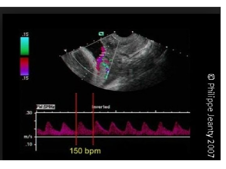
Bleeding is primarily maternal, but fetal bleeding can occur if a fetal vessel is disrupted.
Ultrasound presentation and course 10 and 20 weeks 1 -6% early resolve 90%
The lower uterine segment lengthens from 0. 5 cm at 20 weeks more than 5 cm at term Development of the lower uterine segment.
Placental atrophy may explain why the portion of the placenta that sonographically appeared to cover the cervix resolves.
prediction the best predictor of placenta previa at delivery. distance the placenta extends over the internal cervical os is
An anterior placenta previa Resolve more often and more quickly than posterior placenta previa
Bleeding • initial bleeding episode occurs prior to 30 w onethird one-third of patients becomes symptomatic between 30 and 36 weeks, Or first bleed after 36 weeks About 10 percent of women reach term without bleeding.
prediction of hemorrhage near the os if the placental edge is thick (>1 cm identification of an echo-free space in the placental edge cervical length ≤ 3 cm
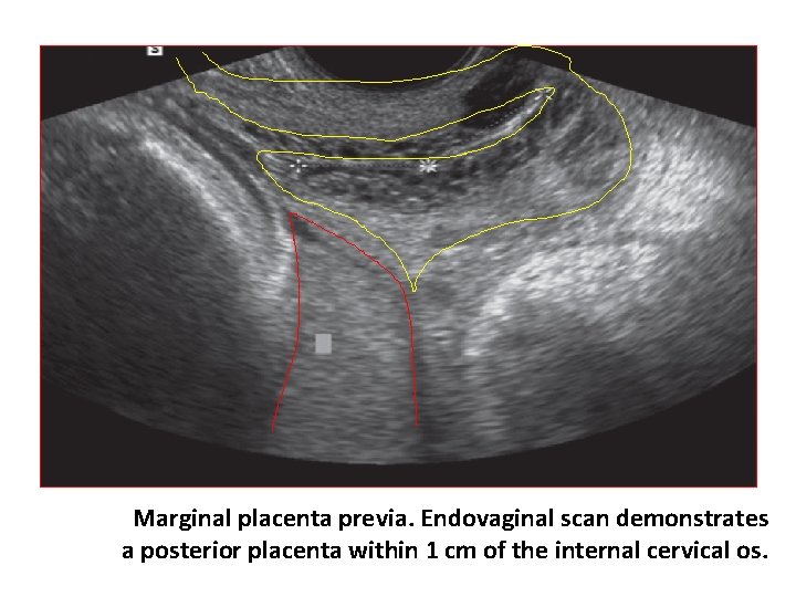
Marginal placenta previa. Endovaginal scan demonstrates a posterior placenta within 1 cm of the internal cervical os.
Placenta accreta complicates 1 to 5 percent of pregnancies with placenta previa and an unscarred uterus. one previous cesarean birth (11 to 25 %), ≥four previous cesarean births (50 to 67 %)
Preterm labor and rupture of the membranes — Antepartum bleeding from any cause is a risk factor for preterm labor and premature rupture of membranes.
Malpresentation predisposes the fetus to assume a noncephalic presentation.
Intrauterine growth restriction An increased risk of intrauterine growth restriction has been reported by several but not all, and remains controversial.
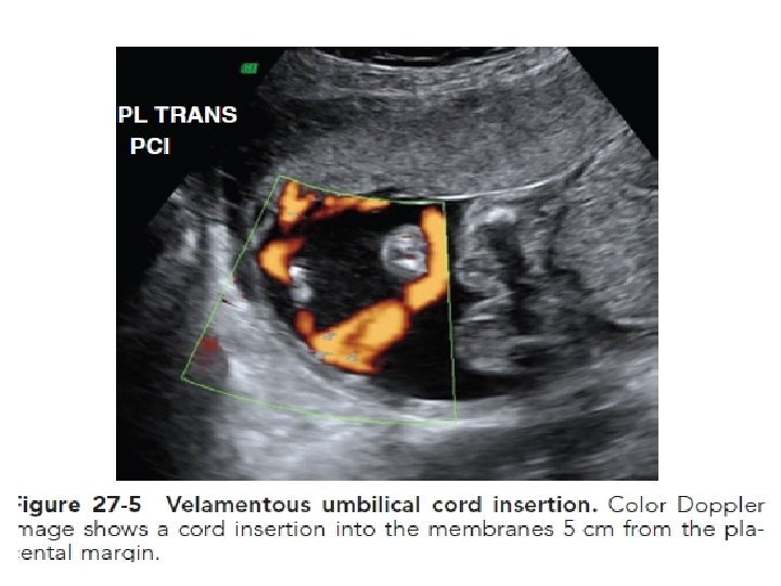
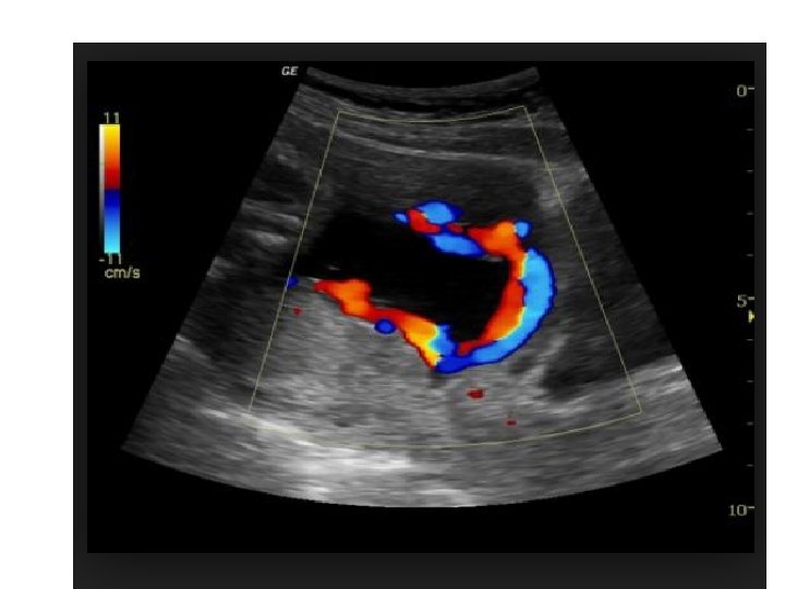
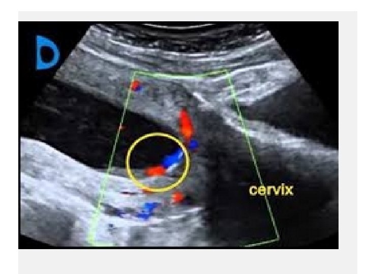
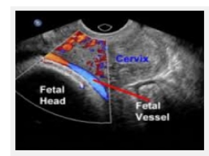
Vasa previa and velamentous umbilical cord — Vasa previa and velamentous umbilical cord insertion are uncommon, but when present they are often associated with placenta previa.
Congenital anomalies — Population-based cohort studies have reported an increase in the overall rate of neonatal congenital anomalies in pregnancies complicated by placenta previa, but no single anomaly or syndrome was associated with the disorder
Amniotic fluid embolism — A large population-based cohort study reported a strong association between placental pathology, such as placenta previa, and amniotic fluid embolism
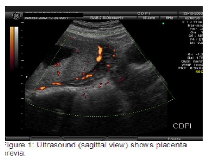
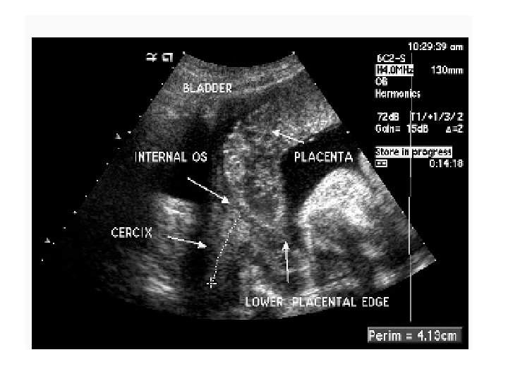
Ultrasonography • Transabdominal ultrasonography is used for initial placental localization. .
over-distended bladder can compress the anterior lower uterine segment against the posterior lower uterine segment to give the appearance of a previa
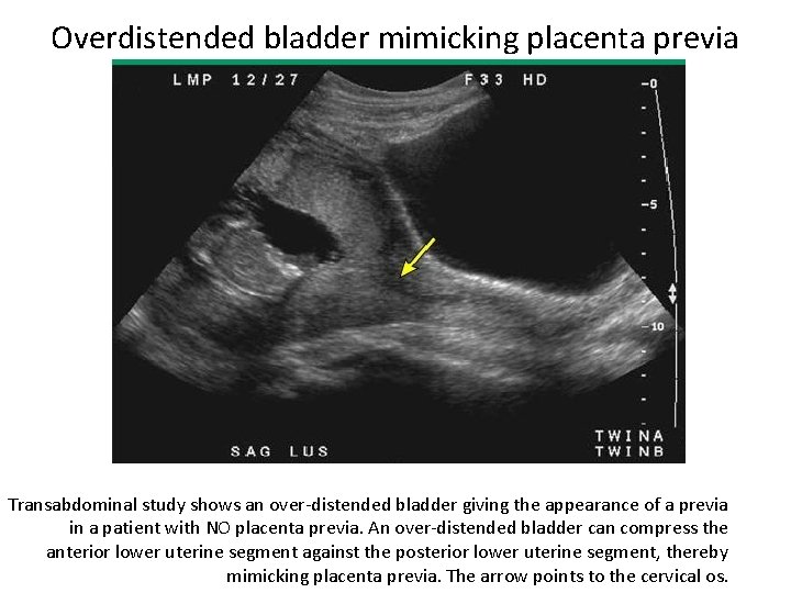
Overdistended bladder mimicking placenta previa Transabdominal study shows an over-distended bladder giving the appearance of a previa in a patient with NO placenta previa. An over-distended bladder can compress the anterior lower uterine segment against the posterior lower uterine segment, thereby mimicking placenta previa. The arrow points to the cervical os.
Specific points. when the lower uterine segment is contracting, which commonly occurs after a woman empties her bladder.
be missed near term if the fetal head is low in the pelvis may be better visualized by placing the patient in Trendelenburg position and/or gently pushing the fetal head cephalad.
transabdominal ultrasound false positive rate of transabdominal ultrasound is high 25 so the diagnosis should be confirmed by transvaginal ultrasound unless the previa is clearly central
TVS can be performed safely since the optimal position of the vaginal probe for visualization of the internal os is 2 to 3 cm away from the cervix the angle between the cervix and vaginal probe is sufficient to prevent the probe from inadvertently slipping into the cervical canal
Translabial (transperineal) an alternative technique that provides excellent images of the cervix and placenta
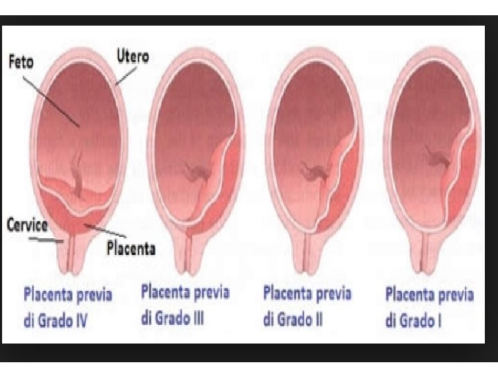
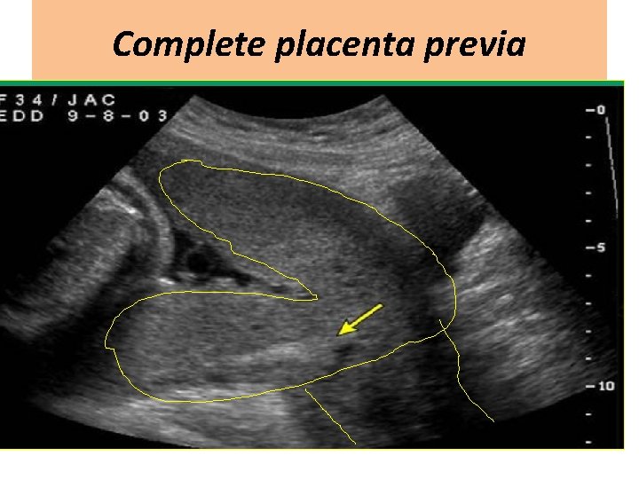
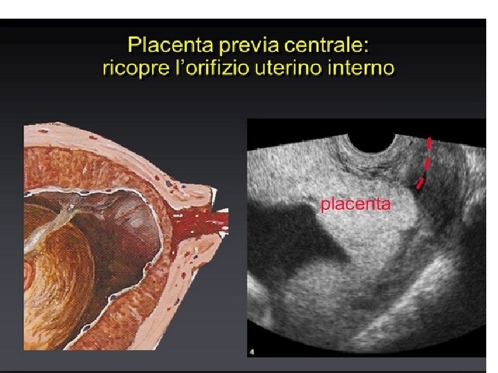
Complete placenta previa
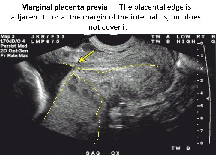
Marginal placenta previa — The placental edge is adjacent to or at the margin of the internal os, but does not cover it
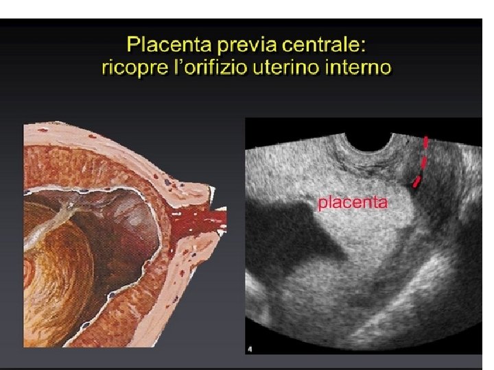
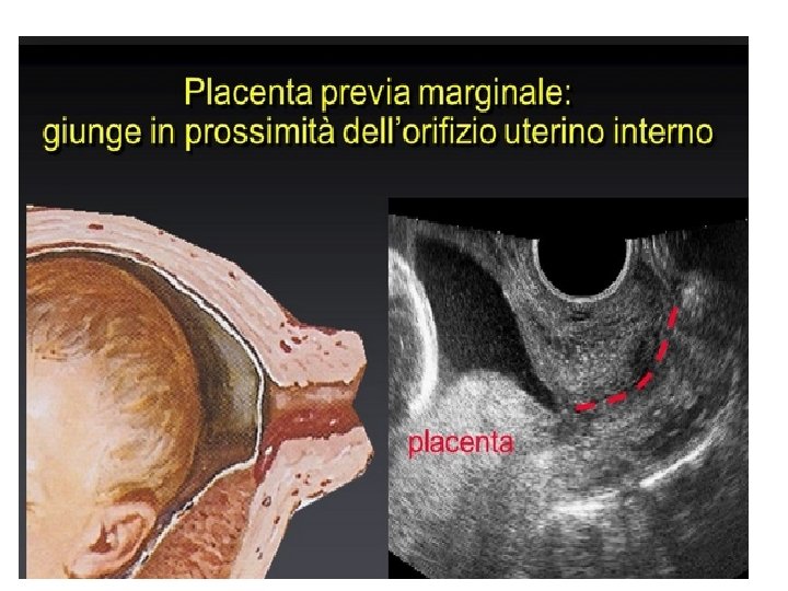
Low placenta A placental edge in close proximity to the internal os. There is no universal standard; a common definition is a placental edge >0 but <2 cm from the os.
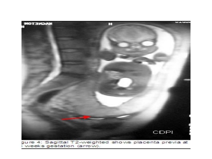
Magnetic resonance imaging well-suited to the assessment high cost, limited availability, and the well-established safety and accuracy of transvaginal sonography
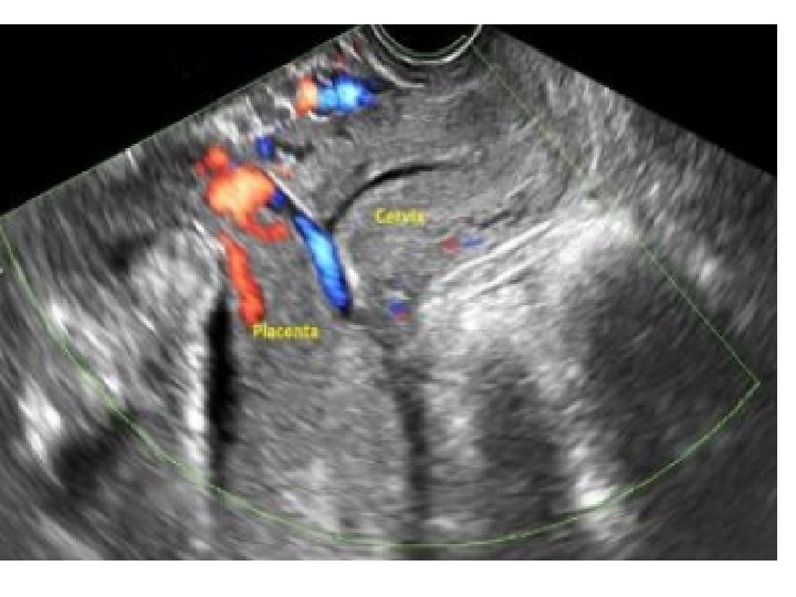
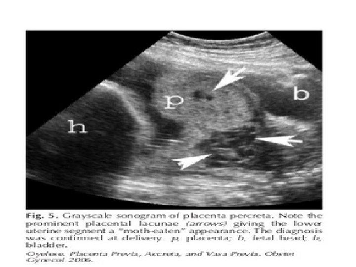
Exclusion of placenta accreta The normal interface between the placenta and bladder is characterized by a hypoechoic boundary that represents the myometrium and the normal retroplacental myometrial vasculature. lacunar flow)
management of pregnancies the clinical setting: asymptomatic women, women who are actively bleeding, women who are stable after one or more episodes of active bleeding.
follow-up transvaginal ultrasound examination • , asymptomatic placenta previa. • — There is no consensus for timing and frequency of ultrasound surveillance of incidentally diagnosed
Follow-up transvaginal ultrasound >0 mm away from the internal cervical os before 20 weeks we do not perform follow-up ultrasound examination as placenta previa at delivery is unlikely.
placentas overlying the internal cervical os by 0 to 20 mm at ≤ 20 weeks , serial transvaginal ultrasound . We begin at 28 weeks at four-week intervals until resolution or delivery.
Placentas overlying the cervical os by ≥ 20 mm are unlikely to resolve thus do not require serial assessment.
Confirm Follow-up scanning for persistence of previa at 33 -35 weeks or with vaginal bleeding Confirm placenta previa within 1 -2 weeks before delivery
avoid intercourse avoid to exercise decrease physical activity
asymptomatic women generally be managed on an outpatient basis until vaginal bleeding .
Delivery uncomplicated placenta previa expert opinion, ACOG at 36 0/7 ths to 37 6/7 ths weeks, without documentation of fetal lung maturity by amniocentesis RCOG 38 -39 W
ACUTE CARE OF BLEEDING PLACENTA PREVIA — admitted to the Labor
Maternal Assessment cardiac monitor blood pressure . Urine output estimation of vaginal blood loss
Fetal — The fetal heart rate monitoring hypoxia or anemia may result in category 2 or 3 fetal heart rate tracings.
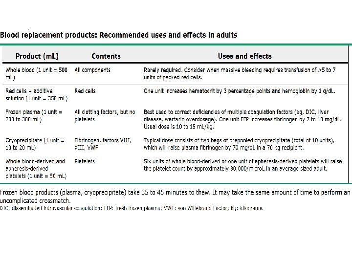
Laboratory cross-matching two to four units of PC suspicion of coexistent abruption should prompt evaluation for coagulopathy: fibrinogen level, APTT PT
A Kleihauer-Betke test on a specimen of vaginal blood can diagnose fetal bleeding from disruption of fetal vessels in placental villi, vasa previa, or a velamentous cord; results in fetal demise or a nonreassuring fetal heart rate
Transfusion guided by the volume of blood loss changes in hemodynamic parameters hemoglobin level. fails to improve after two liters of crystalloid have been rapidly infused.
Tocolysis — in symptomatic to reduce or eliminate uterine contractions, which may promote placental separation and bleeding , indomethacin inhibitory effect on platelet function avoided in women with placenta previa due to the risk of increased blood loss
Is there a place for tocolytics in women who bleed? RCOG do not administer tocolytic drugs to actively bleeding patients. to may use tocolytics administering a course of betamethasone B. mem etic NO
Is there a place for the use of prophylactic tocolytics in women to prevent bleeding? NO INDICATION RCOG
what precautions should be taken against venous thromboembolism for inpatients? mobility should be encouraged use of thromboembolic deterrent stockings adequate hydration. Prophylactic anticoagulation can be hazardous should be taken on an individual basis. should be taken on an individual basis
In the postnatal period , immobility, massive haemorrhage operative delivery . Thromboprophylaxis should be postnatal thromboembolism. Thromboprophylaxis should be considered according to Green-top Guide
Magnesium sulfate for neuroprotection in (24 to 32 weeks) placenta previa in whom a decision has been made to deliver within 24 whom a decision has been made to hours , deliver within 24 hours. Emergency delivery should not be delayed to administer magnesium sulfate.
for neuroprotection , 4 gram intravenous loading dose followed by a 1 gram infusion. discontinued by 24 hours after initiation if delivery has not occurred.
Indications for delivery — Cesarean delivery is indicated if : A nonreassuring fetal heart rate. Life-threatening refractory maternal hemorrhage Significant vaginal bleeding after 34 weeks of gestation
consent form for caesarean section? risks associated with caesarean section in general the specific risks of placenta praevia massive obstetric haemorrhage, massive haemorrhage, blood transfusion hysterectomy
Anesthesia — General anesthesia hemodynamically unstable women fetal status is nonreassuring , regional anesthesia choice in hemodynamically stable women with reassuring fetal heart rate tracings
CONSERVATIVE MANAGEMENT AFTER AN ACUTE BLEED 50 percent of women with a symptomatic previa (any amount of bleeding) were not delivered for at least four weeks mean prolongation of pregnancy of 17 days
Inpatient versus outpatient management • We discharge selected women with placenta previa whose bleeding has stopped for a minimum of 48 hours and who have no other pregnancy complications, although the safety and efficacy of this approach has not been established
candidates for outpatient care should Be able to return to the hospital within 20 minutes to maintain bed rest at home. Understand the risks by outpatient. Have an adult companion available 24 h.
Correction of anemia — Iron supplementation Stool softeners high-fiber diet help to minimize constipation and avoid excess straining that might precipitate bleeding.
Autologous blood donation is safe in stable women who meet usual criteria hemoglobin ≥ 11. 0 g/d. L to >10 g/d. L
Antenatal corticosteroids should be administered to symptomatic between 23 and 34 weeks not administer not to asymptomatic women or to those whose first bleed is after 34 or whose first bleed is after 34
Anti-D immune globulin —The presence of positive anti-D antibody titers on periodic assessment can help ensure that the patient is protected from potential alloimmunization in the setting of recurrent bleeds. Readministration is not necessary if delivery or rebleeding occurs within three weeks of administration, unless a large fetomaternal hemorrhage is detected.
Is there a place for cervical cerclage in these women? we do not advise prophylactic cerclage in these patients.
after 34 weeks with moderate vaginal bleeding Or progressively increasing frequency or volume of bleeding after cessation of an initial bleed, we deliver the patient if she has previously received a course of betamethasone anytime during the pregnancy.
after 34 w and has not received a course of betamethasone If she is clinically stable We would give a course of betamethasone and then perform cesarean delivery in 48 hours, based on limited data that even late in gestation neonatal respiratory problems may be reduced with steroid use , .
Route • cesarean delivery is always evidence of a complete placenta previa and a viable fetus. Vaginal delivery may be fetal demise previable fetus, as long as the mother remains stable.
marginal. . Route vaginal delivery can occasionally be performed high risk of intrapartum hemorrhage; we suggest scheduled cesarean delivery to minimize the risk of emergent delivery and need for transfusion.
Low placenta —vaginal delivery without hemorrhage when the placenta is more than 20 mm from the internal os.
PREGNANCY TERMINATION IN WOMEN WITH PLACENTA PREVIA — Clinicians should discuss with patients the various options for pregnancy termination , hysterotomy, dilation and evacuation, use of abortifacient document the discussion in the medical record.
second or third trimester terminations of pregnancy by administration of systemic abortifacients in women with complete previa The authors concluded that preinduction feticide might help to reduce blood loss in these cases.
Vasa previa: . twice weekly nonstress testing beginning at 28 to 30 weeks, hospitalization at 30 to 32 weeks to detect early evidence of cord compression. Hospitalization ability to perform an emergency cesarean delivery. scheduled delivery at about 35 weeks
RECURRENCE — Placenta previa recurs in 4 to 8 percent of subsequent pregnancies
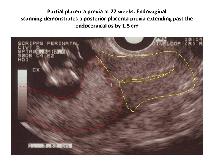
Partial placenta previa at 22 weeks. Endovaginal scanning demonstrates a posterior placenta previa extending past the endocervical os by 1. 5 cm
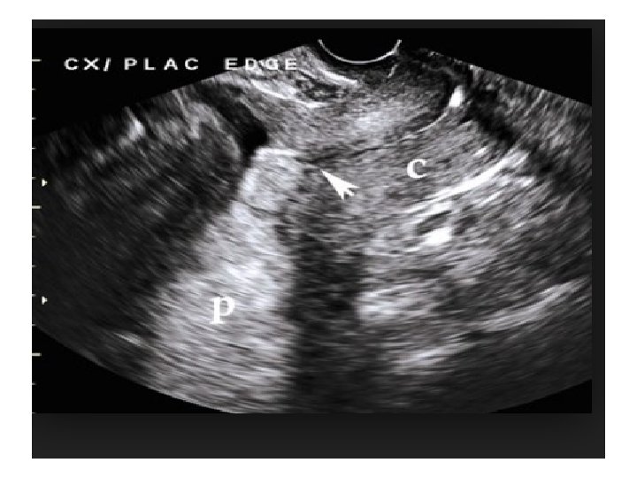
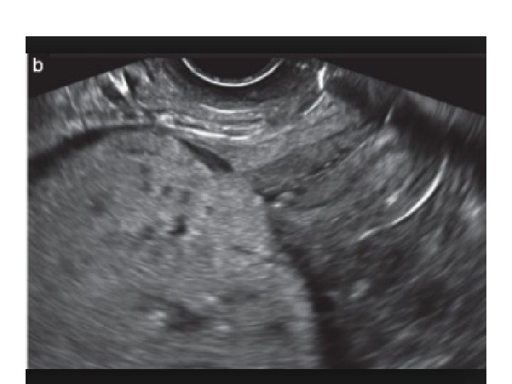


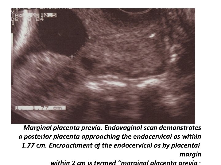
Marginal placenta previa. Endovaginal scan demonstrates a posterior placenta approaching the endocervical os within 1. 77 cm. Encroachment of the endocervical os by placental margin
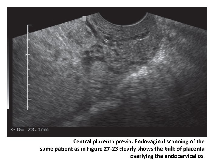
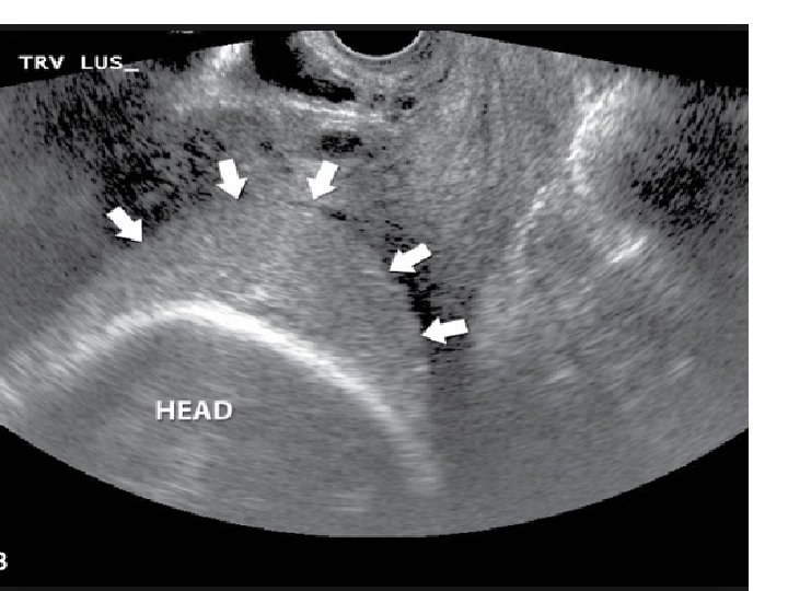
Central placenta previa. Endovaginal scanning of the same patient as in Figure 27 -23 clearly shows the bulk of placenta overlying the endocervical os.
- Slides: 104