Placenta Placenta Fetomaternal organ It connects the growing


 and one umbilical vein")

- Slides: 23
Placenta
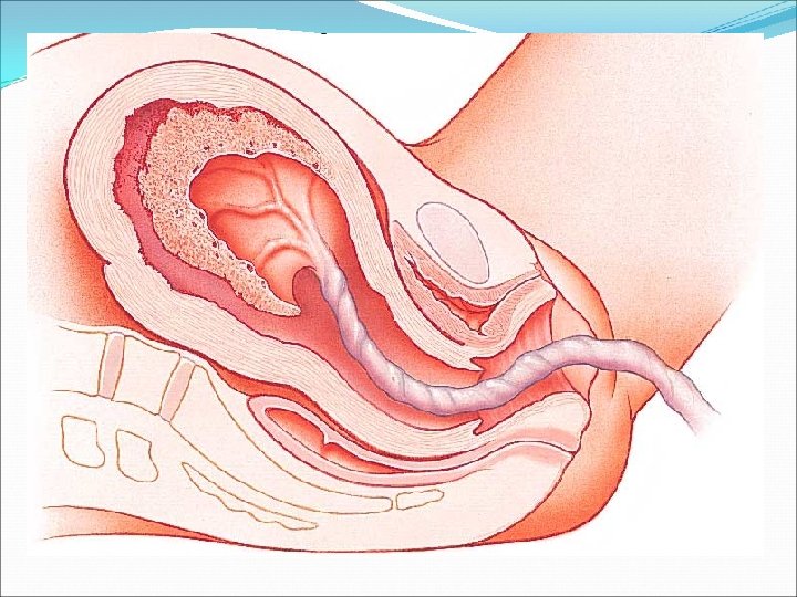
Placenta Feto-maternal organ. It connects the growing fetus to the wall of gravid uterus. Irregular maternal surface -15 -20 small lobulesmaternal cotyledons and a smooth fetal surface covered with amnion. Umbilical cord is attached to the centre of this surface.
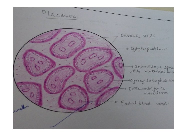
Placenta-chorionic plate on fetal side and basal plate on the maternal side. Stem villi between the two plates. Intervillous space between the stem villi filled with maternal blood.
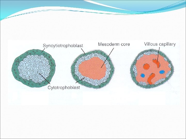
Chorionic plate Within outwards Primary mesoderm with fetal blood vessels. Cytotrophoblast. Synctiotrophoblast. Basal plate Decidua basalis containing maternal blood vessels. Outer layer of synctiotrophoblast. Outer shell of cytotrophoblast. Inner layer of synctiotrophoblast.
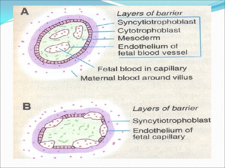
Placental barrier Early stage of pregnancy: 1. Endothelium of fetal blood vessels 2. Basement membrane of fetal blood vessels 3. Embryonic connective tissue 4. Basal lamina of cytotrophoblast 5. Cytotrophoblast 6. Syncytiotrophoblast.
Layers of barrier in placenta nearing to term: 1. The endothelial cells of fetal blood vessels 2. Basement membrane of fetal blood vessels 3. Syncytiotrophoblast cells Thickness reduces from 0. 025 mm to 0. 002 mm
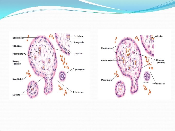
Placenta Maternal blood in the intervillous space Cross section of villus Fetal blood vessel Extra embryonic mesoderm Trophoblast
FUNCTIONS OF PLACENTA 1. Exchange of gaseous and metabolic products 2. Storage organ for glucose in the form of glycogen, calcium and iron in the first few months of pregnancy. 3. Synthesis of hormones - H. C. G, progesterone, oestrogen, placental lactogen. 4. Secretes prostaglandins
Identification points: 1. Cut section of villi’s with inner cytotrophoblast and outer syncytiotrophoblast layer. 2. Villi’s contains fetal blood vessels 3. Intervillous space has maternal blood.
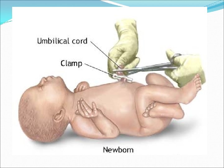
UMBILICAL CORD
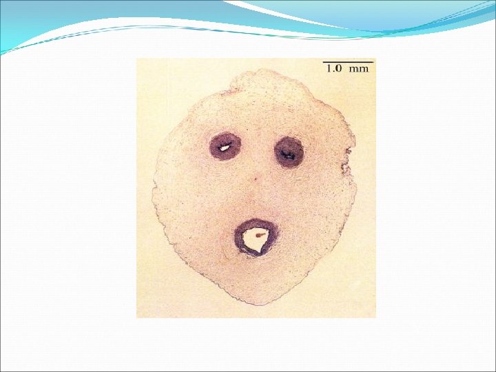
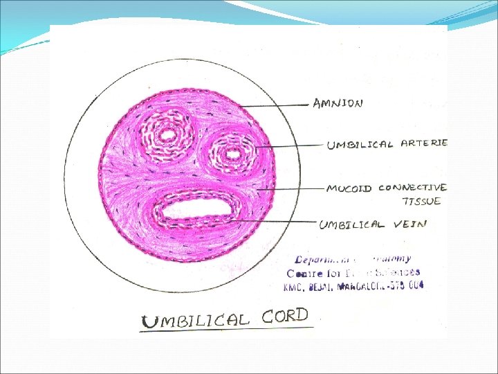
Umbilical cord Amnion Umbilical artery Wharton’s jelly Left umbilical vein
Identification points Presence of 2 umbilical arteries (medium sized artery) and one umbilical vein Presence of collagen fibres surrounding blood vessels and fibroblasts Layer of amnion which has simple sqamous epithelium.
THANK YOU.