Placenta and Fetal membrane Department of Anatomy Inje

Placenta and Fetal membrane Department of Anatomy In-je University College of Medicine

Blastocystic wall의 초기 변화 1. Trophoblast ① 밖- syncytiotrophoblast ② 안- cytotrophoblast 2. Extraembryonic mesoderm의 2 layer 생성 ③ extraembryonic somatic mesoderm ④ extraembryonic splanchnic mesoderm Chorionic cavity ⇒ ①+②+③ = chorion 3. Wall의 표면 전체에 chorionic villi가 덮혀 있다. (8주까지) → 그 후 embryonic pole에 있는 villi는 발생 2개월 말에 더 발달하여 villous chorion이 되어 placenta의 fetal part를 형성 → fetal membrane part는 villi가 없어져서 smooth chorion이 된다. (발생 3개월)
는 임신이 진 행됨에 따라 3 part로 구분")
Decidua 1. Maternal endometrium의 functional layer (=decidua)는 임신이 진 행됨에 따라 3 part로 구분 1. 2. 3. Decidua basalis Decidua capsularis Decidua parietalis ① decidua basalis는 placenta의 maternal wall이 되고, ② 아기주머니가 커지면서 decidua capsularis와 decidua parietalis는 융합되어 smooth chorion 쪽 cholionic sac의 표면을 싸게 된다. 2. Decidual cells ① 태아의 영양 ② Syncytiotrophoblast가 자궁조직에 무한정 침투하는 것을 방지 ③ 호르몬 생산

Decidua 4 weeks

Placenta의 발생 1. 8주; chorionic villi가 chorionic sac전체를 덮음. 2. Chorionic sac의 팽창 → → Decidua capsularis와 인접한 villi가 눌림 Blood flow의 감소 Villi의 퇴화 Smooth chorion 형성 3. 이후 decidua basalis 부위의 villi는 급격히 발달 → villous chorion 형성 4. 그 후 villous chorion → placenta smooth chorion → fetal membrane
 ① Decidua basalis ② Fetal part에 인접해 있다.")
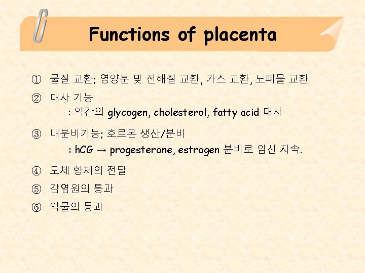
Placenta의 구성 1. Maternal side (endometrium) ① Decidua basalis ② Fetal part에 인접해 있다. 2. Fetal side (chorionic sac) ① 안쪽 면의 amniotic membrane ② Villous chorion (chorion frondosum) ③ Villi들은 intervillous space 속으로 돌출되어 있다. 3. Intervillous space 속에는 150 ml의 maternal blood가 들어있다. ; 80~100개의 spiral endometrial arteries로 피가 들어오고 spiral endometrial veins로 흘러간다. (분당 3~4회씩 교환) ; Maternal blood → spiral arteries → intervillous space → endometrial vv. of decidua basalis

Placenta 5 weeks 22 weeks

Early development of Chorionic villi

Structure of Chorionic villi 10 week Full term Villous tree

Placenta circulation 1. Fetal placental circulation ; 2. Fetus → umbilica aa. → placenta → villi로 들어가기 전에 chorionic plate 에서 수많은 가지를 냄. → villi 안에서 동맥모세혈관-정맥계 형성 → welloxygenated blood → umbilical vein → fetus Maternal placental circulation ; spiral aa. (80~100개) → intervillous space로 cytotrophoblastic shell에 존재하는 틈을 통해 박동성으로 혈액 분사 → 대사물질 및 기체성 산물의 교환 → 자궁속막정맥

Placenta circulation

Full term placenta의 형태 ① ② ③ ④ ⑤ Fetal side는 amniotic membrane으로 덥혀 있고 중심부근에 umbilical cord가 연결된다. Size : 직경= 20 cm, 두께= 3 cm, wt= 500 g 배출된 placeta의 maternal side의 불룩불룩한 부분은 cotyledone이다. 혈액 150 ml 수용 Fetal side Maternal side

Mature placenta Maternal side Fetal side
의 layers = villous wall 1. 20 주까지 ① Syncytiotrophoblast ②")
Placental membrane (placental barrier)의 layers = villous wall 1. 20 주까지 ① Syncytiotrophoblast ② Cytotrophoblast ③ Mesodermal connective tissue core ④ Endothelium of villous capillary 2. 20주 후 ; cytotrophoblast가 사라짐.

Various anomalies of Placenta

Uterine growth during pregnancy nonpregnancy 20 weeks 30 weeks

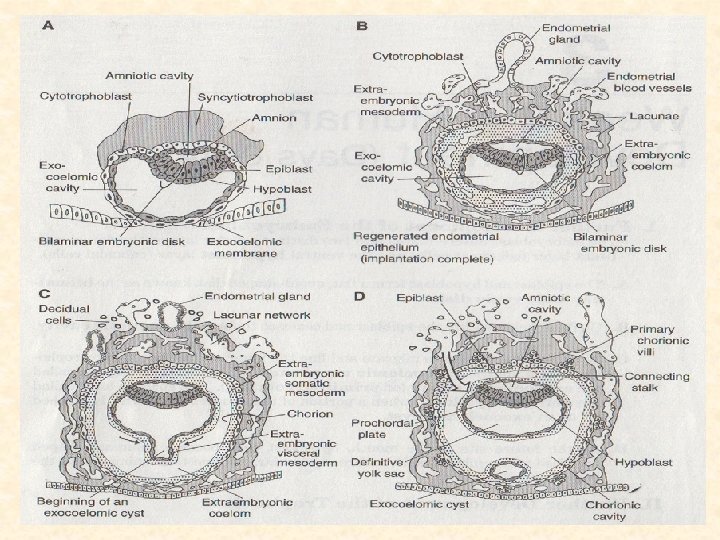
Blastocyst의 3 cavities의 변화 ① Embryonic disc 상부에 있던 amniotic cavity는 embryonic folding의 결과로 embryo 주변을 감싸게 되며 amniotic fluid의 증가로 큰 amniotic cavity가 되어 embryo (fetus)를 싸는 물주머니가 된다. ② Yolk sac은 folding의 결과로 primitive gut을 형성하고 밑부분은 umbilical cord에 포함된 vitelline duct로 남게 된다. → 흔적물 (meckel's diverticulum) ③ Chorionic cavity는 amniotic cavity가 커지면서 소실된다. 즉 extraembryoic mesoderm의 2 layer는 융합된다.
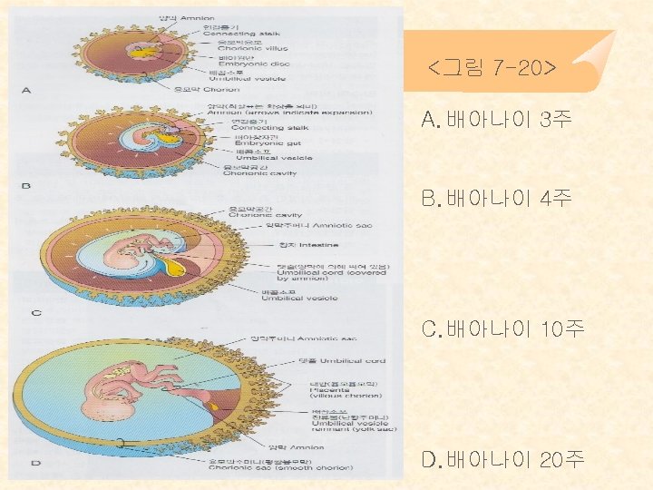

Umbilical cord ① 임신 초기 connecting stalk이 변형된 것이라 보면 된다. ② Placenta의 fetal side 중심부에서 fetus의 umbilicus 사이를 연결하는 끈 조직 → 30~90 cm (av: 55 cm), 직경= 2 cm ③ 구성 표면은 ammiotic membrane wharton’s jelly (embryonic connective tissue) One umbilical veins Two umbilical antery Yolk sac의 흔적 → vitelline duct

Umbilical cord

Knot of Umbilical cord True knot False knot

Anomalies of Umbilical cord Encircling fetal leg Marginal attachment Velamentous insertion omphalocele

Development of Aminon Amniotic cavity

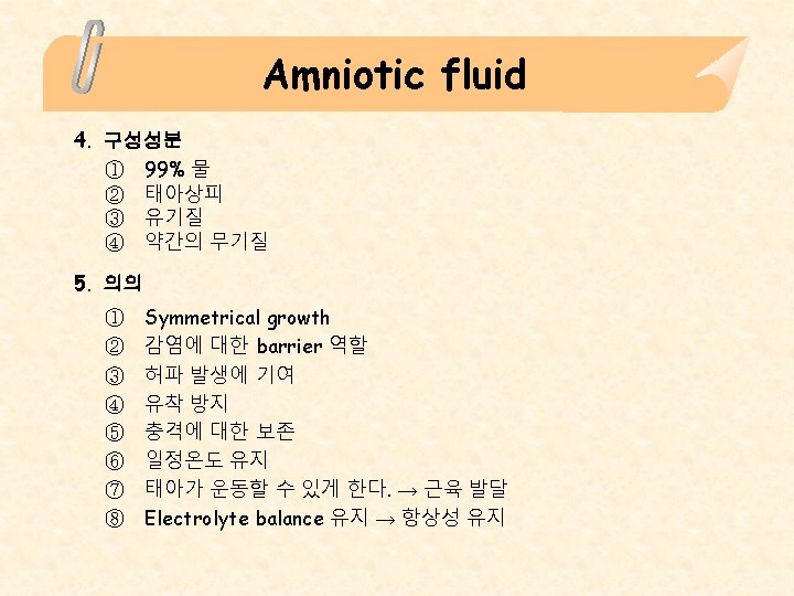
Amniotic fluid 1. 생산 ① 초기에는 소량으로 amniotic cell에서 분비 ② Decidua에서 침투된 체액이 chorion과 amnion을 통해 들어온다. ③ Placenta 형성 뒤에는 intervillous sapce에서 chorionic plate, amnion을 통해 침투된다. ⇒ origin – amnioblast, maternal blood, fetal urine 2. 양 ① 10주; 30 ml ② 20주; 350 ml ③ Full term; 700~1000 ml (av. 800 ml) 3. 순환 ; amniotic fluid내 물은 약 3시간마다 바뀜 ① Amniochorionic membrane을 통해 자궁모세혈관으로 유입 (많은 양) ② Amniotic fluid- fetal blood 사이의 상호교환 ③ Fetus가 amniotic fluid를 삼킴 → 흡수 → 태아혈류 → 모체혈류

Significance of Yolk sac � Transport nutrients during formation of uteroplacental circulation � Aids in the formation of part of gastrointestinal & respiratory system � Site of hemopoiesis until 6 th week � The primitive gut tube is derived from the dorsal part of the yolk sac � Site of origin of primordial germ cells � Fate; Meckel’s diverticulum – 2% of adult
- Slides: 29