Placenta and Amniotic fluid Structure Function and Abnormalities

Placenta and Amniotic fluid. Structure, Function, and Abnormalities

Placenta • Human placenta develops from two sources Fetal component- Chorionic frondosum Maternal component- decidua basalis • Trophoblast cells (syncytiotrophoblast , cytotrophoblast )

Human placenta is • Discoid in shape • Haemochorial • Deciduate

Placenta at Term- Gross Anatomy • • Fleshy Weight-500 gm Diameter- 15 -20 cm Thickness-2. 5 cm Spongy to feel Occupies 30% of the uterine wall Two surfaces- Maternal and fetal 4/5 th of the placenta is of fetal origin and 1/5 is of maternal origin

Fetal surface of the placenta • Covered by smooth and glistening amnion overlying the chorion • Umbilical cord is attached at or near its centre • Branches of the umbilical vessels are visible beneath the amnion as they radiate from the insertion of the cord

Maternal surface of the placenta • Rough and spongy • Maternal blood gives it dull red colour • Remanants of the decidua basalis gives it shaggy appearance • Divided into 15 -20 cotyledons by the septa

• Margins of the placenta are formed by fused chorionic and the basal plate • Placenta is attached to the upper part of the uterine body either at the posterior or anterior wall • After delivery , placenta separates with the line of separation being through decidua spongiosum (intermediate spongy layer of the decidua basalis

Structure of the placenta • Placenta is limited by the amniotic membrane on the fetal side and by the basal plate on the maternal • Between these two lies the intervillous space filled with maternal blood and stem villi with their branches

• Amniotic membranesingle layer of cubical epithelium loosely attached to adjacent chorionic plate and does not take part in placental formation • Chorionic plate- forms the • Ø Ø Ø roof of the placenta From outside inwards consists of Syncitotrophoblast Cytotrophoblast Extraembryonic mesoderm with branches of umbilical vessels

• Basal Plate- forms the floor From outside inwards it consist of Ø Compact and spongy layer of decidua basalis Ø Layer of Nitabuch Ø Cytotrophoblastic shell Ø Syncytiotrophoblast Basal plate is perforated by the spiral arteries allowing entry of maternal blood into intervillous space

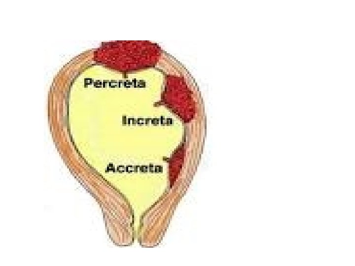
• Layer of Nitabuch - is a fibrinous layer formed at the junction of cytotrohoblastic shell with decidua due to fibrinoid degeneration of syncitotrohoblast • It prevents excessive penetration of the decidua by the trophoblast • Nitabuch membrane is absent in placenta accreta and other morbidly adherent placentas

Intervillous space: Ø Numerous branch villi arising from the stem villi project into this space Ø It is lined internally on all sides by the syncytiotrophoblast and is filled with maternal blood
 Ø Arise from the chorionic plate and extend to the")
Stem (Anchoring villi ) Ø Arise from the chorionic plate and extend to the basal plate Ø Fetal cotyledon (60 -100 ) – derived from one major primary stem villus and is the structural unit of placenta Ø Maternal cotyledon (15 -20 ) contains 3 -5 fetal cotyledons Ø Villus is the functional unit of placenta Ø Total surface of the villi for exchange varies between 4 -14 sq meters

Placental barrier or membrane Maternal and fetal blood are separated by placental membrane or barrier (0. 025 mm thick ) • Endothelial lining of fetal vessels • Connective tissue of the villi • Basement membrane • Cytotrophoblast • Syncytiotrophoblast

Placental Function Ø Transfer of gases , nutrients and waste products , namely • Respiratory function • Nutritive function • Excretory function Ø Endocrine and enzymatic function Ø Barrier function Ø Immulogical function

Factors affecting the transfer between mother and the fetus • Physical properties of the substance- molecular weight, lipid solubility, ionised substances • Area and functional integrity of the placental membrane • Rate of blood flow • Concentration gradient of the substance on either side of the exchange membrane

Mechanism involved in the transfer of substances • Simple diffusion-O 2 and CO 2 • Facilitated diffusion ( carrier mediated ) –glucose , vitamins • Active transfer ( against concentration gradient )-ions • Endocytosis- invagination of cell membrane to form intracellular vesicle • Exocytosis-Release of substances in the vesicles to extracellular space eg Ig. G immunoglobulin

Respiratory function • Although fetal respiratory movement occurs, no active exchange of gases takes place • Intake of oxygen and output of carbon dioxide take place by simple diffusion across the fetal membrane • O 2 delivery to the fetus is at the rate of 8 ml/kg which is achieved by cord blood flow of 160320 ml/min

Excretory function • Waste products from the fetus such as urea, uric acid, cretinine are excreted to the maternal blood by simple diffusion

Nutritive function Fetus obtains its nutrients from the maternal blood • Glucose- transferred to the fetus by facilitated diffusion • Lipids for fetal growth and development has dual origin. They are transferred across the fetal membrane or synthesised in the fetus • Amino acids are transferred by active transport • Water and electrolytes- Na, K , Cl cross by simple diffusion, Ca , P, and Fe cross by active transport • Water soluble vitamins are transferred by active transport but the fat soluble vitamins are transferred slowly

Barrier Function • Placental membrane is thought to be a protective barrier for the fetus against harmful agents in the maternal blood • Substances with large molecular weight or size like insulin or heparin are transferred minimally • Only Ig. G ( not Ig. A or Ig M )antibodies and antigens can cross the placental barrier • Most drugs can cross the placental barrier and some can be teratogenic • Various viruses, bacteria, protozoa can cross the placenta and affect the fetus in utero

Immunological function • Inspite of foreign paternally inherited antigens in the fetus and placenta, there is no graft rejection due to immunological protection provided by the placenta

Endocrine and Enzymatic function • Placenta secretes various hormones – Protein hormones like HCG, human placental lactogen, pregnancy specific beta 1 glycoprotein, , pregnancy associated plasma protein, steroidal hormones like estrogen and progestrone • Enzymes secreted are diamine oxidase-which activates the circulatory pressor amines, oxytocinase which neutralizes oxytocin, phospholipase A 2 which synthesizes arachidonic acid
 • One or more small lobe or cotyledon of")
Placental abnormalities Placenta succenturiata (3%) • One or more small lobe or cotyledon of placenta may be placed at a varying distance from the main placental margin • A leash of vessels connecting the main to the small lobe traverse through the membranes • Accessory lobe is developed from activated villi on the chorionic laeve

Clinical significance. If succenturiate lobe is retained following birth of placenta it may lead to Ø PPH Ø Subinvolution Ø Uterine sepsis Ø Polyp formation Treatment- exploration of the uterus and removal of the lobe

Circumvallate placenta Development • Due to smaller chorionic plate than the basal plate • The chorionic plate does not extend into the placenta margin • The amnion and chorion are folded and rolled back to form a ring leaving a rim of uncovered placental tissue

Morphology • Fetal surface has a central depressed zone surrounded by a usually complete thickened white ring made up of double fold of amnion and chorion • Branching vessels radiate from the cord insertion upto ring only • Area outside the ring is thicker, elevated and rounded

Abnormal Placenta • • Placenta Previa. Placenta acreta. Placenta increta. Placenta percreta.
- Slides: 29