Pituitary Stalk Lesions Omid Gharooei Ahangar Problem list

Pituitary Stalk Lesions Omid Gharooei Ahangar

Problem list • Young healthy woman • Prolactinemia with micro adenoma • Mass of stalk • Ca in ULN • PTH high • Vitamin D low • IGF 1 HIGH

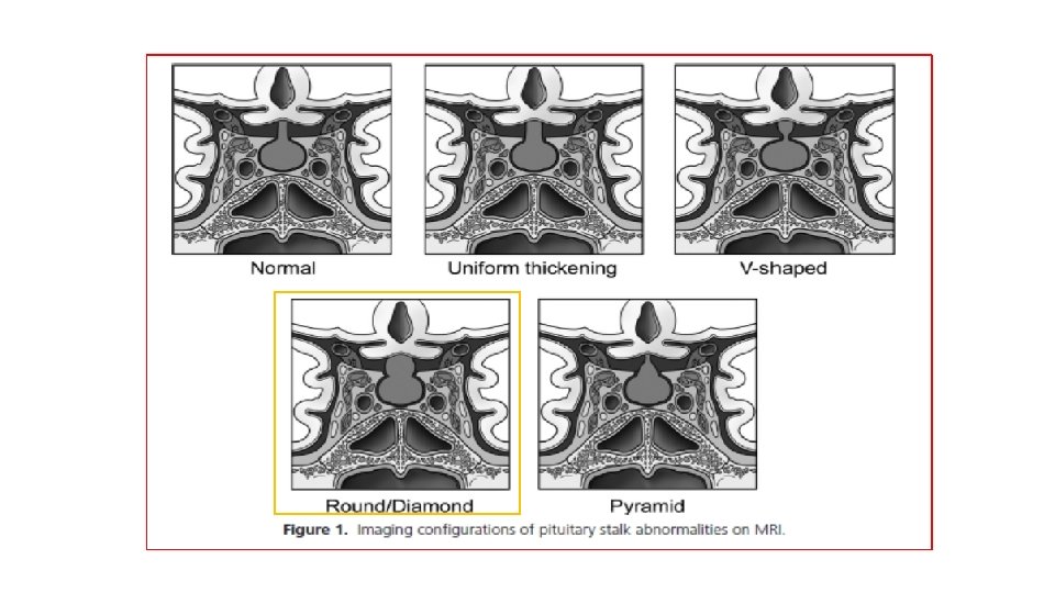
AGENDA • DDX of pituitary stalk lesion • Kinds of stalk lesion shape in MRI • DDX of any shape • What DX close to our patient • What the next plan
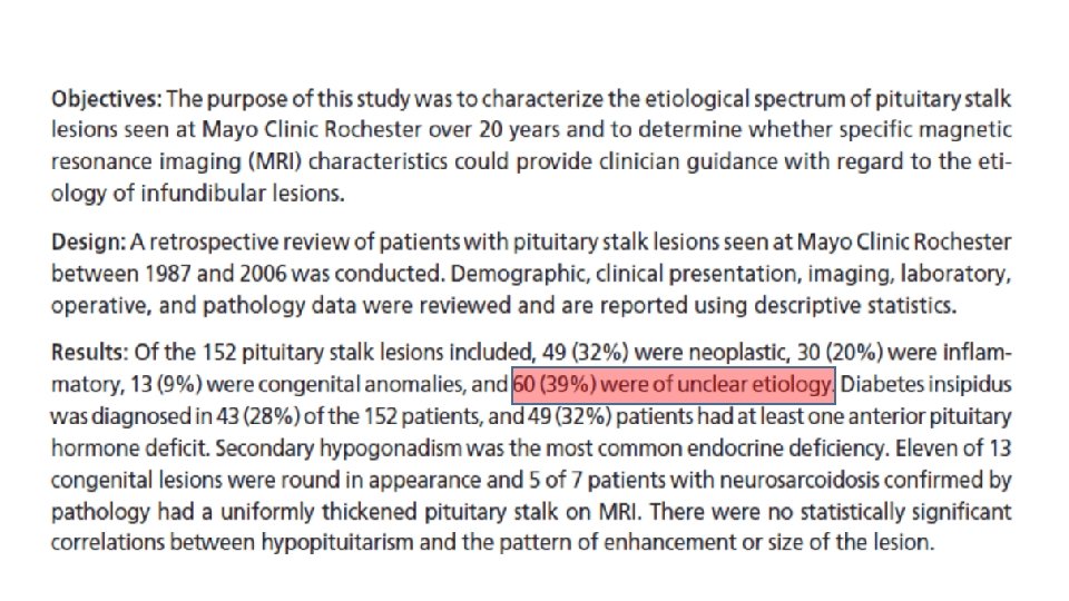

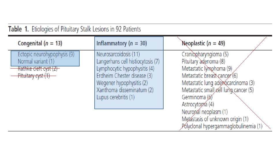
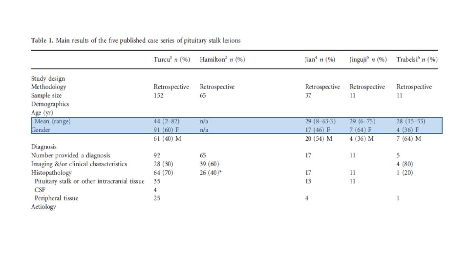
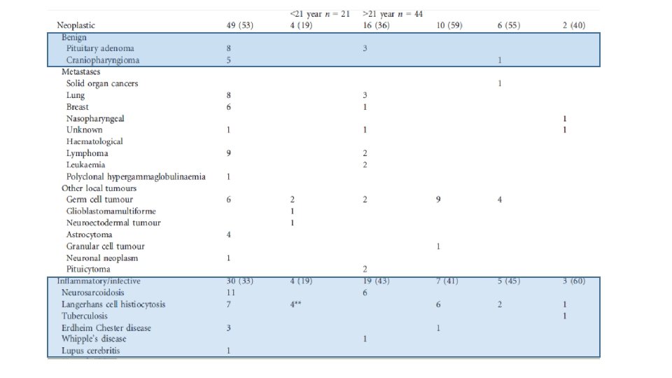
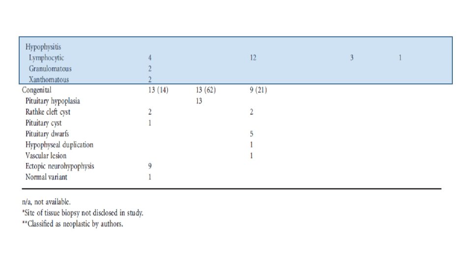
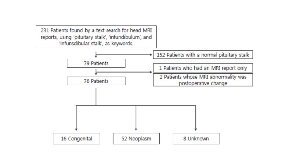
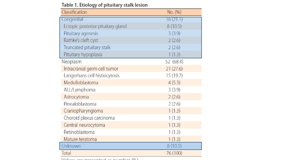
• The etiological spectrum of these lesions is broad and can be divided in 3 categories: • neoplastic • inflammatory/infectious • Congenital.

In our patient • Because of chronicity it is unlikely to be malignant cause so I can not consider it. • Also the lesion was not cystic so DDX of cystic can not be consider. • So DX limited to some congenital or inflammatory cases.
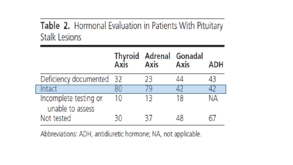
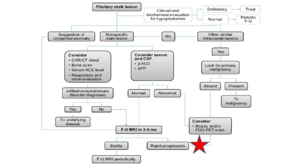

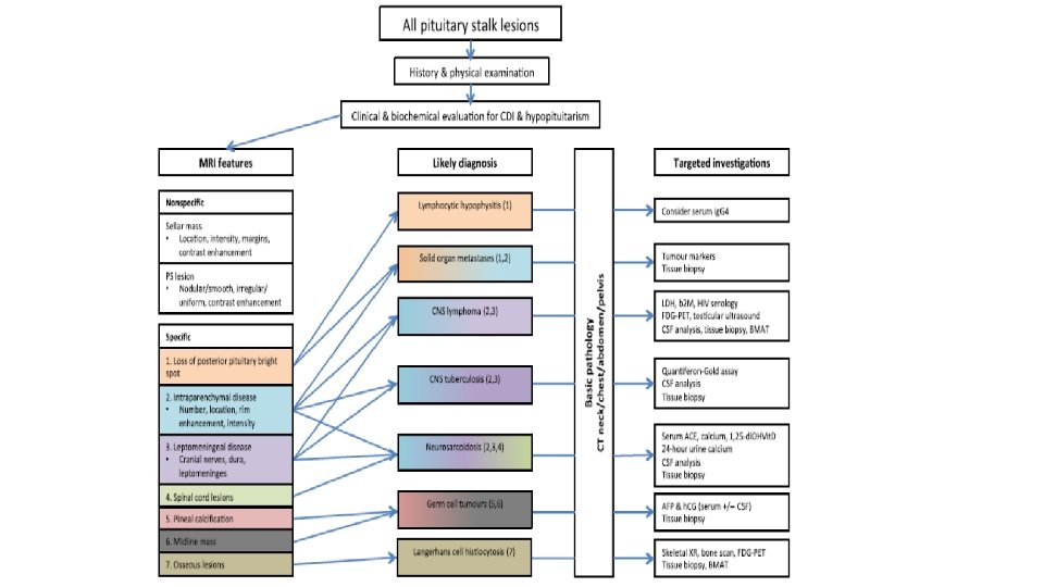
Investigating a pituitary stalk lesion • All patients discovered to have a PS lesion, including incidental cases should undergo the following noninvasive preliminary investigations. • Anterior pituitary hormones (FSH , LH, testosterone (men), estradiol (women), ACTH, cortisol, TSH, f. T 4, prolactin, GH, IGF-1) • Full blood examination, blood film, biochemistry, calcium and phosphate, renal and liver function • Markers of cell turnover (LDH, B 2 M) and inflammation CRP (C-reactive protein), ESR (erythrocyte sedimentation rate) • Serum ACE, 1, 25 dihydroxyvitamin D • Serum AFP (alpha-fetoprotein) and h. CG (human chorionic gonadotropin) • c-ANCA (cytoplasmic antineutrophil cytoplasmic antibody) • Quantiferon Gold in the presence of risk factors for tuberculosis. Urinalysis • CT neck, chest, abdomen and pelvis with contrast

Incidental pituitary stalk lesion • We suggest clinical review and repeat pituitary hormone testing in 3 months, and MRI pituitary in 6 months, and thereafter every 6– 12 months during the first 2– 3 years. • If the PS lesion increases significantly in size (>65 mm in width) or new lesions develop, or if pituitary hormone deficiency arises or the patient’s clinical status changes, then we suggest proceeding to more invasive investigations including consideration of PS biopsy.

BX We suggest PS biopsy when all the following criteria are met: • Isolated PS lesion (especially if width >65 mm) with or without associated pituitary pathology on imaging. (Normal measuring 3. 25± 0. 56 mm in transverse diameter at the optic chiasm and 1. 91± 0. 40 mm at its pituitary insertion) • CDI and/or hypopituitarism, or progressive disease on imaging. • Diagnosis unclear from extensive investigations. • Alternate tissue sites for biopsy not available or accessible. • Pituitary neurosurgical expertise available.

Congenital lesions • Pituitary stalk interruption syndrome is characterized by the following: an ectopic posterior pituitary, a hypoplastic anterior pituitary, and lack of or significant thinning of the pituitary stalk. On MRI, the ectopic posterior pituitary appears as a high-signal nodule in the area of the infundibular recess of the third ventricle. GH deficiency is a common feature. • septooptic dysplasia, is characterized by midline forebrain abnormalities, optic nerve hypoplasia, and hypopituitarism. • Duplication of the pituitary, including the stalk has been reported. These cases are often associated with midline facial abnormalities and many of these patients die in infancy.

What about calcium?
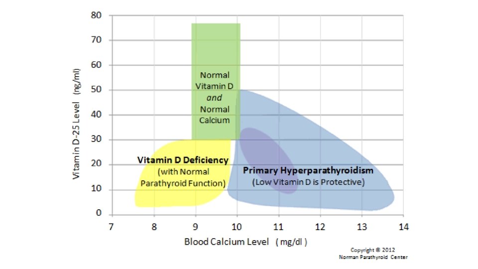

• Here is the most important fact on this page: • since vitamin D is required for humans to absorb calcium in their intestines, a low vitamin D cannot ever be the cause of high blood calcium. This fact is not debatable. • Thus, if you have a low vitamin D and your calcium is above 10. 0, then the high calcium in your blood must have come from somewhere else other than your diet (it came from your bones). Thus, if you have a low vitamin D, and a calcium level above 10. 1, then you are hyperparathyroidism almost guaranteed to have primary

What is next step? • I am suggested BX of lesion Because of size and progression • Correction of vitamin D • Repeated IPTH after correction • Urine 24 hours for hypercalciuria • GTT for elevated IGF-1 • Consider MEN_1

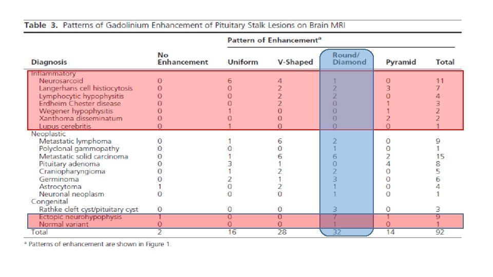
Lymphocytic hypophysitis • Although it most commonly affects the anterior pituitary, and rarely involves the posterior pituitary and PS. • A probable diagnosis of lymphocytic hypophysitis can be made on the basis of clinical, laboratory and imaging findings, however histopathology is required for a definitive diagnosis. • Current literature supports the use of supraphysiological doses of glucocorticoids when there is significant enlargement of the pituitary or PS, CDI or hypopituitarism.

Neurosarcoidosis • neurosarcoidosis occurs in 5– 15% of cases and usually in the context of widespread disease. • The pituitary, PS or hypothalamus are affected in 18% of patients with neurosarcoidosis. • Neurosarcoidosis requires a tissue sample for diagnosis. Serum ACE is a useful preliminary investigation (specificity 95%, sensitivity 24– 76%). • Chest imaging is useful as it is abnormal in up to 90% of patients with systemic sarcoidosis. • The presence of ACE in the CSF is highly specific (94– 95%), but lacks sensitivity (24– 55%) and therefore is not recommended as a first-line investigation.
- Slides: 33