Pituitary Disorders Objectives Anatomy of hypothalamus and pituitary

Pituitary Disorders

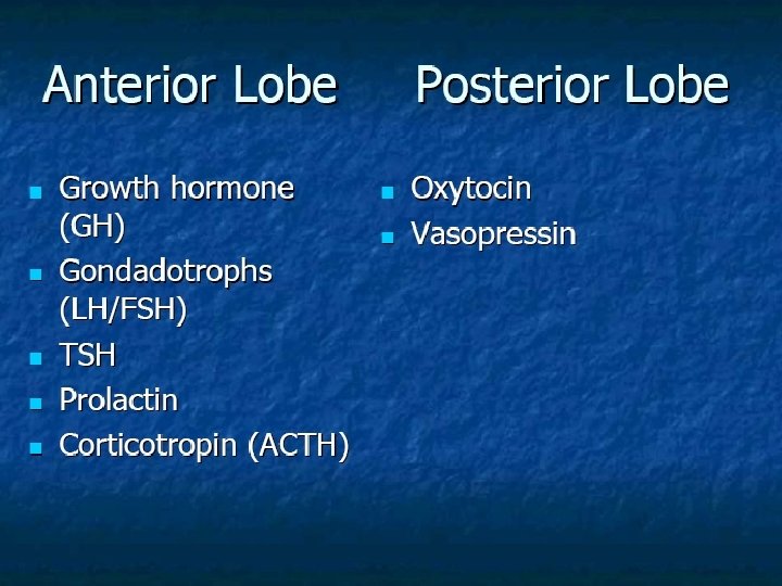
Objectives Anatomy of hypothalamus and pituitary Function of hypothalamus and pituitary Hormones: Anterior pituitary with related disorders Posterior pituitary with related disorders

Hypothalamic-Pituitary Axis The hypothalamus is the coordinator of Endocrine system Received signals from cortical brain, autonomic function, environment cues like light and temperature It affects function of thyroid gland, adrenal, gonads, growth, milk production and water balance

Hypothalamus-pituitary Axis nonendocrine functions such as temperature regulation, the activity of the autonomic nervous system, and control of appetite.

Endocrine system

Hypothalamus At the base of the brain, below third ventricle, above pituitary gland optic chiasm Hypothalamus is connected to the pituitary gland by pituitary stalk which connect median eminence to the pituitary gland Multiple nuclei in anterior part producing hormones to anterior pituitary Paraventricular and supraoptic nuclei produce ADH to control poster pituitary function

Hypothalamus

Function of hypothalamus Terminals of hypothalamic neurones are in the median eminence carrying the hormones through capillary plexus to the pituitary gland Release all the hormones to control the pituitary function beside neuroendocrine function

Hypothalamus

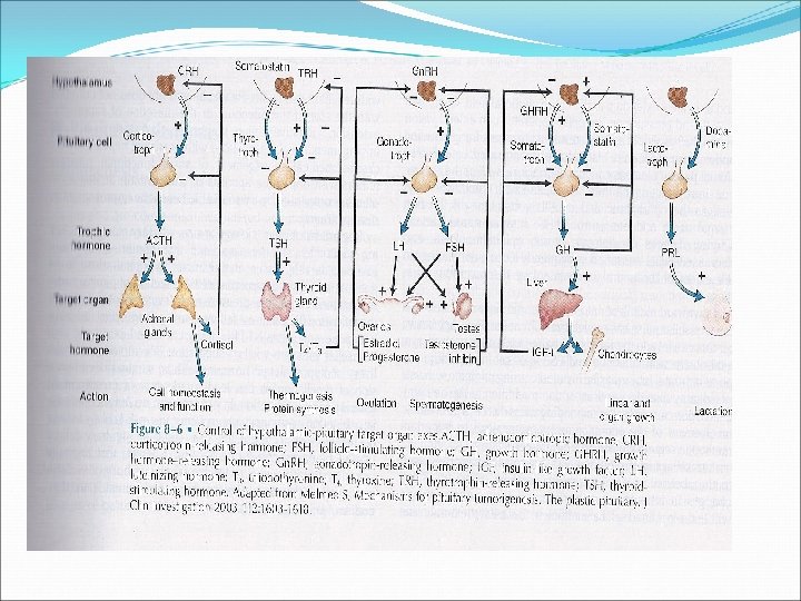
Major hypothalamic hormones and their effect on anterior pituitary hormones Hypothalamic stimlatory hormones Pituitary hormones Corticotropin-releasing hormone - 41 amino acids; released from paraventricular neurons as well as supraoptic and arcuate nuclei and Adrenocorticotropic hormone - basophilic corticotrophs represent 20 percent of cells in anterior pituitary; ACTH is product of proopiomelanocortin (POMC) gene limbic system Growth hormone-releasing hormone - two forms, 40 and 44 amino acids Growth hormone - acidophilic somatotrophs represent 50 percent of cells in anterior pituitary Gonadotropin-releasing hormone - 10 amino Luteinizing hormone and follicle-stimulating hormone gonadotrophs represent about 15 percent of anterior acids; mostly released from preoptic neurons pituitary cells Thyrotropin-releasing hormone - three amino acids; released from anterior hypothalamic area Thyroid-stimulating hormone - thyrotropes represent about five percent of anterior pituitary cells Prolactin-releasing factors - include serotonin, Prolactin - lactotrophs represent 10 to 30 percent of acetylcholine, opiates, and estrogens anterior pituitary cells Hypothalamic inhibitory hormones Somatostatin - 14 amino acids Inhibits the release of growth hormone Prolactin-inhibiting factors - includes dopamine Major prolactin control is inhibitory

Hormonal regulation

Growth hormone Polypeptide hormone Somatotrophs of anterior pituitary Action is mediated by IGF-I Half life is 20 -50 mins Has a binding protein: GHBPs Pulsatile secretion: variable level in the blood Binds to its receptor on cell- surface: cytokine receptor Lack intrinsic enzyme activity Has similar receptor structure to others: leptin, IL-2, PRL Controlled by HP and peripheral factors GHRH stimulates it, somatostatin inhibits

Growth hormone ↑↑ GH: Physiologic: sleep, exercise, stress, fasting Pathologic: Liver cirrhosis, AN, CRF, starvation Pharmacologic: Estrogen, ACTH, ADH, GHRH, Ghrelin dopamine agonist, K infusion, serotonin arginin ↓↓ GH: Physiologic: ↑glucose, ↑ FFAs, Pharmacologic: Somatostatin, GH, GC, PG Pathologic: ↑ or ↓ T 4, Obesity
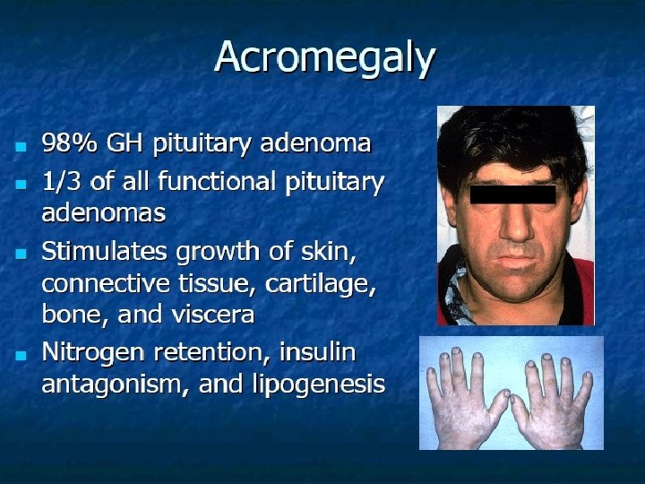
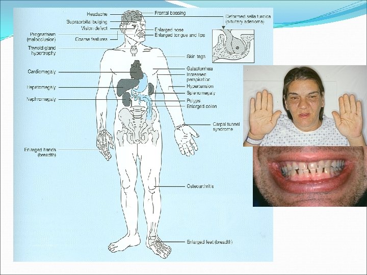
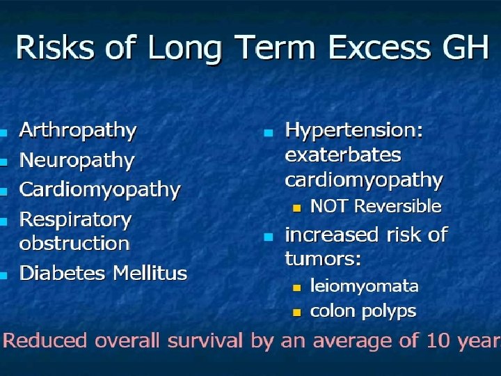

Diagnosis of acromegaly GH, IGF-I Oral glucose tolerance testing To assess excess GH secretion

Acromegaly-Diagnosis Oral glucose tolerance test Measure GH and IGF-I : high

Growth hormone deficiency Diagnosis in children and adult

Diagnosis of GH-deficiency and management GH, IGF-I level Dynamic testing: clonidine stimulation test, glucagon stimulation, exercise testing, arginine-GHRH, insulin tolerance testing X-ray of hands: delayed bone age In Adult: Insulin tolerance testing, MRI pituitary to rule out pituitary adenoma Management: GH replacement

Cortisol under ACTH Stable circadian rhythm Altered by: Physical stress Psychological stress CNS and pituitary disorder liver and renal failure

Cushing’s Disease High ACTH leading More common in females 3 -8 times than in men Generally not common : 5 -25 per million
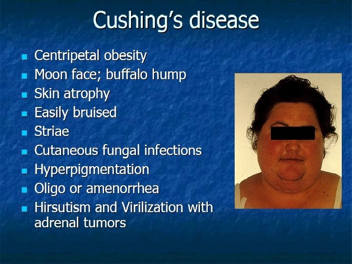
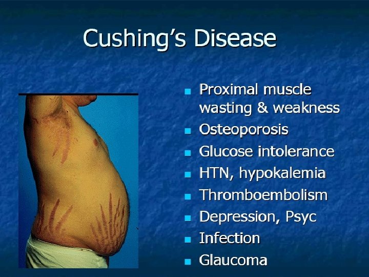

Cushing’s disease

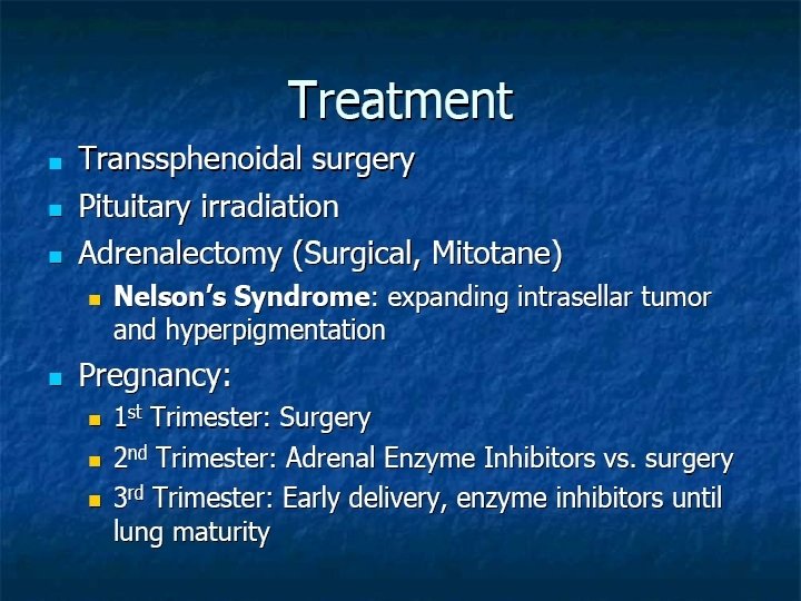
Diagnosis of cushing’s disease 24 hr urine free cortisol Overnight 1 mg dexamethasone suppression testing ACTH level and Pm cortisol MRI pituitary: for pituitary adenoma

hyperprolactinemia
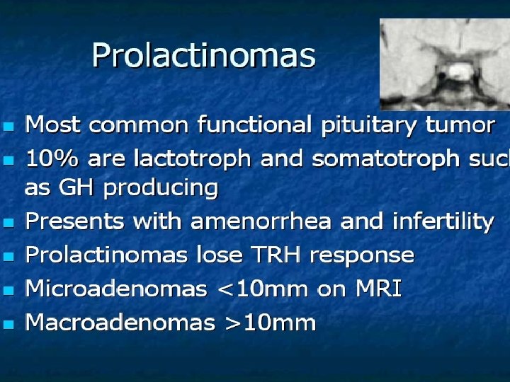

Symptoms Premenopausal women: Hypogonadism, amenorrhoea or olighomenorrhoea, infertility, Galactorrhoea Men: hypogonadortropic hypogonadism, decreased libido, infertility, galactorrhoea, Gynecomastia

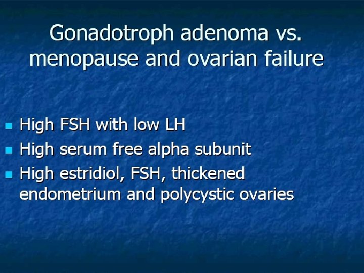
LH and FSH-secreting adenoma
- Slides: 41