
Pitfalls in Fetal Echocardiography Jung Yun Choi Department







- Slides: 29
Pitfalls in Fetal Echocardiography Jung Yun Choi Department of Pediatrics Seoul National Univ. Children’s Hospital
Pitfalls of Fetal Echocardiography • Pitfalls due to technical factors • Pitfalls related to interpretation
Pitfalls due to technical factors Structural Image Doppler Spectral Image Color Doppler image
Pitfalls in Structural Image Echo drop-out Artifact : Shadowing Imperfect resolution Slice thickness
Echo drop-out; Shodowing; Artifact
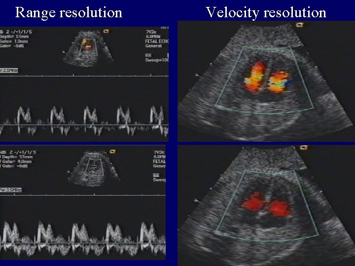
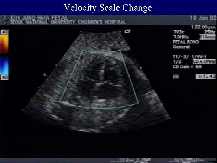
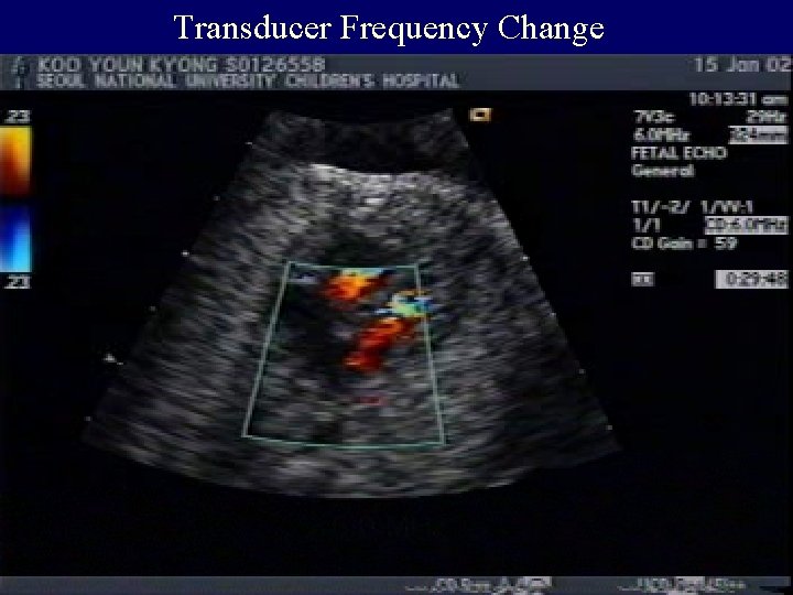
Pitfalls in Color Doppler image Low frame rate Poor range resolution Poor velocity resolution
Range resolution Velocity resolution
Resolution of color Doppler study
Velocity Scale Change
Transducer Frequency Change
Pitfalls in assessing size Normal size Large heart Small heart
Normal size • What is normal ? Mean ± 2 SD • Growth parameters : gestational period, body weight, head circumference, height, etc • Heart parameters : dimension, area, volume
Large Heart • Right or left or both sides • Causes – cardiac malformation – valvular or ventricular dysfunction – secondary to fetal diseases • Effects on fetus; long-term effects
Small Heart • Anatomic definition: diameter / area below - 2 SD ? • Functional definition : too small to be a ventricle • Growth parameters : body weight > gestational age • Any potential of catch up growth ?
How small? • Too small: hardly seen, no potential of growth • Questionable • Not too small: -2 ~ -3 SD, may have a potential
Too Small Heart • Causes : HLHS, HRHS • How to assess : PFO, ductus, coronary fistula • Counsel : single ventricle and Fontan
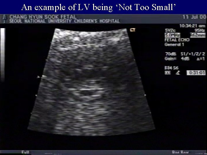
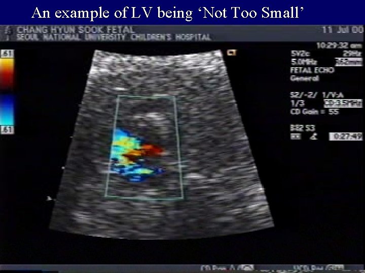
Not Too Small • Causes: unknown, COA, AS, MS, PS etc • How to assess: complete study on heart / vessel • Counsel : cautious optimism
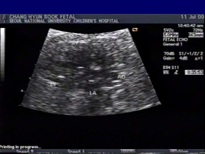
An example of LV being ‘Not Too Small’
An example of LV being ‘Not Too Small’
Pitfall in Natural History • Newly diagnosed in the 3 rd trimester • Cardiac malformations become worse • Cardiac defects / diseases become better
Newly diagnosed in the 3 rd trimester • Cardiac malformation • Cardiomyopathy • Cardiac tumor • Secondary cardiac disease
Natural History May get worse May get better May stay the same
Pitfalls in Arrhythmia • Technical pitfalls: difficult to obtain signal • Interpretation: requires thorough knowledge • Common mistake – transient bradycardia – intermittent premature contraction
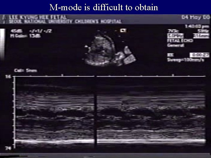
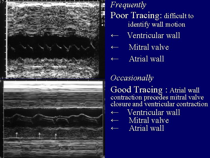
M-mode is difficult to obtain
Frequently Poor Tracing: difficult to identify wall motion ← Ventricular wall ← Mitral valve ← Atrial wall Occasionally Good Tracing : Atrial wall contraction precedes mitral valve closure and ventricular contraction ← Ventricular wall ← Mitral valve ← Atrial wall
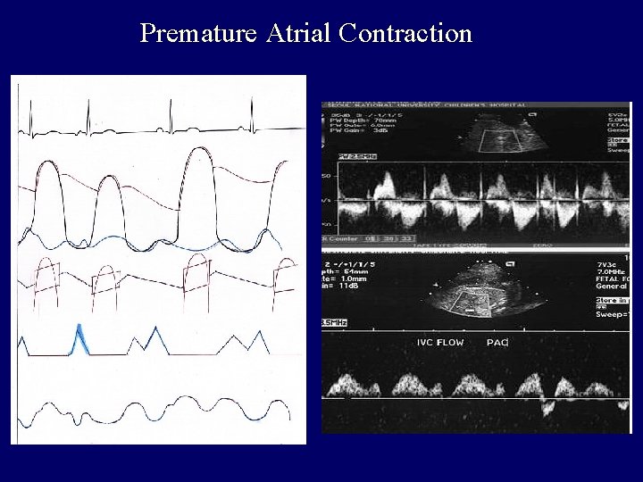
Premature Atrial Contraction
Brief bradycardia is common
Intermittent premature contraction is frequent