Pigmented Choroidal Lesions UHNM Choroidal Naevus clinic Dr


 and no other risk factors")


 Tumour within 3")


- Slides: 27
Pigmented Choroidal Lesions – UHNM Choroidal Naevus clinic Dr Andrew Brown November 2016
• Differentiating between benign and malignant choroidal lesions • When to refer urgently • When to refer routinely • How to refer • UHNM choroidal Naevus Clinic
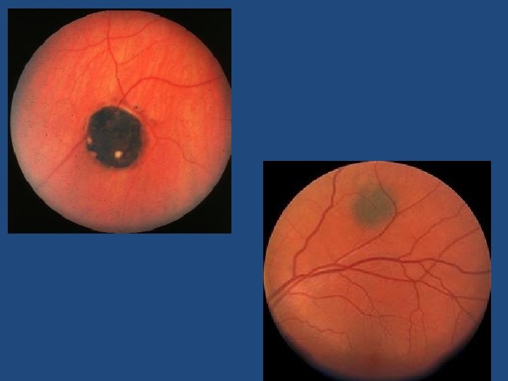
Problem • Naevi are present in 4. 6 -7. 9% of the Caucasian population – common • Malignant transformation of naevi occurs in approximately 1 in 8, 845 – rare • Can be difficult to tell apart
Definitions • Naevus – Thickness < 2 mm (USB) and no other risk factors • Melanoma – Thickness > 2. 5 mm (USB) and 2 other risk factors • Indeterminate – Fitting into neither of the above categories
From: Size Overlap between Benign Melanocytic Choroidal Nevi and Choroidal Malignant Melanomas Invest. Ophthalmol. Vis. Sci. . 2008; 49(7): 2823 -2828. doi: 10. 1167/iovs. 07 -1603
• Lesions smaller than 3 mm unlikely to be melanoma • Lesions larger than 6 mm unlikely to be naevus • 1 disc diameter approx 1. 5 mm • Lesions less than 1. 5 mm thick unlikely to be melanoma • Lesions greater than 2 mm thick unlikely to be naevus • Impossible to determine thickness to this degree of accuracy clinically
Learning point • Flat lesions 3 mm or less – observe in community (digital imaging preferable) • Lesions 6 mm or more urgent referral • Lesions 3 mm – 6 mm asses risk factors to determine urgency of referral • Lesions that are raised clinically require referral regardless of diameter • Initial assessment must include pupil dilation
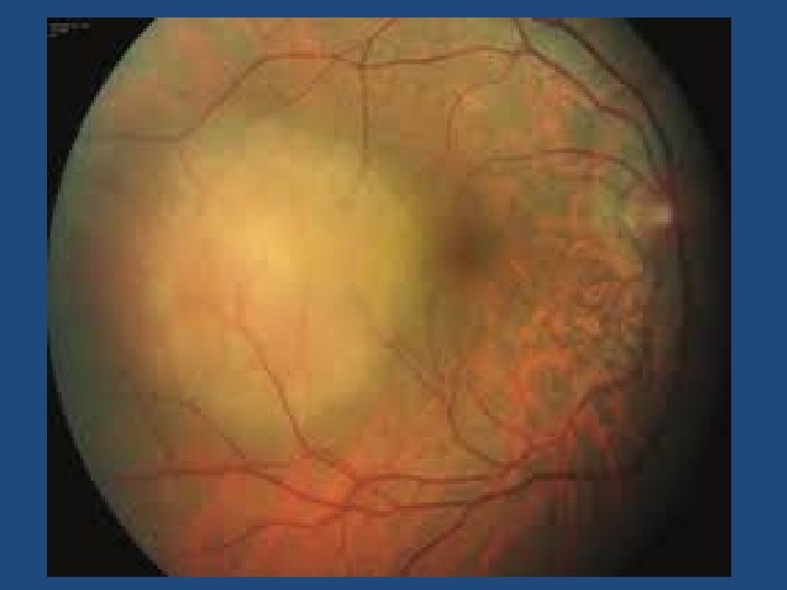
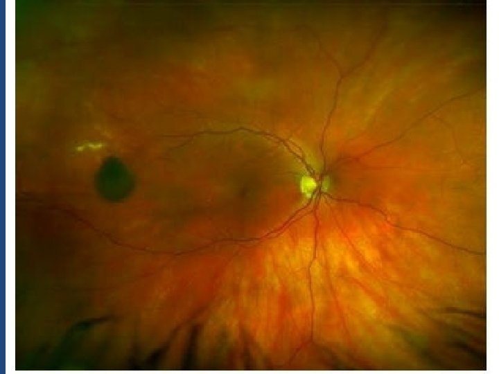
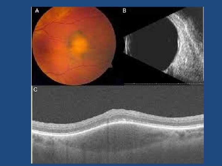
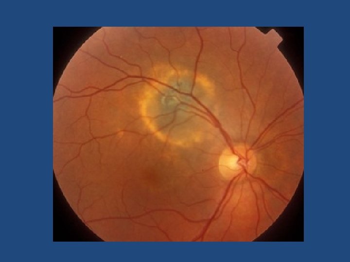
Risk factors • • • Symptoms Sub-retinal fluid Orange pigment (autofluoresent) Tumour within 3 mm optic Disc Absence of Halo Amelanotic (p=0. 002) (p<0. 001) (p=0. 009)
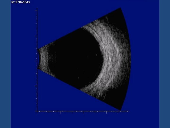
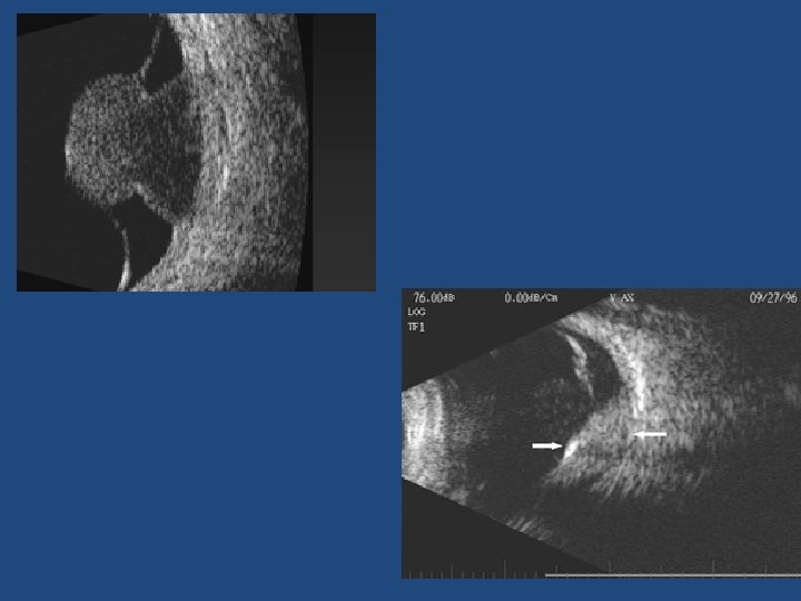
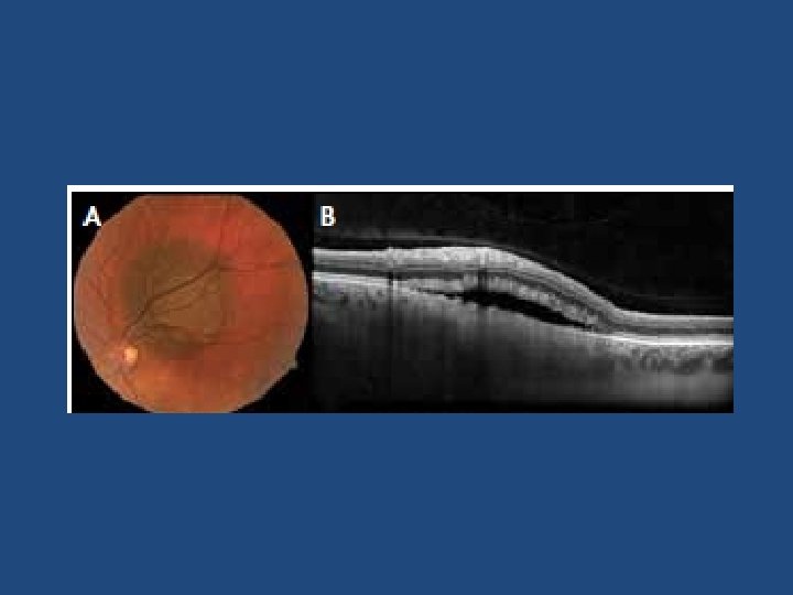
Other risk factors • Ultrasonographic hollowness • Thickness > 1. 5 mm • Suspicious OCT findings (p<0. 001)
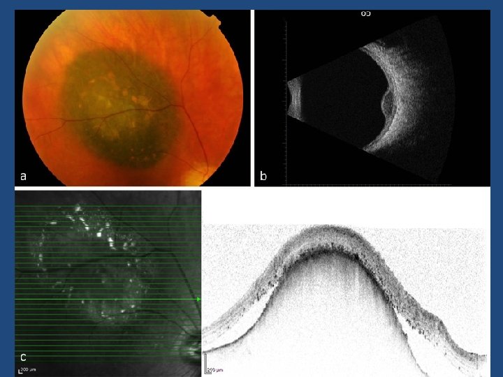
Suspicious OCT findings • • • Intra retinal oedema Shaggy photoreceptors Loss of ELM Loss of IS/OS junction (p=0. 003) (p=0. 005) (p=0. 008) (p=0. 02)
Risk Factors • 1 risk factor = 38% chance of growth • 2 risk factors = 50% chance of growth • Risk factors only relevant for flat lesions larger than 3 mm largest diameter
Learning Point – Lesions 3 -6 mm size • 0 -1 risk factor – routine referral • 2 or more risk factors – urgent referral
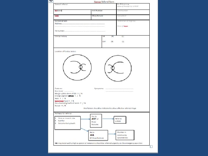
Community Referral Guidelines Lesions over 3 mm diameter Any lesion with documented growth Any raised lesion, regardless of size Risk factors should be indicated to allow effective referral triage • Any lesion with a high suspicion of melanoma should be referred urgently via the emergency eye clinic • •
UHNM Clinic • • Measurement of visual acuity Dilation and Retinal examination of new patients B ultrasound Colour photographs OCT autofluorescence OCT-EDI Optos wide field imaging Optos autofluoresence
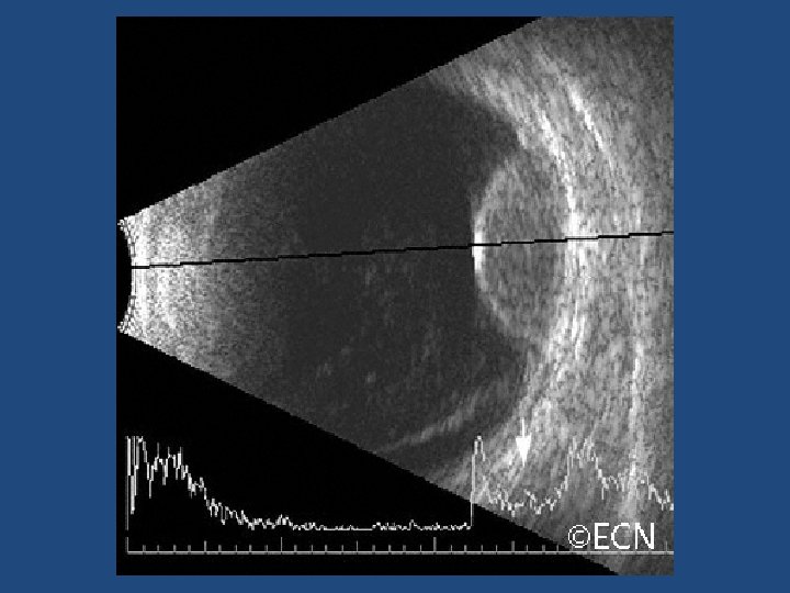
Ultrasound vs OCT-EDI • OCT-EDI measurements are approximately 50% less thick than USB measurements (i. e. a 3 mm lesion on USB will measure 1. 5 mm on OCT-EDI) • USB cannot detect/measure lesions <0. 75 mm thick – these can be measured with OCT-EDI • OCT-EDI less helpful as tumours enlarge (>3 mm) • The lesion must be at least 1. 5 mm thick on USB measurement before meaningful comments on echogenicity can be made • USB echogenicity is determined by the density of cellularity of the tissue comprising the lesion (the denser the tissue the lesser the internal reflectivity) • OCT-EDI appearance is determined by the amount of pigment in the lesion and does not correlate to ultrasonic internal reflectivity • OCT-EDI offers a better estimate of the true dimensions of the lesion compared to USB
Thank You Questions?