PHYSIOLOHY OF BRAIN STEM Dr Hayam Gad Associate


PHYSIOLOHY OF BRAIN STEM Dr. Hayam Gad Associate Professor of Physiology, College of Medicine, KSU

Learning Objectives § Components of Brain stem § Important structures in brain stem § Functions of the Brain Stem § Signs & Symptoms of brain stem lesion § Brain stem function tests

THE BRAIN STEM The brain stem is the lower part of the brain, adjoining and structurally continuous with the spinal cord.

Components of Brain stem § Mid Brain § Pons § Medulla Oblongata

§ The midbrain, pons and medulla connect to the cerebellum via the superior, middle and inferior peduncles respectively.

Midbrain The midbrain is divided into three parts: 1 - The Tectum 2 - The Tegmentum 3 - Cerebral Peduncles
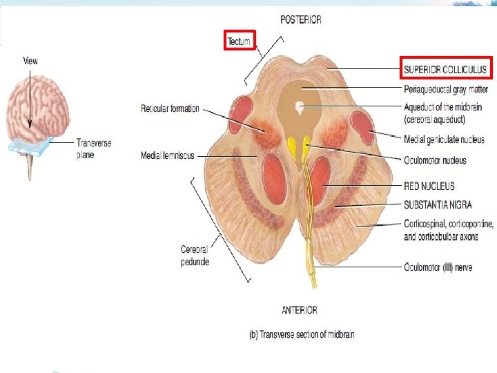
 includes: a) The superior colliculus § It")
1 - The Tectum ("roof" in latin) includes: a) The superior colliculus § It constitutes center for visual reflexes § It sends its superior brachium to the lateral geniculate body of the thalamus.
 The inferior colliculus § It is associated with auditory pathway § It sends")
b) The inferior colliculus § It is associated with auditory pathway § It sends its inferior brachium to the medial geniculate body of the thalamus. § The cerebral aqueduct runs through the midbrain, beneath the colloculi.

2 - The Tegmentum Ventral to the cerebral aqueduct. Several nuclei, tracts and the reticular formation is contained here. 3 - The ventral side is 3 comprised of paired Cerebral Peduncles. These transmit axons of UMN.

Midbrain internal structures Periaqueductal Gray: Around the cerebral aqueduct, contains neurons involved in the pain desensitization pathway.
 nucleus. Nerve (CN IV) nucleus. Nucleus This")
Occulomotor Trochlear Red Nerve (CN III) nucleus. Nerve (CN IV) nucleus. Nucleus This is a motor nucleus that sends a descending tract to the lower motor neurons.

Substantia Nigra: a concentration of neurons in the ventral portion of midbrain that is involved in motor function.

Central Tegmental Tract: Directly anterior to the floor of the 4 th ventricle, this is a pathway by which many tracts project up to the cortex and down to the spinal cord.

Reticular Formation: A large area that is involved in various important functions of the midbrain: RIt contains LMN RIt is involved in the pain desensitization pathway RIt is involved in the arousal and consciousness systems RIt contains the locus ceruleus, which is involved in intensive alertness modulation and in autonomic
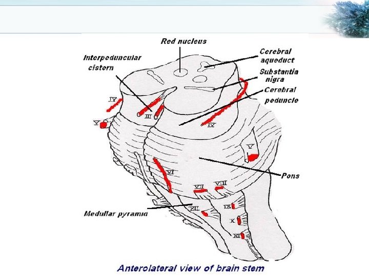
 emerges. o")
Pons o At the level of the midpons, trigeminal nerve (CN V) emerges. o Between the basal pons, cranial nerve 6 (abducens), 7 (facial) & 8 (vestibulo-cochlear) emerge (medial to lateral).

Ventral view Medulla Ø The most medial part of the medulla is the anterior median fissure. Ø Moving laterally on each side are the pyramids. They contain the fibers of the corticospinal (pyramidal) tract as they head inferiorly to synapse on lower motor neuronal cell bodies within the ventral horn of the spinal cord.

Ø The anterolateral sulcus is lateral to the pyramids. Ø Emerging from the anterolateral sulci are the hypoglossal nerve (CN XII) rootlets. Ø Lateral to the anterolateral sulci are the olives containing underlying inferior olivary nuclei and afferent fibers). Ø Lateral (and dorsal) to the olives are the rootlets for glossopharyngeal (IX) & vagus (X) cranial nerves.

Dorsal view v. The most medial part of the medulla is the posterior median fissure. v. Moving laterally on each side is the fasciculus gracilis. v. Lateral to that is the fasciculus cuneatus. v. Superior to each of these, are the gracile and cuneate tubercles, respectively. Underlying these are their respective nuclei.

v. In the midline is the vagal trigone and superior to that is the hypoglossal trigone. Underlying each of these are motor nuclei for the respective cranial nerves.

Functions of the Brain Stem Though small, brain stem is an extremely important part of the brain: 1. Conduct functions. 2. Provides the origin of the cranial nerves (CN III-XII). 3. Conjugate eye movement. 4. Integrative functions.

1. Conduct functions All information related from the body to the cerebrum and cerebellum and vice versa, must traverse the brain stem.
 The ascending sensory pathways coming from the body to the brain includes: §")
a) The ascending sensory pathways coming from the body to the brain includes: § The spinothalamin tract for pain and temperature sensation. § The dorsal column, fasciculus gracilis, and cuneatus for touch, proprioceptive and pressure sensation.
 Descending tracts § Corticospinal tract (UMN): runs through crus cerebri, basal part of")
b) Descending tracts § Corticospinal tract (UMN): runs through crus cerebri, basal part of pons and medullary pyramids; 7090 % of fibers cross in pyramidal decussation to form the lateral corticospinal tract, synapse on LMN in ventral horn of spinal cord. § Upper motor neurons that originate in brain stem's vestibular, red, and reticular nuclei, which also descend and synapse in the spinal cord.

2. The brain stem provides the main motor and sensory innervation to the face and neck via the cranial nerves (CN IIIXII). The fibers of cranial nerve nuclei except for olfactory & optic nerve either originating from, or terminating in, the cranial nerve nuclei in brain stem.
 •")
Origin & functions of the cranial nerves From midbrain • CN III (oculomotor) • CN IV (trochlear) Both moves eyes; CN III constricts the pupils, accommodates.
: Chews and feels front of the head. •")
From pons • CN V (trigeminal): Chews and feels front of the head. • CN VI (abducens): Moves eyes. • CN VII (facial): Moves the face, tastes, salivates, cries. • CN VIII (acoustic): Hears, regulates balance.
: Tastes, salivates, swallows, monitors carotid body and sinus.")
From medulla • CN IX (glossopharyngeal): Tastes, salivates, swallows, monitors carotid body and sinus. • CN X (vagus): Tastes, swallows, lifts palate, talks, communication to and from thoraco-abdominal viscera. • CN XI (accessory): Turns head, lifts shoulder. • CN XII (hypoglossal): Moves tongue.

Classification of the cranial nerves according to functions • Sensory CN I, CN II, CN VIII • Motor CN III, CN IV, CN VI, CN XII • Mixed CN V, CN VII, CN IX, CN X

3. Conjugate eye movement. It refers to motor coordination of the eyes that allows for bilateral fixation on a single object The frontal eye field (FEF) projects to the opposite side at the midbrain-pontine junction, and then innervates the paramedian pontine reticular formation (PPRF). From there, projections directly innervate the lateral rectus (contralateral to FEF) and the medial rectus muscle (ipsilateral to FEF). The left FEF command to trigger conjugate eye movements to the right.
 through")
4. Integrative functions Ø It controls consciousness & sleep cycle (alertness and arousal) through reticular formation. Ø It has got center for cardiovascular, respiratory & autonomic nervous system. Ø It has centers for cough, gag, swallow, and vomit. Ø Sense of body balance (Vestibular functions)

o Substantia which is a part of the basal ganglia is present in midbrain and is involved in control of movement. o Midbrain also contain red nucleus which regulate the motor activity through cerebellum.

o Inferior and superior colliculi are situated on the dorsal surface of the midbrain and is involved in auditory & visual processing required for head movements. o Pain sensitivity control: Periaqueductal grey matter of mesencephalon is an area which is rich in endogenous opioid and is important in modulation of painful stimuli.

Functional organization of the Brain Stem o. Ventral layer of brainstem is motor in function. o. Middle layer is sensory in function & contains medial lemniscus which conveys sensory information from dorsal column.

Function of Midbrain • • Nerve pathway to cerebral hemispheres. Auditory and Visual reflex centers. Cranial Nerves: CN III - Oculomotor [motor]. (Related to eye movement). • CN IV - Trochlear [motor]. (Superior oblique muscle of the eye which rotates the eye down and out).
 deficits: Ipsilateral CN III,")
Signs & Symptoms of midbrain lesion • Cranial Nerve (CN) deficits: Ipsilateral CN III, CN IV palsy and ptosis (drooping). • Pupils: Size: Midposition to dilated. Reactivity: Sluggish to fixed. • Movement: Abnormal extensor. • Respiratory: Hyperventilating. • Loss of consciousness (LOC): Varies

Functions of pons • Respiratory Center. • Cranial Nerves: § CN V - Trigeminal [motor and sensory]. (Skin of face, tongue, teeth; muscle of mastication). § CN VI - Abducens [motor]. (Lateral rectus muscle of eye which rotates eye outward). § CN VII - Facial [motor and sensory]. (Muscles of facial expression). § CN VIII - Acoustic [sensory]. (Hearing)

Symptoms and signs of lesion in pons • Pupils size: Pinpoint • LOC: Semi-coma • Movement: Abnormal extensor. • Respiratory: -Apneustic (Abnormal respiration marked by sustained inhalation). -Hyperventilation. • CN Deficits: CN V, CN VII, CN VIII.

Functions of medulla oblongata • • Crossing of motor tracts. Cardiac Center. Respiratory Center. Vasomotor Center (nerves having muscular control of the blood vessel walls) • Centers for cough, gag, swallow, and vomit. • Cranial Nerves:
![• CN IX - Glossopharyneal [mixed]. (Muscles & mucous membranes of pharynx, the](http://slidetodoc.com/presentation_image_h/12be6d800135e23b6ad580b7d4f9517a/image-42.jpg "• CN IX - Glossopharyneal [mixed]. (Muscles & mucous membranes of pharynx, the")
• CN IX - Glossopharyneal [mixed]. (Muscles & mucous membranes of pharynx, the constricted openings from the mouth & the oral pharynx and the posterior third of tongue). • CN X - Vagus [mixed]. (Pharynx, larynx, heart, lungs, stomach). • CN XI - Accessory [motor]. (Rotation of the head and shoulder). • CN XII - Hypoglossal [motor]. (Intrinsic muscles of the tongue).

Signs and symptoms of lesion in medulla • Movement: Ipsilateral paralysis. • Pupils: Size: Dilated. Reactivity: Fixed. • Respiratory: Abnormal breathing patterns • CN Palsies: Inability to control movement. Absent cough, gag. • LOC: Comatose.

Brain Stem Function Tests FTo test reticular formation Alertness, Consciousness & Sleep. FCorticospinal tract Motor power, reflexes FPain response Facial grimacing on firm pressure over the supra orbital ridge. FTo test respiratory center look for the normal pattern of respiration

FTo test cardiovascular center Look for normal circulatory function FTo test brainstem reflexes: • Pupilary and corneal reflexes. • Vestibulo-ocular reflex: Injection of iced water into the ear will produce eyes movement. • Oculo-cephalic reflex: Eyes will be fixed when head is moved in one or another directions. • Gag reflex.


- Slides: 47