
PHYSIOLOGY Sensory Systems Gustatory Receptors n Taste or



- Slides: 80
PHYSIOLOGY Sensory Systems
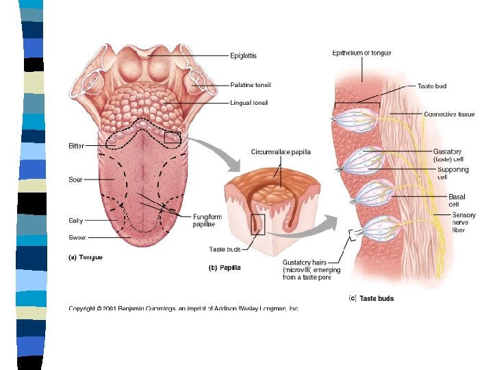
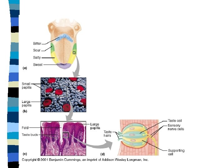
Gustatory Receptors n Taste or Gustation – The sensation following the stimulation of oral chemoreceptors – Chemoreceptors are surrounded by supporting cells • Chemoreceptors are shed every 10 -14 days and are renewed by division of the supporting cells.
Tastes n Four basic tastes – Sweet • Glucose, fructose, amino acids – Sour • H+ concentrations – Salty • Na+ concentration – Bitter • Quinine, caffeine, nicotine, strychinine, etc. – Umami • Produced by compounds like monosodium glutamate • Not a classic taste
Gustatory Transduction n Chemicals enter the pores of taste buds and react with the gustatory hairs n Chemicals may open sodium gates directly or may stimulate membrane receptors and G proteins and the second messenger system
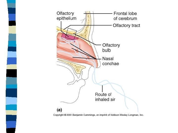
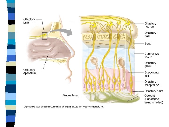
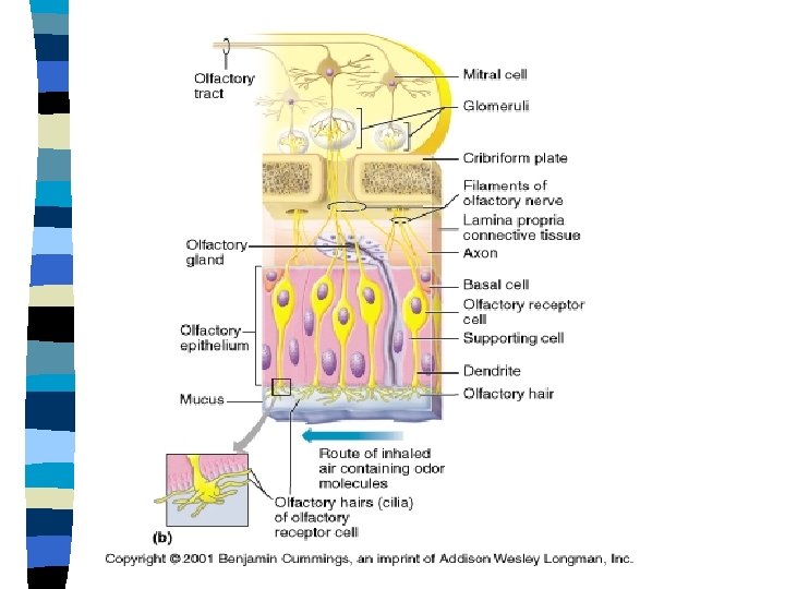
Olfaction n Olfactory cells lie in a specialized region in the roof of the nasal cavity – The olfactory epithelium n Odors combine to produce depolarization and impulse activity – 80% of taste is smell – Olfactory neurons are bipolar neurons
Olfactory Receptors n Supporting cells secrete mucus n Continual degeneration and replacement of neurons – Every 60 days – Basal cells differentiate into olfactory neurons
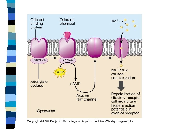
Olfaction Humans can detect about 104 different smells n Odiferous compounds are mainly organic n – Containing 3 -20 carbon atoms – Odiferous compounds reach the olfactory epithelium, aided by sniffing – The molecules must dissolve in the mucus layer (water soluble) to react with the receptors on the olfactory cilia
Odorant receptors n One receptor per olfactory neuron – 1000 different receptors – c. AMP system is used for smells
Glomeruli n Olfactory neurons synapse with the olfactory bulb in regions called glomeruli – From the olfactory bulb to the temporal lobe – Each olfactory neuron synapses with only one glomerulus – Each glomerulus receives input from several thousand olfactory neurons in the epithelium – Each glomeruli receives input from neurons expressing the same receptor
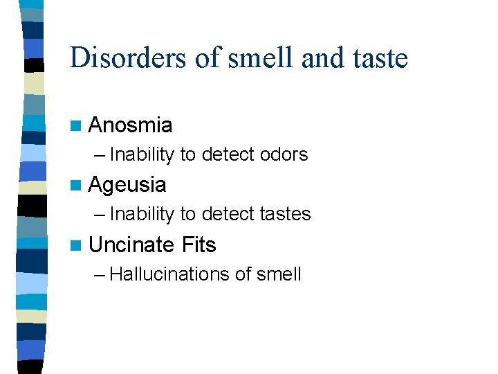
Disorders of smell and taste n Anosmia – Inability to detect odors n Ageusia – Inability to detect tastes n Uncinate Fits – Hallucinations of smell
Vision
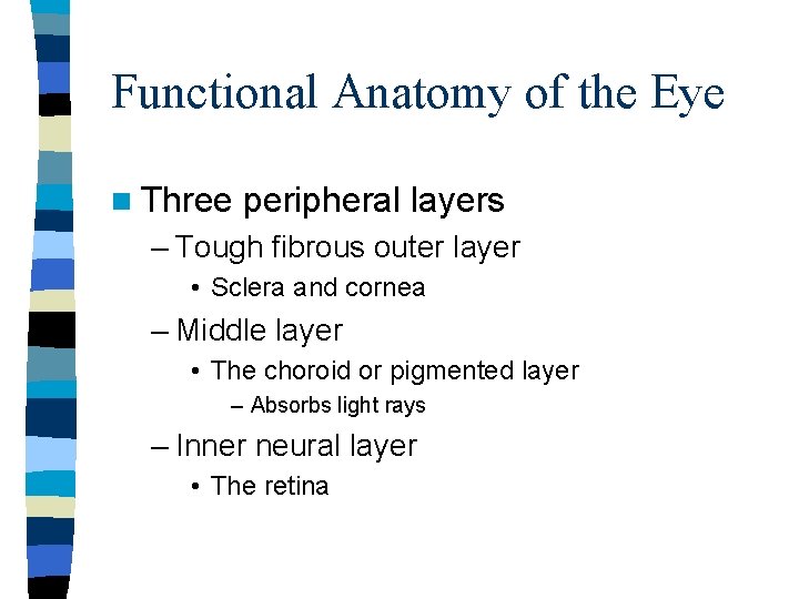
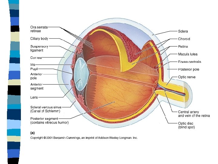
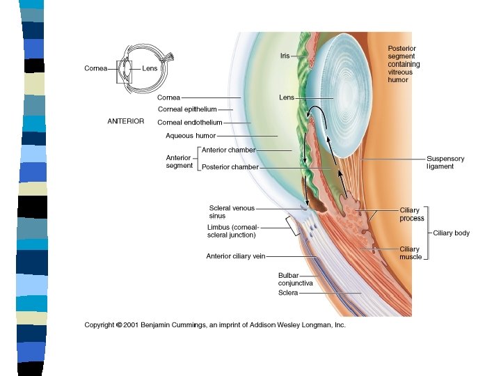
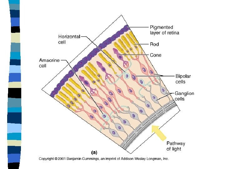
Functional Anatomy of the Eye n Three peripheral layers – Tough fibrous outer layer • Sclera and cornea – Middle layer • The choroid or pigmented layer – Absorbs light rays – Inner neural layer • The retina
Vitreous Humor n In the posterior chamber of the eye – Used to • Maintain the shape of the eye • Holds the retina in place – Produced in the fetal stage of development
Aqueous Humor n Produced by the ciliary muscles into the anterior chamber of the eye n Drains into the canal of Schlemm or Scleral Venous Sinus – ½ teaspoon is produced per day and this much drains per day – Clog of the canal may cause Glaucoma
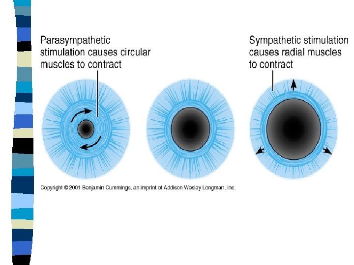
Constriction of the Pupil n Miosis – Results in a better depth of focus – Light rays pass only through the central part of the lens n Sympathetic Nervous System – Dilator control – Mydriasis n Parasympathetic Nervous System – Constrictor control n Pupils are consensual
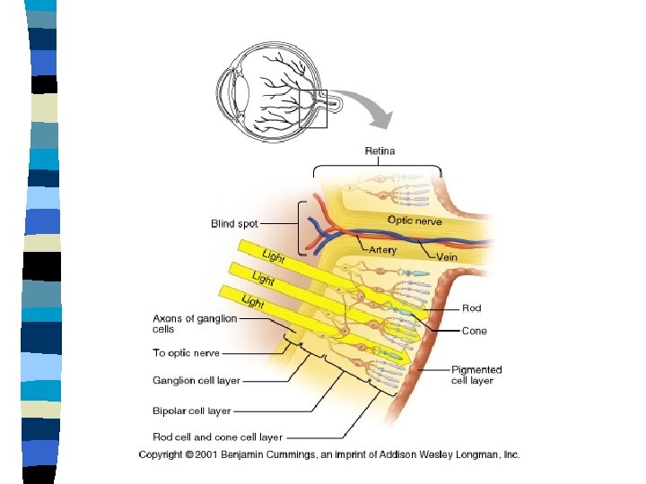
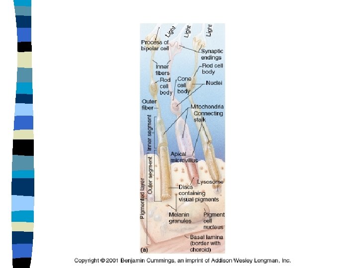
Rods and Cones n Rods – More numerous than cones by a ratio of 20: 1 – Function well in low light – Nighttime vision n Cones – High-acuity vision – Color vision during the daytime – High levels of light
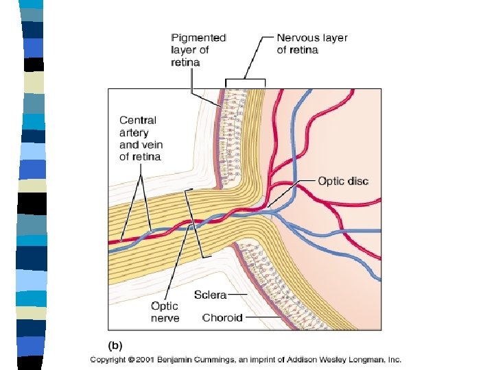
Eye n Optic Disc – Axons of the ganglion cells all form the optic nerve – The optic nerve leaves the eye at the optic disc – No rods or cones at the optic disc • Blind spot
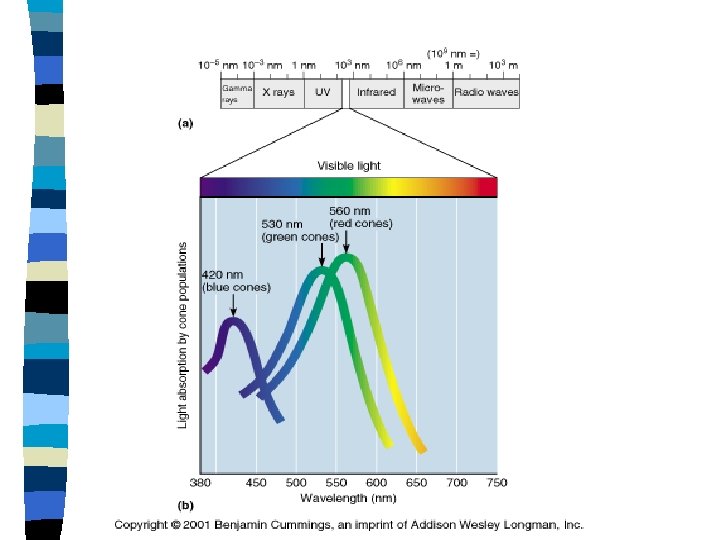
Light Each cone contains visual pigments that are excited by different wavelengths of light n Visual pigment n – Bound to cell membranes of dendrites – The transducers that convert light energy into a change in membrane potential n Rods – Visual pigment is rhodopsin
Cones n Red, green, blue, yellow(? ) cones n Each cone type is stimulated by a range of light wavelengths but is most sensitive to a particular wavelength n Colorblindness – Lack of cones – X-chromosome
Convergence n The eye muscles pull eyes so that both eyes see one fused image
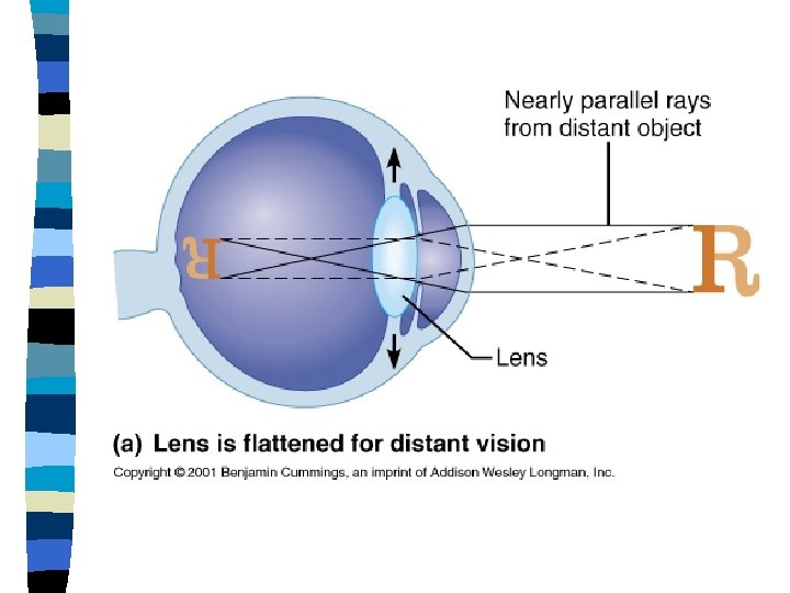
Accommodation The process by which the eye adjusts the shape of the lens to keep objects in focus n Presbyopia n – Hardening of the lens with age due to addition of layers to the lens n Focused at Infinity – The lens is pulled flat by tension in the ligaments n Close Up – The lens rounds up after the ciliary muscles contract and the suspensory ligaments relax
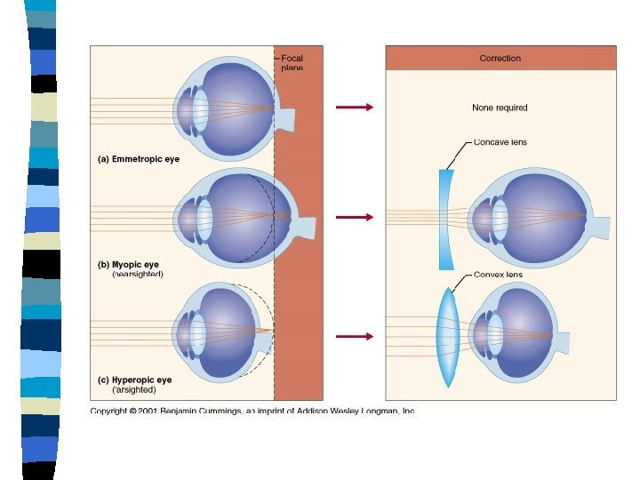
Lenses n Concave – Light bends outward n Convex – Light bends inward
Lens Focuses Light on the Retina n Light passes through the cornea and lens prior to striking the retina – Light must refract n Focal Point – The single point where the rays converge n Focal Length – Distance from the center of a lens to its focal point
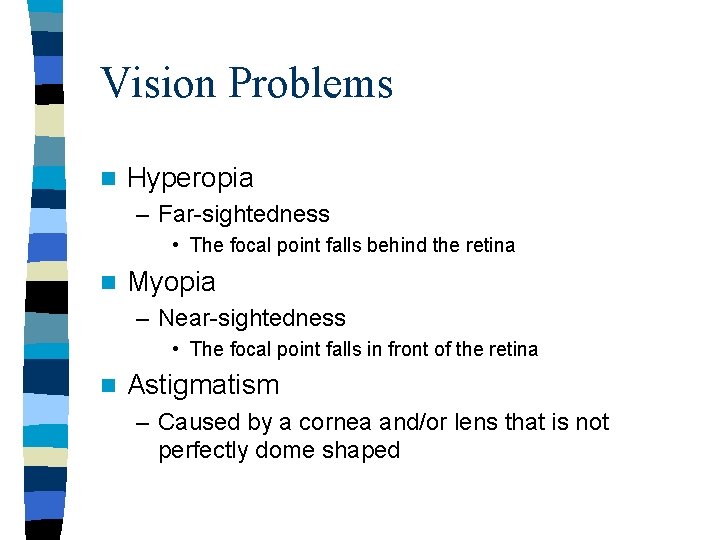
Vision Problems n Hyperopia – Far-sightedness • The focal point falls behind the retina n Myopia – Near-sightedness • The focal point falls in front of the retina n Astigmatism – Caused by a cornea and/or lens that is not perfectly dome shaped
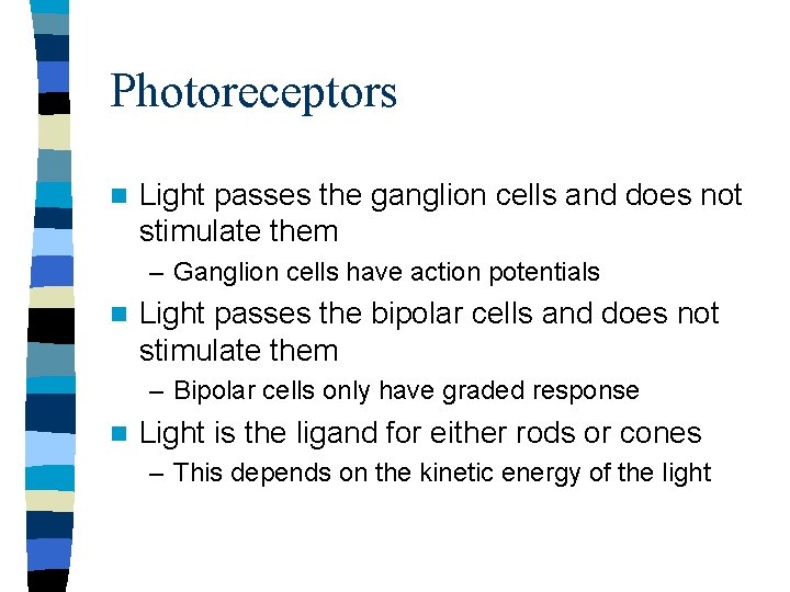
Photoreceptors n Light passes the ganglion cells and does not stimulate them – Ganglion cells have action potentials n Light passes the bipolar cells and does not stimulate them – Bipolar cells only have graded response n Light is the ligand for either rods or cones – This depends on the kinetic energy of the light
Photoreceptors n Photoreceptors in the retina transduce light energy into electrical signals n The Fovea Centralis – The point on which light focuses
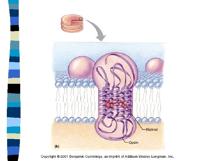
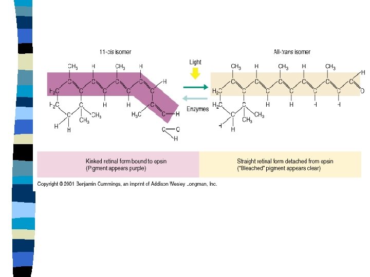
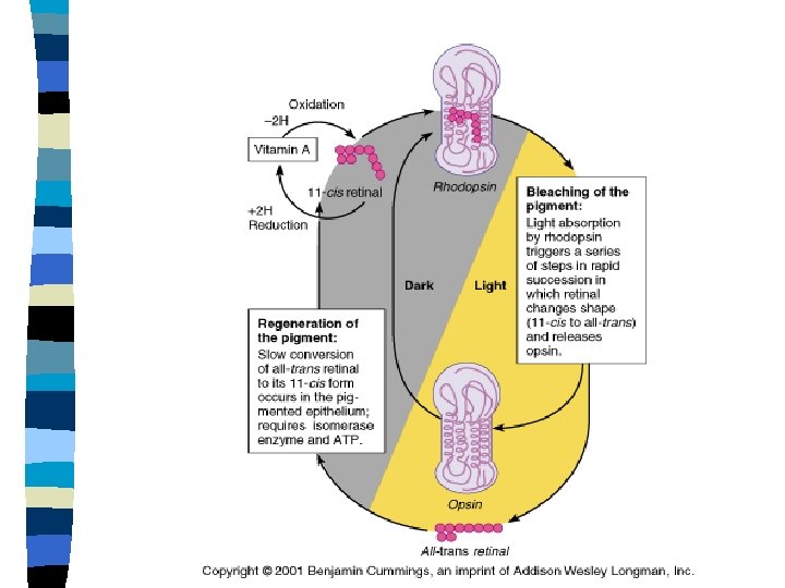
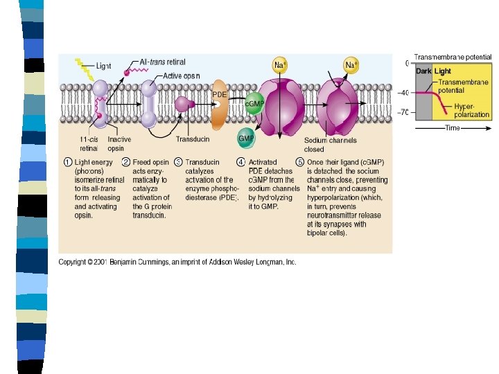
Phototransduction n Rhodopsin – Opsin plus 11 cis retinal • Purple and “kinked” in shape – Visual pigment for rods – When activated by as little as one photon of light the 11 cis retinal can be bleached n Bleaching – Light Changes 11 cis retinal to all trans retinal • All trans retinal is clear and a “straight” chain
Phototransduction n When a rod is in darkness – Rhodopsin is not active – cyclic. GMP levels in the rod are high – Sodium channels are open – Depolarization of the rod
Phototransduction Kinetic Energy of light transforms 11 cis retinal to all trans retinal n All trans retinal and Opsin separate n Opsin moves horizontally in the membrane and binds with transducin n – Transducin is a G protein Transducin binds to phosphodiesterase n PDE converts c. GMP to GMP n Sodium gates close n
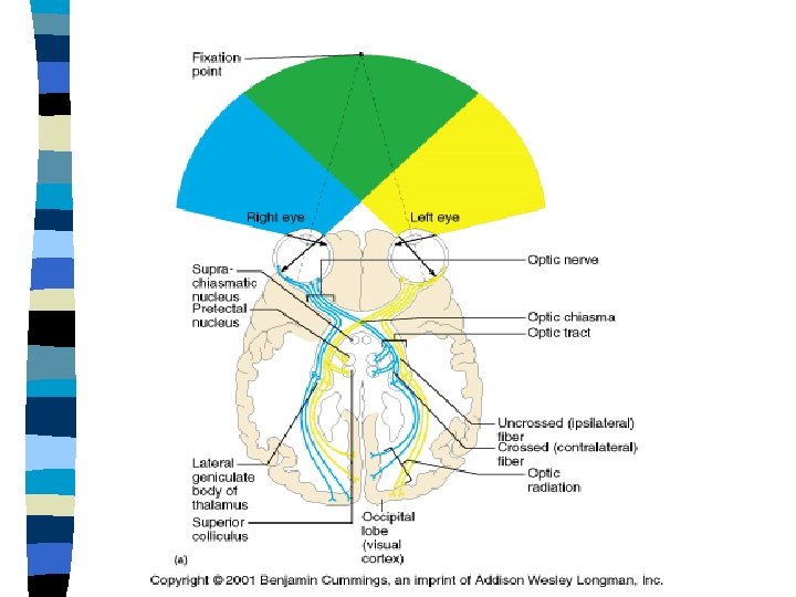
Binocular Vision n Visual Field – Each ganglion cell receives signals from a particular area of the retina n Binocular Zone – – Where the visual fields overlap Provides 3 -D Vision Medial aspect crosses over Lateral aspect stays on same side of the brain
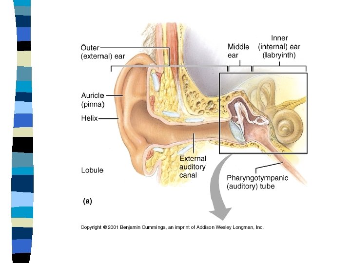
Ear n Outer Ear – Pinna • Collects sound waves – Ear Canal • Sends sound waves to tympanic membrane – Tympanic Membrane • Ear Drum • Vibrates at the same frequency and amplitude as the original wave
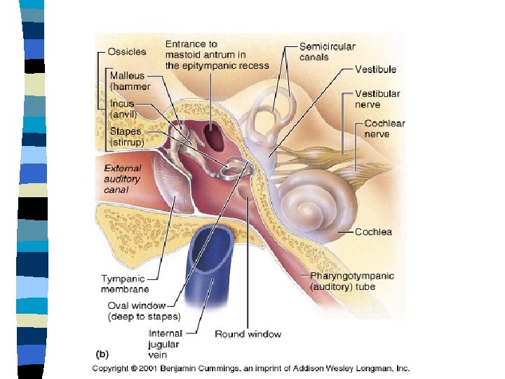
Middle Ear n Eustachian Tube – Normally collapsed – Opens transiently to equlibrate middle ear pressure and atmospheric pressure n Ossicles – Used to amplify the original sound wave by as much as 20 X on the oval window • Malleus • Incus • Stapes
Sound n Frequency – The number of waves that pass a particular point in a second – The longer the wave lengths the lower the frequency • The units of frequency is Hertz – The higher the frequency the higher the pitch of the sound
Sound n Amplitude – The height of the wave – Amplitude is measured in decibels – The higher the amplitude the louder the sound
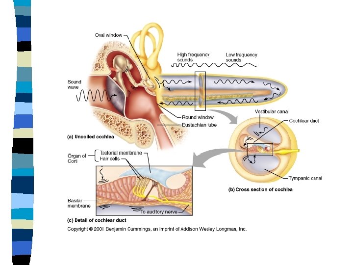
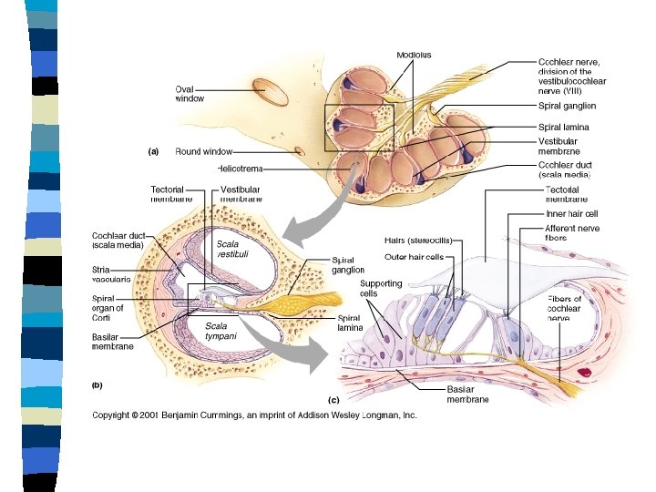
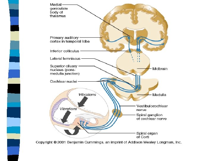
Inner Ear n Cochlea – Scala vestibuli • Top canal • Filled with Perilymph – Scala Media • Middle canal • Cochlear duct – Contains neurons for hearing • Filled with Endolymph • Organ of Corti – Scala tympani • Bottom canal • Filled with Perilymph
Cochlear Duct n Tectorial Membrane – Dendritic hairs are embedded in the tectorial membrane n Basilar Membrane – Supporting cells are embedded in the basilar membrane – Supporting cells surround auditory neurons
Sound Transduction n Sounds waves become mechanical vibrations, then fluid waves, then chemical signals and finally action potentials
Phonotransduction n First Transduction – Sound waves strike the tympanic membrane and become vibrations – The sound wave energy is transferred to the three bones of the middle ear, which vibrate
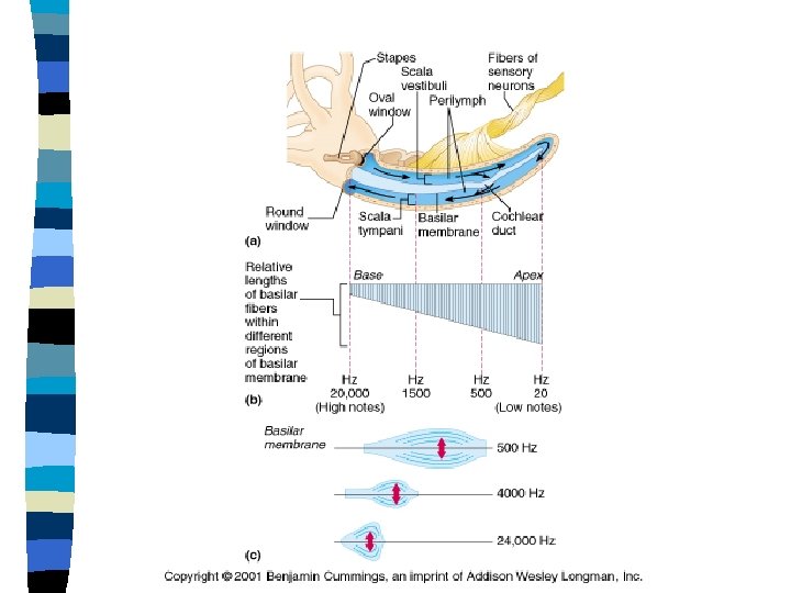
Phonotransduction n Second – The stapes is attached to the membrane of the oval window – The Stapes strikes the oval window and increases the force of the original wave 20 X – Vibrations of the oval window creates waves in the perilymph at the same frequency and amplitude as the original sound wave n Third – The fluid waves push on the flexible tectorial and basilar membranes of the cochlear duct. – Hair cells bend and release neurotransmitter
Phonotransduction n Fourth – Neurotransmitter is released, creating action potentials that travel through the cochlear nerve to the brain – Energy from the waves transfers across the cochlear duct is dissipated at the round window
Organ of Corti n The bending or shearing of the neurons indicates pitch and loudness – The bending of the neurons in the first third of the neuron signals high pitch sounds to the brain – The bending of neurons in the first and second third of the neuron signals medium pitch sounds – The bending of neurons in the first, second and third part of the cochlea signals a low pitch sound to the brain
The Organ of Corti n The higher the amplitude of the wave the more the kinetic energy – The high amplitude waves cause a greater shearing force which opens more sodium gates – The more sodium gates that open the more the action potentials • This creates a louder sound
Equilibrium
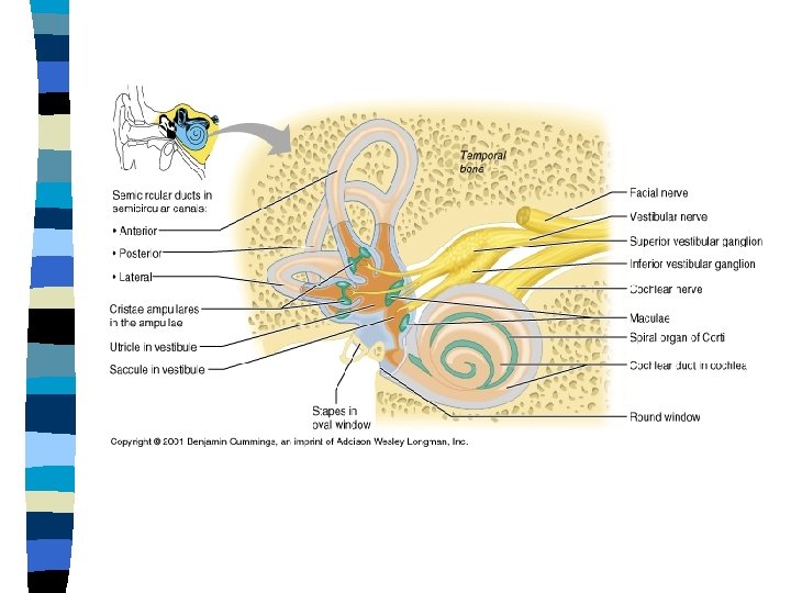
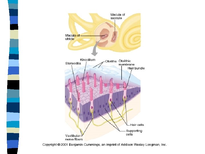
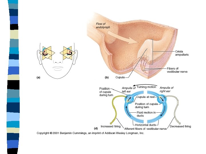
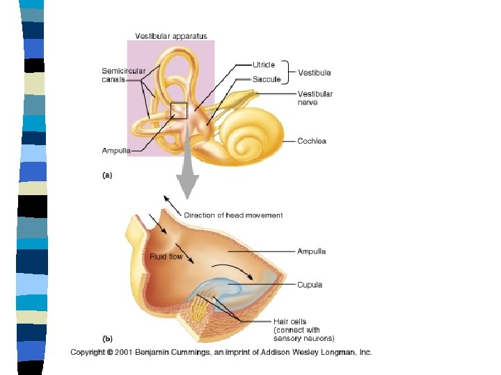
Equilibrium n Static Equilibrium – Little to no movements – Uses the vestibular region of the inner ear n Dynamic Equilibrium – Greater body movements – Uses the semicircular canals
Static Equilibrium The Vestibular Apparatus senses Linear Acceleration n Vestibular Apparatus n – Two saclike otolith organs • The utricle and the saccule – The sensory receptors of the utricle and saccule • The macula consists of a gelatinous mass known as the otolith membrane • Otolithic crystals are embedded in the membrane – Made of calcium carbonate crystals – Shearing or bending of the dendrites sends signals to the brain
Dynamic Equilibrium n Semicircular Canals sense rotational acceleration n Endolymph within the semicircular canals are in three different planes n Endolymph moves and moves the gelatinous cupula to activate receptor cells