Physiology of Vision Lecture2 Accommodation Pupillary Light Reflex


")

 side")


")
 – Changes in the pupil – Convergence of")


")


 Dioptre (s) = 1 Focal length (m) Diopteric power if the eye:")

 Near point (cm) Amplitude of Accomodation")


 is subject to bright light, a direct light")


 Pupils constrict in response: to accomodation reflex but not to")


 • Secondary association area, (areas 18, 19)")
 • On medial aspect of each occipital lobe •")


 • In parietal & temporal lobes •")


- Slides: 45
Physiology of Vision Lecture-2 - Accommodation & Pupillary Light Reflex Dr. Salah Elmalik
The Physiology of Vision Objectives: At the end of this lecture the student should be able to: • Describe visual acuity & depth perception • To know visual pathway and field of vision • Describe the process of accommodation reflex and its pathway, • Identify and describe pupillary light reflex , its pathway and relate these to clinical situations as Argyl Robertson pupil • Identify the lateral geniculate body and visual cortex functions.
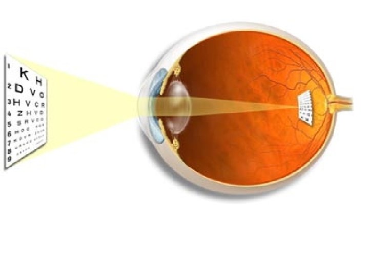
VISUAL ACUITY Definition : - • Degree to which details of objects are perceived. Visual threshold: • is minimal amount of light that elicit sensation of light • ü ü ü Snellen s chart Normal acuity =6/6 A person of 6/12 has less vision than normal vision (d/D Patient/normal)
Visual Pathway ( Pathway from Retina to the Visual Centers in the Brain ) • Photoreceptors : Rods and Cones synapse on Bipolar Cells , which in turn , synapse on Ganglion Cells. • Axons of Ganglion Cells constitute the Optic Nerve. These axons converge at the Optic disc , which is also called Blind Spot ( Why ? ). • Passing through the Blind Spot they leave the eye , constituting the Optic Nerve. 20/02/42 5
Visual Pathway 1. Optic nerve 2. Optic chiasm 3. Optic tract 4. Lateral geniculate body (nucleus) 5. Optic radiation 6. Visual cortex
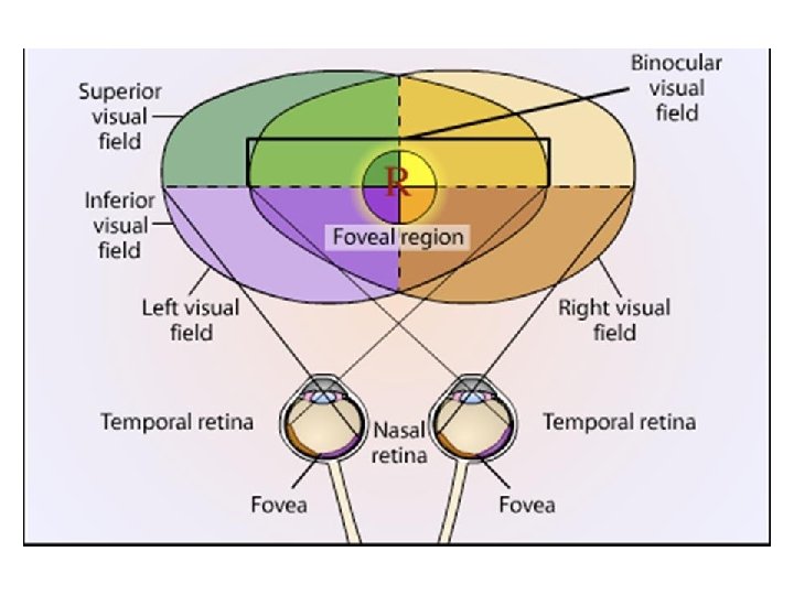
• • • Optic nerve fibers from the medial ( nasal ) side of retinae decussate in the Optic Chiasma. Therefore an Optic Chiasma lesion ( e, g, Pituitary Tumor ) will cause vision loss from the both lateral halves of the Field of Vision Optic nerve fibers from the lateral ( temporal ) parts of the retinae do not decussate. Therefore , each optic tract carries fibers from the both the temporal side of the ipsilateral retina + nasal side of the contralateral retina. Therefore , a lesion in optic tract will cause loss of vision from the ipsilateral nasal field of vision + contralateral temporal field of vision.
Accomodation
Focusing on a nearby object
Accomodation Definition: Modification of the refractive power of the eye (curvature of the lens) to view a nearby object Goal: Clear vision of a nearby object
Accomodation- cont. – Lens changes (accomodation) – Changes in the pupil – Convergence of the eyes The near response
The Near Respose
Image Focusing • Lens accommodation Parallel light rays from distant light source Fully accommodated Fully relaxed (unaccommodated) Focal Distance
Mechanism of accomodation Ciliary muscle. . Contraction: Relaxation of the suspensory ligament Lens more convex Increase diopteric power of the eye Near object focused on the retina
Mechanism of accomodation- cont uscl. Contraction of the suspensory ligament Lens less convex (Flat) Decrease diopteric power of the eye Far object focussed on the retina
Accommodation When the cilary muscles are relaxed, the zonalus pulls tight and keeps the lens flattened for distant vision The elastic lens is attached to the circular cilary muscles by the zonalus which is made of inelastic fibres When the cilary muscles contract, it releases the tension on the zonalus and the elastic lens returns to a more rounded shape suitable for near vision
Distant Vision: Ciliary Muscle Relaxed Suspensory Ligaments Under Tension Lens is Flattened Focus on Distant Objects Accommodation: Ciliary Muscle Contracts Reduced Tension on Suspensory Ligaments Lens becomes Round Focus on Near Objects
ACCOMMODATION TO NEAR OBJECTS THE CILIARY MUSCLE CONTRACTS THE TENSION ON THE SUSPENSORY LIGAMENTS DECREASES. THE LENS BECOMES MORE GLOBULAR IN SHAPE ACCOMMODATION OCCURS TO A CLOSER OBJECT BEING VIEWED. 19
NEAR OBJECT FOCUS POINT ON THE RETINA • THE PROBLEM OF BRINGING DIVERGING RAYS FROM CLOSE OBJECTS TO A FOCUS ON THE RETINA CAN BE SOLVED BY INCREASING THE CURVATURE OR REFRACTIVE POWER OF THE LENS. • THIS IS ACCOMMODATION
Diopter (D) Dioptre (s) = 1 Focal length (m) Diopteric power if the eye: Cornea ………… 40 -45 D Lens …………… 15 -20 D Accomodation …. +12 D
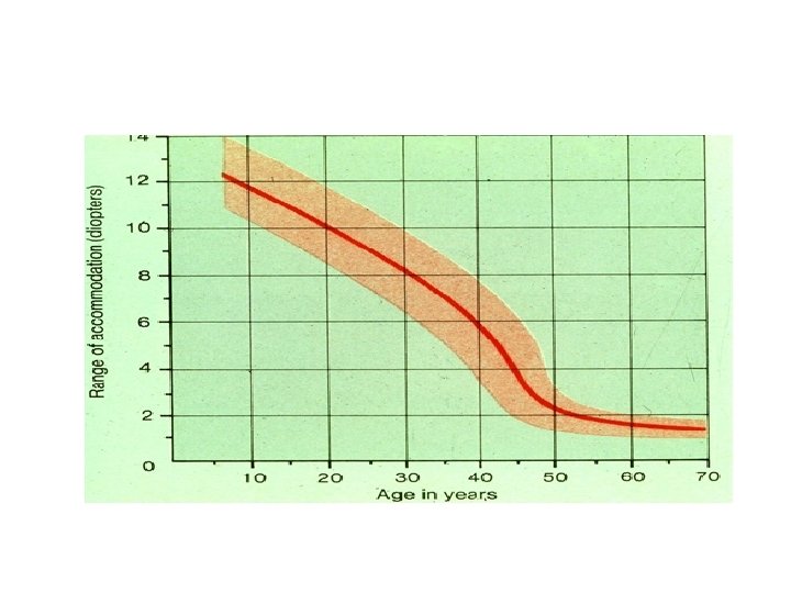
Amplitude of Accomodation Definition The additional diopters added by increasing the convexity of the lens Near point: The nearest point to the eye where an object can be seen clearly Presbyobia: Loss of lens elasticity in old age >>loss of accomodation
Near point and amplitude of accomodation Age (yrs) Near point (cm) Amplitude of Accomodation 10 9. 0 11. 0 20 10. 0 30 12. 5 8. 0 40 18 5. 5 60 83 1. 2 70 100 1. 0
The accomodation Reflex Afferent: Retina optic nerve optic chiasma optic tract lateral geniculate body visual cortex Efferent: Occuluomotor nucleus (parasympathetic) ciliary ganglion ciliary muscle circular pupillary muscle
PRESBYOPIA FOCUS POINT BEYOND THE RETINA IMAGE APPEARS BLURRED LENS IS HARDENED AND CANNOT BECOME MORE SPHERICAL TO REFRACT LIGHT FROM THE CLOSER OBJECT
The light reflex
Light Reflex When an eye (Left) is subject to bright light, a direct light reflex occurs(constriction of the pupil) as well as a consensual (indirect) reflex of the other (Right) pupil Diameter of pupil: varies from 1. 5 mm to 8 mm (Quantity of light changes X 30 fold)
Direct and consensual light reflex
Direct reflex on right Consensual reflex on left
Constriction of the pupil The pupil constricts in response to: • The accomodation Reflex • The light reflex
Argyll Robertson pupils (Neurosyphilis) Pupils constrict in response: to accomodation reflex but not to the light reflex
Lateral Geniculate Body; LGB FUNCTION OF LGB: 1 - Acts as a relay station for visual information from optic tract to cortex. 2 - Acts as gate controls signal transmission to visual cortex i. e control how much signals reach visual cortex N. B/- It receives gating control signals from two major sources: (1) Corticofugal fibers returning in a backward direction from the primary visual cortex to the lateral geniculate nucleus (2) Reticular areas of the mesencephalon. Both of these are inhibitory and, when stimulated, can turn off transmission through selected portions of the dorsal lateral geniculate nucleus
Lateral Geniculate Body; LGB 1 -Th magnocellular pathway, from layers 1 and 2 which have large cells and are called magnocellular. , carries signals for detection of movement, depth, and flicker. 2. The parvocellular pathway, from layers 3, 4, 5, 6 which have small cells and are called parvocellular, carries signals for color vision, texture, shape, and fine detail
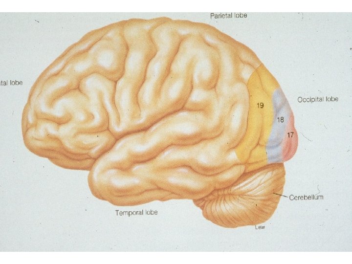
Cortical Visual areas • Primary (area 17) • Secondary association area, (areas 18, 19)
Primary Visual area (Area 17) • On medial aspect of each occipital lobe • Its neurons arranged in the form of columns forming 6 distinct layers • Fovea has broad presentation
Visual Projections to Area 17
Role of Area 17 • Perception of visible objects without knowing the meaning of these objects
Secondary Visual Processing: Association Areas (18 &19) • In parietal & temporal lobes • Interpretation of visual stimuli • Dealing with complex perception of patterns & forms & responsible for object recognition
Retinotopic Organization & Processing of visual information
Determination of Distance of an Object from the Eye—“Depth Perception” • • • A person normally perceives distance by three major means: (1) the sizes of the images of known objects on the retina (2) the phenomenon of moving parallax : when the person moves his head to one side or the other, the images of close-by objects move rapidly across the retinas, while the images of distant objects remain almost completely stationary • (3) the phenomenon of stereopsis or Binocular Vision