Physiology of Vision By Dr Abdel Aziz M


Physiology of Vision By Dr. Abdel Aziz M. Hussein Lecturer of Physiology Member of American Society of Physiology

Colour Vision

Colour Vision

Colour Vision • It is the ability of the eye to perceive the different types and characters of colours. Types of colours: Primary colours • They are red, green, and blue • When they are mixed together in the same proportion they give white colour • When mixed by different proportion they give other colours Complementary colours These colours when mixed together they give white colour e. g. deep blue and yellow; red and cyano

Colour Vision Characters of colours: 1. Hue means the wave length e. g. red light (wavelength 723 -647 nm), green light (575 -492 nm), and blue light (492 -450 nm). 2. Intensity or saturation means the purity of the colour i. e. it is pure or mixed with other colours 3. Brightness means the amount of light in the colour
 theories One the level of receptors or")
Mechanism of Colour Vision l 2 1) theories One the level of receptors or retina → retinal mechanism → Young-Helmholtz Theory 2) The other at the level of pathway →neural mechanism → Herring Theory

Young-Helmholtz Theory • It is the most accepted theory of colour vision • It postulates that there are 3 kinds of cones

Young-Helmholtz Theory • Equal stimulation of the 3 cone systems produce white sensation • Unequal stimulation produce another color e. g. . yellow colour is perceived when red cone stimulated by 83%, green cone by 83%, and blue cone by 0%.

Binocular Vision

Binocular Vision • It is the ability to see an object with 2 eyes without double vision (diplopia). Requirements 1. The visual fields must overlap to a great extent 2. Intact neuromuscular apparatus to allow image to fall on the corresponding points

Corresponding Points

Binocular Vision Requirements of binocular vision 3. A nearly normal refractive power in both eyes 4. Intact visual cortex where fusion of images occur

Binocular Vision Advantages of binocular vision 1. It increases the visual field horizontally from 160 to 200 degrees

Binocular Vision Advantages of binocular vision 2. It corrects the minor defects in one eye by the other eye e. g. blind spot of one eye is corrected by the other eye

Binocular Vision Advantages of binocular vision 3. It is important for stereoscopic vision 4. It improves the depth perception


Depth Perception • It is the ability to known the relative distance of the objects from the eyes

Depth Perception 1. The relative sizes of objects 2. The occlusion of part of a distant object by a nearer on 3. The fade of the colours and details of distant objects 4. Parallel lines appear to converge with distance. 5. Movement parallax
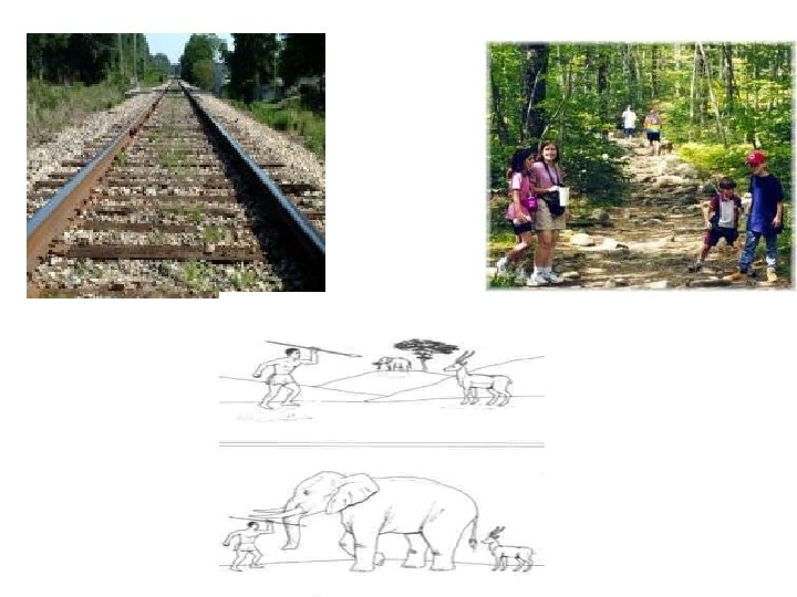

Movement Parallex
 movement Physiology of Special")
Eyeball Movements Sudden jerky movement Smooth pursuit movement Postural (vestibular) movement Physiology of Special senses, Abdelaziz Hussein Convergence movements 22

Control of Eyeball Movements Frontal eye filed area Visual association area Physiology of Special senses, Abdelaziz Hussein 23

Visual Pathway
 → bipolar cells (1 st order neuron)")
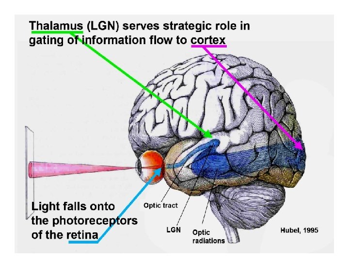
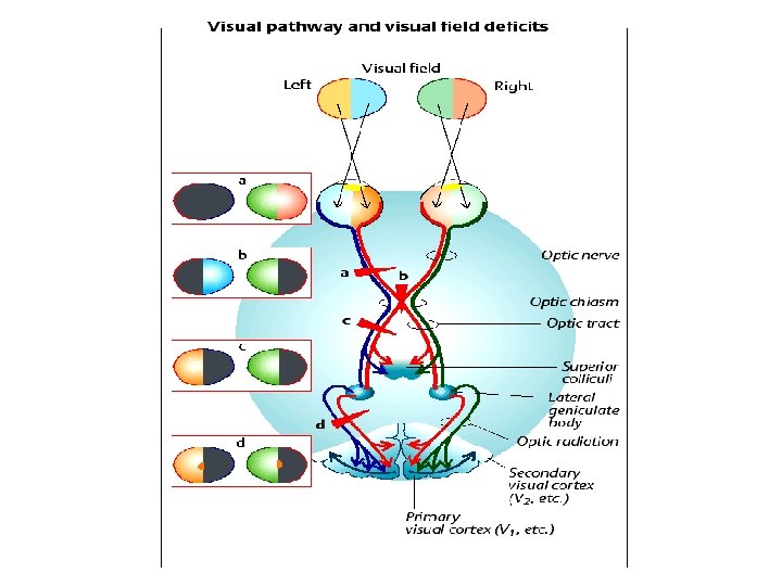
Visual Pathway • Photoreceptors (rods and cones) → bipolar cells (1 st order neuron) → ganglion cells (2 nd order neuron) → their axons form the optic nerve → optic chiasma, where the nasal fibers cross to the opposite side while the temporal fibers pass in the same side • optic tract (ipsilateral temporal fibers + contralateral nasal fibers of retina) → lateral geniculate body (3 rd order neuron) in thalamus • optic radiation → visual cortex in the occipital lobe (1 ry and 2 ry visual areas).

Visual Pathway

Role of Different Parts of Visual Pathway in Visual Perception

Retinal Cells • Analysis of the visual image occurs early in the retina

Role of Bipolar Cells • They constitute direct pathway between photoreceptors and ganglion cells • 2 types; 1. Depolarizing bipolar (on-bipolar) cells 2. Hyperpolarizing bipolar (off-bipolar) cells

Lateral Cells • Horizontal cells represent lateral inhibitory pathway in the retina • Amacrine Cells help in the analysis of the visual signals before leaving retina

Ganglion Cells • 1. 6 millions cells • respond to stimulation by a full action potential i. e. depolarization • 3 Types of Ganglion cells:
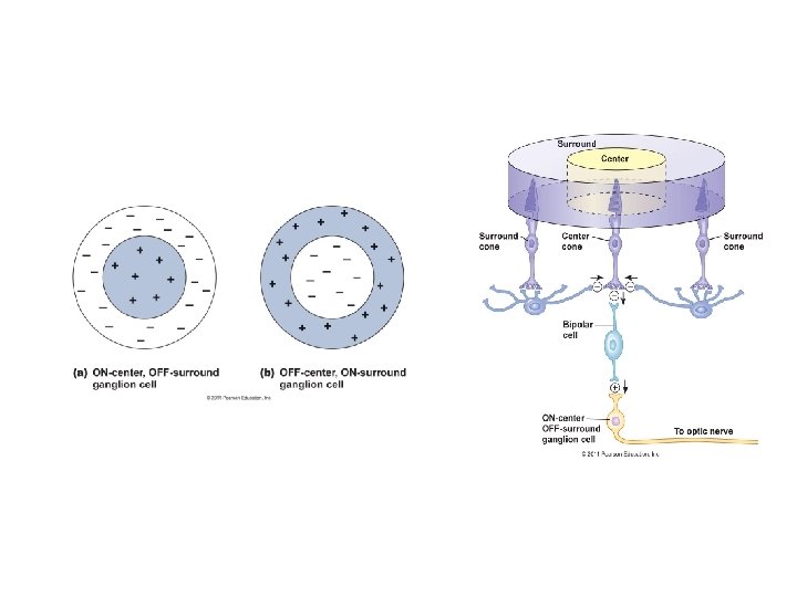

Ganglion Cells • Functions of Ganglion Cells 1. Detection of 2 point discrimination in the visual scene 2. Detection of the contrast in the visual scene 3. Detection of the movement and its orientation in the visual scene 4. Colour analysis
")
Lateral Geniculate Body (LGB)

Lateral Geniculate Body
 1. It plays a part in fusion of")
Functions of Lateral Geniculate Body (LGB) 1. It plays a part in fusion of retinal images from the 2 eyes. 2. It plays a part in stereoscopic vision by comparing the visual images from both eyes and detection of minimal differences. 3. Magnocellular neurons are concerned with perception of white and black, shape and motion. 4. Parvocellular neurons are concerned with perception of color vision and accurate point-point spatial information.

Visual Cortical Areas
 Site • Surrounds the calcarine fissure on the")
1 ry Visual Area (area 17) Site • Surrounds the calcarine fissure on the occipital lobe Representation of retina in area 17
")
1 ry Visual Area (area 17)

Role of 1 ry Visual area in Visual Perception 1. Detection of lines and borders 2. Detection of the orientation of lines and borders 3. Analysis of colors 4. Fusion of the images from the 2 eyes 5. Perception of luministy
 Site: • Occipital lobe around")
2 ry Visual Areas ( areas 18 and 19) Site: • Occipital lobe around 1 ry visual area and extend to parietal & temporal lobes • Areas 18 is called area V-2, more distant 2 ry visual areas are assigned V-3, V-4 and so no.
 Functions: Area 18 • It")
2 ry Visual Areas ( areas 18 and 19) Functions: Area 18 • It is also known as visuopsychic area which is concerned with; a. Recognition the nature of the objects and correlates their colours b. Interpretation of visual sensations c. Localization of object in space i. e. depth perception Lesion → visual agnosia
 Functions: Area 19 • It")
2 ry Visual Areas ( areas 18 and 19) Functions: Area 19 • It is also known as the occipital eye field area. a. It shares area 18 its functions. b. It controls the different types of eyeball movements.

TEST YOURSELF • 1. If the principal focal distance of a lens is 0. 5 m, its refractive power is • (A) 25 diopter s • (B) 75 diopters • (C) 1. 0 diopter • (D) 2 diopters • (E) 10 diopters
 increased tension on the lens")
TEST YOURSELF • 2. Visual accommodation involves • (A) increased tension on the lens ligaments • (B) a decrease in the curvature of the lens • (C) relaxation of the sphincter muscle of the iris • (D) contraction of the ciliary muscle • (E) increased intraocular pressure

TEST YOURSELF 5. The following events that occur in rods in response to light are listed in random sequence: 1. Activation of transducin 2. Decreased release of synaptic transmitter 3. Structural changes in rhodopsin 4. Closure of Na+ channels 5. Decrease in intracellular c. GMP What is the sequence in which they normally occur? (A) 2, 1, 3, 5, 4 (B) 1, 2, 3, 5, 4 (C) 5, 3, 1, 4, 2 (D) 3, 1, 5, 4, 2 (E) 3, 1, 4, 5, 2

THANKS
- Slides: 49