Physiology of Vision By Dr Abdel Aziz M


Physiology of Vision By Dr. Abdel Aziz M. Hussein Lecturer of Physiology Member of American Society of Physiology

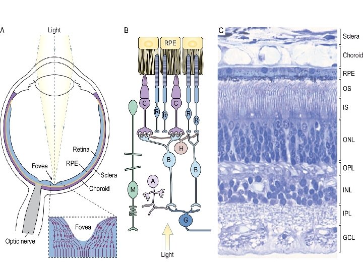
The Retina • The retina is the innermost photosensitive layer of the eyeball. • It contains about 55 types of cells and 70% of total sensory receptors • Histologically, it is formed of 10 layers • Physiologically, 4 layers have special importance

The Retina

The Retina

Functional Layers Pigmented Epithelium Photoreceptors Bipolar cells Ganglion cells

Pigmented Epithelium

Contains large amounts of melanin pigments

Produces a sticky ECF matrix

continual renewal of the outer segment of the rods and cones.

Breakdown and Resynthesis of the Photopigments

Stores Large Amount of Vit. A

Photoreceptor Layer

Rods and Cones

Outer Segment

Outer Segment

Inner Segment Outer segment Inner segment
 Cones 7 millions Cone")
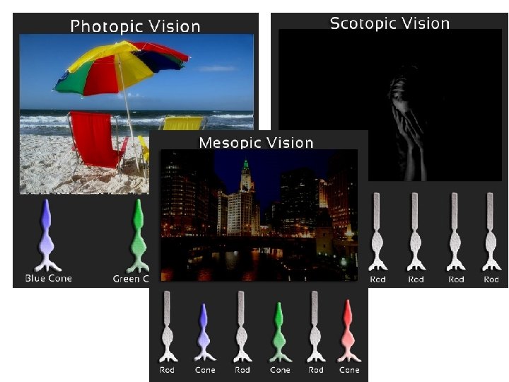
Rods and Cones Rod 120 millions Rod One pigment (rhodopsin) Cones 7 millions Cone 3 pigments (iodopsin)
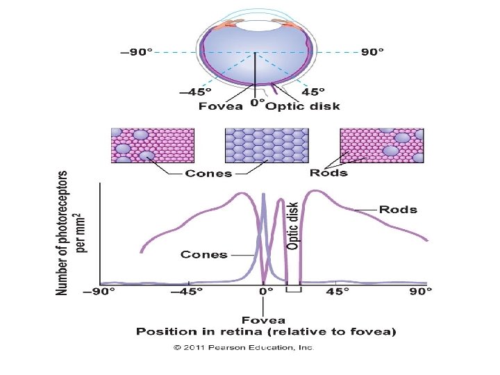

Distribution of Rods and Cones cones are central Rods are peripheral

Rods and Cones Convergence

Rods and Cones Functions Rod Cone more sensitive to light less sensitive to light Less accurate more accurate Cannot perceive colours Can perceive colours Night vision Day vision

Ganglion and Bipolar cells

Rods and Cones Convergence Bipolar cells 1 st order neuron Ganglion cells 2 nd order neuron

Lateral Cells

Blood Supply of the Retina

Blood Supply to the Retina Central retinal artery Supply inner layer of retina Choroidal blood vessels Supply pigmented epithelium and photoreceptors
 In")
Blood Supply to the Retinal detachment (occurs between the pigmented epithelium and photoreceptors) In this case the retina can resist degeneration for days due to its own blood supply (from central retinal artery) and diffusion of fluids across the detachment gap. If the retina is not replaced rapidly within few days degeneration of the retina occurs.

Retinal Detachment

Special Areas of the Retina

Special Areas of the Retina • Fundus oculi is the part of the retina which can be seen by ophthalmoscope. Fovea centralis Optic disc

Fovea Centralis Fovea centralis

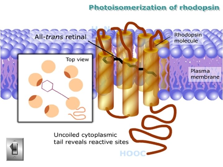
Phototransduction

Phototransduction 1. Decomposition of photosensitive pigment in rods & cones 2. Excitation of the photoreceptors by activated rhodopsin and generation of photoreceptor potential 3. Termination of excitation 4. Regeneration of photopigments

Decomposition of Pigment

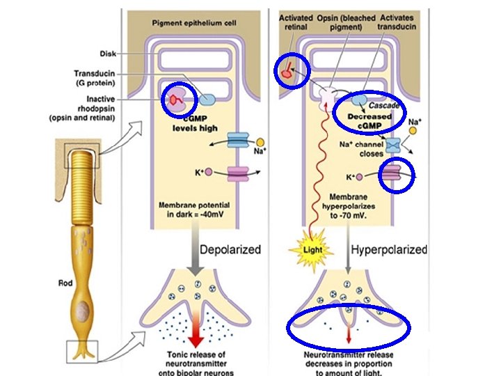
Photoreceptor Potential In Dark

Photoreceptor Potential In Light

Termination of Excitation • Within a sec the activated rhodopsin is inactivated by the enzyme rhodopsin kinase

Regeneration of Pigment

Summary of Generation of Photoreceptor Potential

THANKS
- Slides: 45