Physiology of vision BINESH TYAGI SFE H Main

“ Physiology of vision ” BINESH TYAGI SFE H
 �")
Main mechanisms involved: � Incidence of light beam � Transduction (initiation of vision) � Transmission of visual sensation � Visual perceptions
")
Visible spectrum (397 nm- 723 nm)

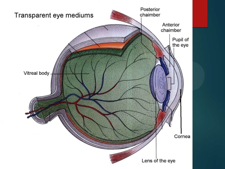
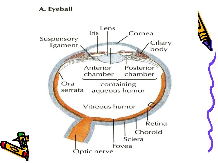
Optic system of eyeball �Cornea -allows light to enter the eyeball. �Aqueous humor -fills anterior and posterior chambers. �Crystalline lens –transparent, elastic biconvex lens. �Vitreous body -a transparent gel enclosed by vitreous membrane, fills eyeball behind lens. �Retina

Structure of visual analyser • Receptor part – rods & cones of the retina • Conducting pathways- optic nerve, optic chiasm, optic tracts, lateral geniculate body • Visual cortex – occipital lobe around calcarine fissure

 Lens (19")
Refractive mediums of the eye • • • Cornea (40 -43 D) Lens (19 -33 D) Vitreous body (0 D) Light conduction & refraction Refractive power of the eye is 59 -73 D

Construction of image on the retina of the reduced eye • Real • Reduced • Turned upside down

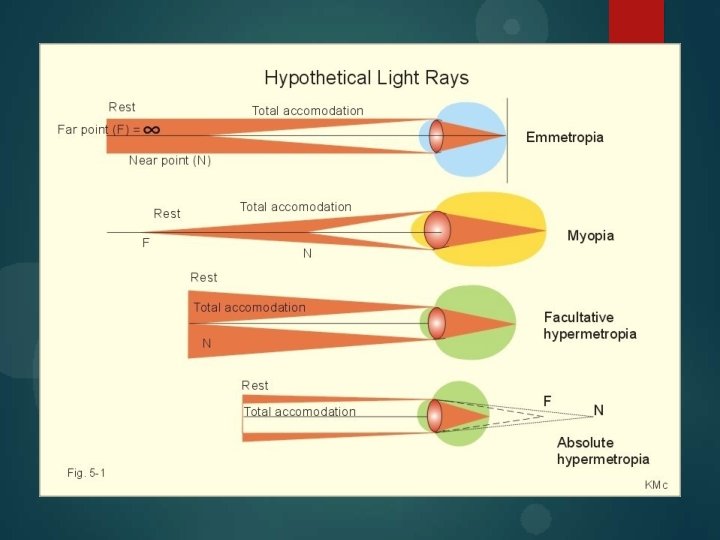
Emmetropia Hypermetropia corrected with convex lens Myopia corrected with concave lens

Aberrations and astigmatism � Spherical aberration light rays pass through peripheral parts of the eye lens and are not focused sharply. -because of more refractive power in central part of lens. Due to this effect object loose clear contour. � Chromatic aberration Unequal deviation of light rays of different wavelengths. - focusing of different colours at different distances behind lens. -object get rainbow contour. �Diffractive aberration occur in case of small object interfere to light rays in clear mediums of eyeball (for instance foreign body). - point object looks like rounded by gray and white circles. � Astigmatism is an errors of refraction in which light rays do not all come to a common focal point. Oblong shape of cornea or lens causes it. So, cylindrical lenses may correct this defect of refraction.

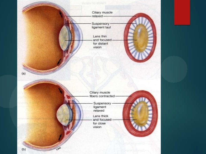
Accommodation and its regulation �Accommodation is adjustment of eye lens for various distances. Relaxation of ciliary muscle cause decrease of refractive power of eye lens and provides clear vision for long distance. �Parasympathetic influence to ciliary muscle controls it. In case of parasympathetic stimulation of ciliary muscle, it contracts, lens ligament relax, lens get more spherical, refractive power increases and eye can see clear near objects.

Age peculiarities � In newborn anatomical axis of eyeball is shorter, comparing to adults. – complex neural pathways not developed. � In infant- Structure and visual development go side by delicate balance between visual stimulation in right amount at right time and development of brain. –eye protects the immature brain from level of stimulation that it cannot cope with initially by- short size, hypermetropia, immature fovea, absence of binocular stereoscopic vision, limited peripheral field of view. Also excess sleeping most of the time, slow communication due to absence of fatty myelin sheath contributes. � In old age lens of eye loose elasticity. So this condition, when lens become non-accommodating, called presbiopia. Corrected by bifocal glasses with upper segment focused for far-seeing and lower segment focusing for near-seeing.

Initiation of vision Inadequate stimuli • Produce glowing sensation called. PHOSPHENES • Pressure phosphenes • Movement phosphenes • Electrical phosphenes • Radiation phosphenes Adequate stimuli • Formed by the visible part of electromagnetic radiation spectrum

Physiological peculiarities of pigmented layer and photoreceptors. � Light falls on retina on inner side i. e. on inner limiting membrane. � Central portion of macula called fovea centralis. This is composed entirely of cones. It is a minute area of 1 mm in center of retina. It provides acute and detailed vision. � Pigmented layer of retina contains black pigment, i. e. melanin. It prevents light reflection through the globe of eyeball and stores vitamin A.

Photochemical reactions in retina � Outer segment of photoreceptors contain photochemicals. Inner segment contains nucleus, synaptic body and other organelles. Photochemicals are lightsensitive chemicals that decompose on exposure to light and excite nerve fibers leading from eye to central nervous system. � Rhodopsin is present in rods. Scotopsin and 11 -cis-retinal compose it. Iodopsin is photochemical pigment of cones. Photopsin and 11 -cisretinal compose it.

� Rhodopsin cycle: rhodopsin under the influence of light converts to prelumirhodopsin – metaphodopsin I - metaphodopsin II – opsin – rhodopsin. Metarhodopsin II converts also to alltransretinal (vitamin A) – (isomerase’s action) – II cisretinal – rhodopsin. (Activate transducin)

Rhodopsin Metarhodopsin II Activation of transducin Activation of phoshpdiasterase Decreased intracellular c. GMP Hyperpolarization

Neurophysiology of vision � Genesis of visual impulse in photoreceptors � Processing and transmission in retina � Processing and transmission in visual pathway � Analysis in visual cortex � 3 -part system hypothesis of visual perception

Physiological activities in the retinal cells Neurotransmitters � Glutamine – by rods and cones. � Amacrine cells produce 5 different inhibitory transmitters, GABA, glycine, dopamine, Ach, Indolamine. � Cholinesterase by muller , Horizontal, Amacrine and Ganglion cells. � Carbonic anhydrase from cones and RPE.

Cellular activities � Horizontal cells: Enhance visual contrast by lateral inhibition, hence processing of spatial information. -when a minute stroke of light strikes the retina, the central most area is excited while the area around is inhibited. � Bipolar cells: stimulated by hyperpolarization of photoreceptors. • Two different types, provide opposing excitatory (depolarizing bipolar cells) and inhibitory signals in response to stimulation by light. • Second mechanism of lateral inhibition by centre-surround antagonism. (on-cell and off cells)

� Amacrine cells: • Negative feedback arrangement to subsequent response to be projected onto ganglion cells. • Receive information at the synapse of bipolar cell axon and ganglion cell dendrites. • Temporal processing of this information at other end of bipolar cells. • TYPES of function 1. Direct pathway for rod vision 2. Onset-activated cells 3. Offset-activated cells 4. Illumination change sensitive cells 5. Direction sensitive cells

� Ganglion cells: transmit signal as action potential to the brain. • Three groups • W-ganglion- small, 40% of total, broad fields in retina, excitation from rods, detect direction movement anywhere in the field. • X-ganglion- medium diameter, 55% of total, small field, colour vision. Sustained response. • Y- ganglion cells- largest, 5%, very broad dendritic field, respond to rapid eye movement or rapid change in light intensity. Transient response.

Transmission of vision � Impulses from retina pass to optic nerve – optic chiasma (fibers from nasal halves of retina cross to opposite side) – optic tracts – synapse in lateral genicular body – geniculocalcarine fibers – pass through optic radiation or geniculocalcarine tract – primary visual cortex in calcarine fissure or medial aspect of occipital lobe.

Lateral geniculate body � � Functions: relay station, to gate the transmission of signals. Layer 1, 2: • large cells, called magnocellular pathways • Input from Y-ganglion cells � • Very rapid conduction • Colour blind system Layer 3 -6: • Parvocellular • Input from X- ganglion cells • Colour vision • Moderate velocity.

Other connections of optic tract � � � In addition to lateral genicular body, fibers from optic tract also pass to: - suprachiasmatic nucleus of hypothalamus for controlling circadian rhythms; - pretectal nuclei – for control of fixation of eyes on objects of importance and for pupillary light reflex; - superior colliculus – for control of bilateral simultaneous movements of two eyes; - pulvinar – forms secondary visual pathway. Corpus callosum causes exchange of visual information between right and left hemispheres.

Pupillary reflexes � When light pass into eye, pupil contracts. In darkness pupil dilates. This is Direct pupillary light reflex, which helps to adaptation to light conditions. � Reflex arc: light receptors - optic nerve- optic tract pretectal area - Edinger-Westphal nucleus parasympathetic fibers of n. oculomotorius (from n. trigeminus) - n. ciliaris - m. sphincter pupillae decrease of pupillary diameter. � Consensual pupillary light reflex: reaction of eye pupil to light irritation of opposite eye. It is possible due to diverging of nerve fibers from one pretectal nucleus to both Edinger-Westphal nuclei.

Analysis of visual impulse in visual cortex Primary visual cortex • Brodmann’s area 17/ visual area 1 • Grossly striated appearance Secondary visual cortex • Includes visual association areas • Lie ant, sup, inf to 1’ visual cortex • Brodmann area 18, 19/visual area II, III

� Primary visual cortex – 6 distinct layers • Layers I, III- thin, contain pyramidal cells • Layer IV- thickest, contains stellate cells • • • -further subdivided in a, b, c alpha and c beta Layer V, VI relatively thin Connections • Geniculate afferents- rapidly conducted signals from Y-ganglional cells terminate in layer IV c alpha • From X ganglion cells to layer IVa and IVc • Subcortical connections – • upper part of layer VI to magnocellular layers • -lower part of layer VI project to parvocellular layers. • Corticocortical connections- • From layers II and III

Receptive fields of visual cortex � 1. 2. Three receptive field types Simple cells- mainly in layer IV • Respond to bars of light, lines or edges only when oriented in particular direction • Most effective orientation of stimulus is k/a receptive field axis orientation • Parallel bands of On and Off areas Complex cells – above and below layer IV • Preferred orientation of linear stimulus • No on and off regions • Less dependent upon location of stimulus • Plays role specially when stimulus is moving • SO SIMPLE AND COMPLEX CELLS TOGETHER ARE K/A FEATURE DETECTORS.

3. Hypercomplex cells – • In cortical layers II and III • All features of complex cells but require the line stimulus to be of specific length • So specific role in shape and angle detection

� colour blobs • Primary areas for deciphering colours • Special column like areas interspersed among the primary visual columns. • Respond specifically to colour signals

Three part system hypothesis of visual perceptions � First systemmovement, location and spatial organization � Second systemcolor perception � Third systemshapes perception

Visual perceptions � • • Sensations from stimulation of retina by light. Four types. Light sense - Faculty to perceive light in all gradations of intensity. -light minimum Form sense - to perceive the shape of object in outer world. -max at fovea/ cones. Contrast sense- ability to perceive slight changes in luminance between regions not separated by sharp borders. Colour sense -ability to distinguish between different colours as excited by light of different wavelengths.

Theories of colour perception �According to Young-Helmholtz theory there are three types of cones for three fundamental colours: cones for red colour contain erythrolab; cones for green colour contain chlorolab; cones for blue colour contain cyanolab. �Do not explain why dichromats see yellow. �Do not explain complementary colour after images.

According to Hering theory there are couples of opponent colours: Spectrally opponents viz Green – red; yellow – blue. Spectrally non opponent white – black. Subcortical neurons perceive it due to on- and offcenters mechanism. Both theories combined explain the colour vision system. Trichromatic operates at receptor level and the signals are recorded into the opponent process form by higher level neural system of colour vision processing.

Disorders of colour perception �. There are three fundamental colours: Red, Green and Blue. � Clinical presentations: • Total colour blindness • Partial colour blindness • Red–green • Dichromacy (protanopia and deuteranopia) • Anomalous trichromacy (protanomaly and deuteranomaly) • Blue–yellow • Dichromacy (tritanopia) • Anomalous trichromacy (tritanomaly)

Gr, • . . a-z, eltrank ' " "' ' " you. . r . 9
- Slides: 41