Physiology of Reproduction Dr Ashraf Fouda Damietta General


 :")


 appear in the syncytium, increase in size and fuse together")


- Slides: 46
Physiology of Reproduction Dr. Ashraf Fouda Damietta General Hospital
Pregnancy occurs when a mature liberated ovum is fertilized by a mature capacitated spermatozoon
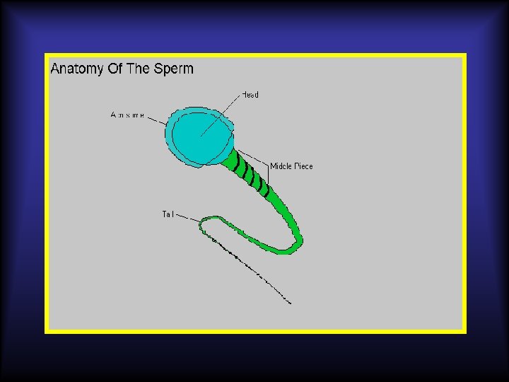
The Sperm: • The spermatozoa leave the testis carrying 23 chromosomes but not yet capable of fertilization. • Their maturation is completed through their journey in the 6 meters of the epididymis and when mixed with the seminal plasma from the epididymis, seminal
The Sperm: After semen is ejaculated, the sperms reach the cervix by their own motility within seconds leaving behind the seminal plasma in the
The Sperm: At time of ovulation, the cervical mucous is in the most favourable condition for sperm penetration and capacitation as: 1. It becomes more copious, less viscous and its macromolecules arrange in parallel chains providing channels for sperms passage. 2. Its contents from glucose and
The Sperm: • The sperms ascent through the uterine cavity and Fallopian tubes to reach the site of fertilization in the ampulla by: 1. Its own motility, and by 2. Uterine and tubal peristalsis which is aggravated by the
The Sperm: • The sperms reach the tube within 30 -40 minutes • But they are capable of fertilization after 2 -6 hours. • This period is needed for sperm capacitation.
Capacitation of sperms • Is the process after which the sperm becomes able to penetrate the zona pellucida, that surrounding the ovum and fertilize it. • The cervical and tubal secretions are mainly responsible for this capacitation.
Capacitation of sperms • Capacitation is believed to be due to : 1. Increase in the DNA concentration in the nucleus, 2. Increase permeability of the coat of sperm head to allow more release of hyaluronidase.
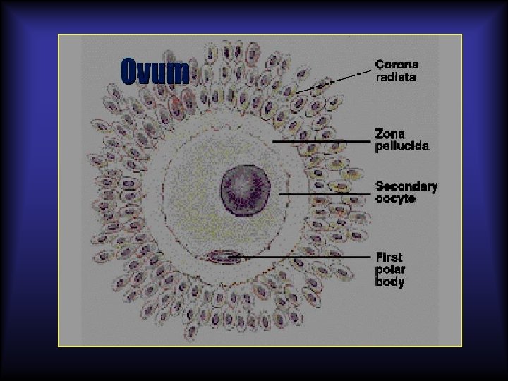
The ovum: The ovum leaves the ovary after rupture of the Graafian follicle, carrying 23 chromosomes and surrounded by the zona pellucida and corona radiata.
The ovum: The ovum is picked up by the fimbrial end of the Fallopian tubes and moved towards the ampulla by the : 1. Ciliary movement of the cells and
Fertilization: • Millions of sperms ejaculated in the vagina, but only hundreds of thousands reach the outer portion of the tubes. • Only few succeed to penetrate the zona pellucida, and only one spermatozoon enters the ovum transversing the
Fertilization: • After penetration of the ovum by a sperm, the zona pellucida resists penetration by another sperms due to alteration of its electrical potential. • The pronucleus of both ovum and sperm unite together to form the zygote (46
Zygote
Sex Determination: * The mature ovum carries 22 autosomes and one X chromosome, while the mature sperm carries 22 autosomes and either an X or Y chromosome. * If the fertilizing sperm is carrying X chromosome the baby will be a female (46 XX), if it is carrying Y chromosome the baby will be a
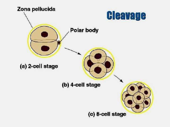
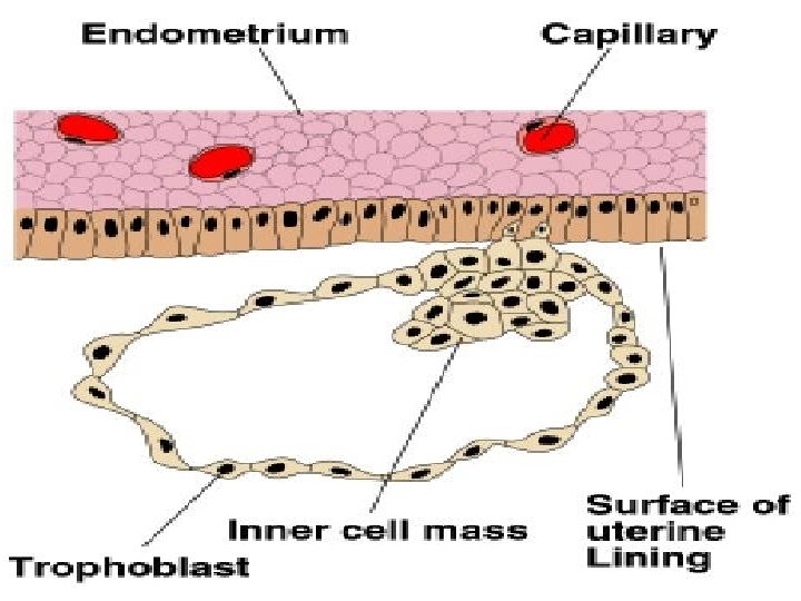
Cleavage and blastocyst formation: On its way to the uterine cavity, the fertilized ovum (zygote) divides into 2, 4, 8 then 16 cells (blastomeres).
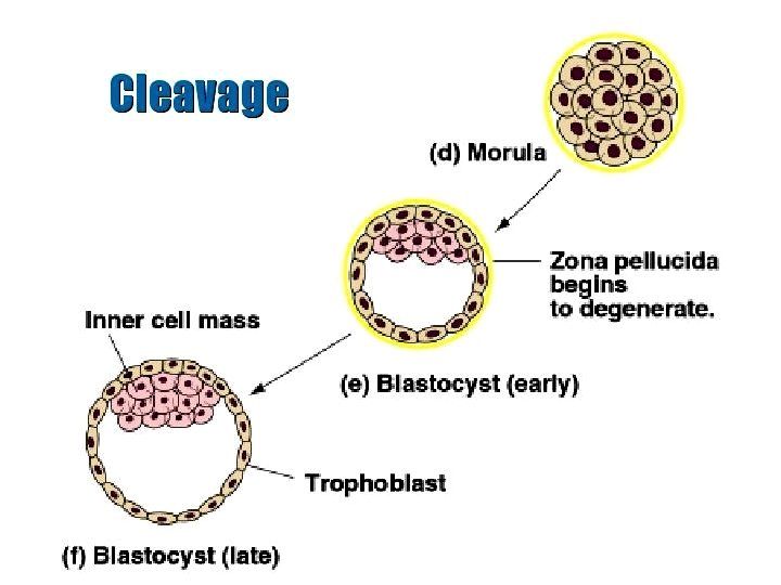
Cleavage and blastocyst formation: • This cleavage starts within 24 hours of fertilization and occurs nearly every 12 hours repeatedly • The resultant 16 cells mass is called morula which reaches the uterine cavity after about 4 days from fertilization.
Cleavage and blastocyst formation: • A cavity appears within the morula converting it into a cystic structure called blastocyst. • The cells become arranged into an : 1. Inner mass (embryoblast) which will form all the tissues of the embryo, and an
Cleavage and blastocyst formation: The blastocyst remains free in the uterine cavity for 3 -4 days, during which it is nourished by the secretion of the endometrium (uterine
Implantation (nidation) :
The decidua: • It is the thickened vascular endometrium of the pregnant uterus. • The glands become enlarged, tortuous and filled with secretion. • The stromal cells become large
The decidua, like secretory endometrium, consists of three layers: 1. The superficial compact layer, 2. The intermediate spongy
The decidua • The separation of placenta occurs through the spongy layer • While the endometrium regenerates again from the basal layer.
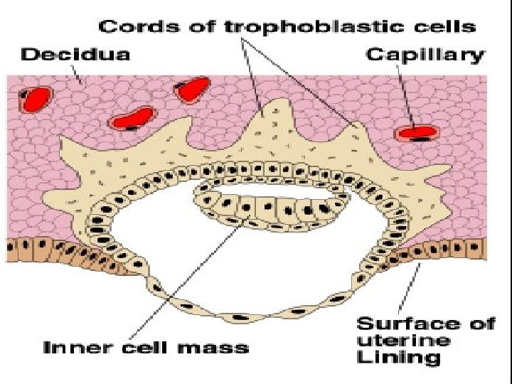
The decidua The trophoblast of the blastocyst invades the decidua to be implanted in: -The posterior surface of the upper uterine segment in about 2/3 of cases, -The anterior surface of the upper uterine segment in about 1/3 of cases.
The decidua After implantation the decidua becomes differentiated into: 1. Decidua basalis; under the site of implantation. 2. Decidua capsularis; covering the ovum. 3. Decidua parietalis or vera; lining the rest of the uterine cavity.
The decidua
The decidua • As the conceptus enlarges and fills the uterine cavity the decidua capsularis fuses with the decidua parietalis. • This occurs nearly at the end of 12 weeks.
The decidua has the following functions: 1. It is the site of implantation. 2. It resists more invasion of the trophoblast. 3. It nourishes the early implanted ovum by its glycogen and lipid
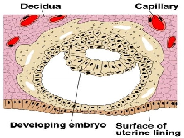
Chorion: After implantation, the trophoblast differentiates into 2 layers: a. An outer one called syncytium (syncytiotrophoblast) which is multinucleated cells without cell boundaries, b. An inner one called Langhan’s layer (Cytotrophoblast) which is cuboidal cells with simple cytoplasm. • A third layer of mesoderm appears inner to the cytotrophoblast.
Chorion: • The trophoblast and the lining mesoderm together form the chorion. • Mesodermal tissue ( connecting stalk) connects the inner cell mass to the chorion and will form the umbilical cord later on.
Chorion: • Spaces (lacunae) appear in the syncytium, increase in size and fuse together to form the " chorio-decidual space" or " intervillus space". • Erosion of the decidual blood vessels by the trophoblast allows blood to circulate in this space.
Chorion: • The outer syncytium and inner Langhan’s cells form buds surrounding the developing ovum called primary villi. • When the mesoderm invades the center of the primary villi they are called secondary villi. • When blood vessels (branches from the umbilical vessels) develop inside
Primary villous Secondary villous
Transverse section of tertiary villous
Chorion: • At first, the chorionic villi surround the developing ovum. • After the 12 th week, the villi opposite the decidua capsularis atrophy leaving the chorion laeve which forms the outer layer of the foetal membrane and is attached to the margin of the placenta.
• The villi opposite the decidua basalis grow and branch to form the chorion frondosum and together with the decidua basalis will form the placenta. • Some of these villi attach to the decidua basalis ( the basal plate) called the "anchoring villi", other hang freely in the
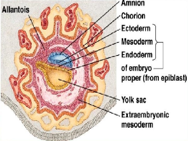
Amnion: After implantation, 2 cavities appear in the inner cell mass; the amniotic cavity and yolk sac and in between these 2 cavities the mesoderm develops.
Thank you