Physiology of Proprioception in Balance Dr Salah Elmalik

Physiology of Proprioception in Balance Dr. Salah Elmalik

Objectives q. By the end of this lecture students are expected to: q. Identify the major sensory receptors & pathways q. Describe the components, processes and functions of the sensory pathways q. Appreciate the dorsal column system in conscious proprioception q. Describe the spinocerebellar tract pathway in unconscious proprioception q. Differentiate between sensory and motor ataxia

Somatic Receptors Somatic receptors are specialized structure present at the peripheral terminations of afferent fiberes. Receptors are detectors and transducers which transduce different form of energy into action potential They are found in many parts of the body including the skin (cutaneous receptors), skeletal muscles, bones and joints (proprioceptors) They differ from specific receptors that mediate the special senses of vision, hearing, smell, taste and equilibrium.
: • ❶ Exteroceptors: Exteroceptors")
Classification of Sensory Receptors-1 A/Based on their location (Sherrington 1906): • ❶ Exteroceptors: Exteroceptors concerned with the external environment • Found on the surface of the body • E. g. touch and temperature receptors • ❷ Interoceptors: Interoceptors concerned with the internal environment e. g. chemoreceptors , osmoreceptors. • ❸ Proprioceptors: Proprioceptors concerned with potion of the body in the space. • Are found in joint, tendons and muscles.

Classification of Sensory Receptors-2 B/Based on their adequate stimulus v (Adequate stimulus is the particular form of energy to which the receptors is most sensitive) Mechanoreceptors : which detect mechanical compression or stretching of the receptor or of tissues adjacent to the receptor eg proprioceptors Thermoreceptors which detect changes in temperature, some receptors detecting cold and others warmth. Chemoreceptors, which detect taste in the mouth, smell in the nose, oxygen level in the arterial blood, osmolality of the body fluids, carbon dioxide concentration, and perhaps other factors that make up the chemistry of the body. e. g chemo R in carotid bodies Electromagnetic receptors, receptors which detect light on the retina of the eye eg rods and cones. Nociceptors (pain receptors) , which detect damage occurring in the tissues, whether physical damage or chemical damage eg free nerve endings

Classification of Sensory Receptors-3 C. Based on their speed of adaptation Adaptation means when a continuous sensory stimulus is applied, the receptor responds at a high impulse rate at first and then at a progressively slower rate until finally the rate of action potentials decreases to very few or often to none at all. Accordingly receptors can be classified Into: Slowly adapting (SA) or tonic receptors: receptors Muscle spindle, joint receptors, baroreceptors. Pain receptors do not adapt at all. Rabidly adapting (RA) or phasic receptors: receptors eg meissner’s corpuscles(touch), pacinian corpuscles(vibration)
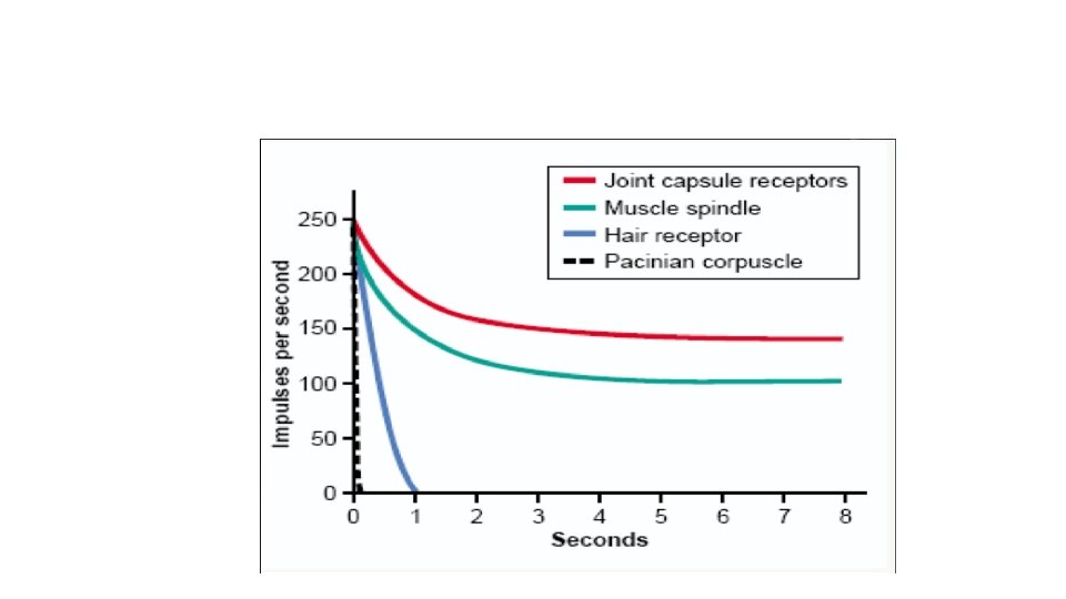

Mechanisms by which Receptors Adapt Is different for each type of receptor In the eye , the rods and cones adapt by changing the concentrations of their light-sensitive chemicals. The pacinian corpuscle is a viscoelastic structure so that after stimulation within few hundredths of a second, the fluid within the corpuscle redistributes, so that the receptor potential is no longer elicited. The second mechanism of adaptation of the pacinian corpuscle, but a much slower one, results from accommodation, which occurs in the nerve fiber itself. This probably results from progressive “inactivation” of the sodium channels in the nerve fiber membrane
 • Stimuli (mechanical, thermal, chemical)")
Activation of Sensory Receptors: Generation of Receptor Potential (RP) • Stimuli (mechanical, thermal, chemical) cause deformation in the sensory receptors • This causes influx of positive ions and generation of RP • RP induces a local circuit of current flow that spreads along nerve fiber and generates APs when threshold is reached

Relation Between Stimulus Strength & Receptor Potential Amplitude Receptor potential is directly related to stimulus strength

Generation of a Receptor Potential PAIN RECEPTOR Slowly adapting receptors detect presence of stimulus and its continuous strength Rapidly adapting receptors detect stimulus

Examples of RA and SA Receptors Muscle spindles & nociceptors are other examples of SA 13 receptors

What Are the Stimulus Features That Are Mediated by Sensory Receptors? Sensory receptors mediate 4 features of a stimulus: Modality: is what we perceive after a stimulus Many sensory modalities: vision, hearing, smell, taste, touch and temperature Each modality has many sub-modalities (e. g. taste can be sweet, bitter, sour, salty), Temperature sub-modalities: cold and heat Intensity: Intensity depends on the stimulus strength and is encoded by action potential frequency. Location: the site on the body or space where the stimulus originated. Duration: time from onset to offset of a stimulus. If persists for long time, the perceived intensity diminishes (adaptation)
 Αα (thickly myelinated) Aβ (intermediate m. ) Aδ")
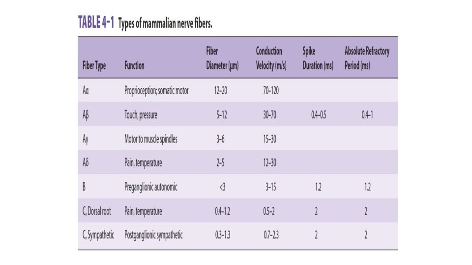
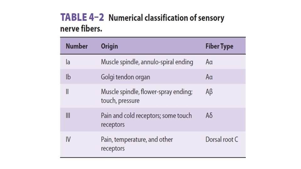
Classification of Nerve fibers Myelinated (A-fiber) Αα (thickly myelinated) Aβ (intermediate m. ) Aδ (thinly myelinated) Unmyelinated (Cfiber)

Ascending Sensory Tracts There are several ascending sensory systems Each system carries different types of sensations or MODALITIES: touch, proprioception, pain, temperature, … etc. , Main ascending sensory pathways ❶ Spinothalamic pathway: pathway carries signals of pain, temperature, crude pressure, and crude touch. ❷ Dorsal column pathway: pathway carries signals of fine touch, pressure, vibration, and proprioception. ❸ Posterior (dorsal) spinocerebellar pathway ❹ Anterior (ventral) spinocerebellar pathway The latter pathways (3 &4) carry subconscious proprioception.

What is Proprioception ? Proprioception stems from the Latin word proprius which means "one's own“ or "individual" It is the sense of one`s own body position It is also called proprioceptive/position sense It is the awareness of body position and of movements of body parts It can be divided into: Static proprioception: conscious perception of the orientation of the different parts of the body with respect to one another, Dynamic proprioception: proprioception rate of movement sense (also called kinesthesia)

Types of Proprioception There are two types of proprioception: ❶ Conscious proprioception: It reaches the level of sensory cerebral cortex (cerebrum) via the dorsal column-medial lemniscus pathway ❷ Unconscious proprioception: is communicated to the cerebellum primarily via: The dorsal spino-cerebellar tract (d. SCT) The ventral spino-cerebellar tract (v. SCT) • These are main ascending sensory pathways for proprioception

Role of Proprioception informs us about: The location of a body part in relation to other parts The rate of movement of a body part when it is moving The degree to which our muscle are being contracted or stretched The amount of tension created in our tendons The head orientation in relation to the ground and in response to movement • Proprioceptive information is carried from periphery to the CNS by proprioceptors and other somatic receptors

Types of Proprioceptors ❶ Muscle spindles *Detect how much a muscle is stretched ❷ Golgi tendon organs * Detect tension of a muscle on its tendon *Provide information about the strength of contraction & tension ❸ Joint Kinesthetic receptors *Are mechanoreceptors in the joint capsules; they detect angle and movement of the joints. Cutaneous & deep receptors also contribute to proprioception Cutaneous receptors Golgi. Torgan ⱡ

Muscle Spindles & Golgi Tendon Organs ❶ Muscle spindles They detect changes in the length of muscle. They convey length information to the CNS via group I and II afferent neurons This information is important for determining the position of body part ❷ Golgi tendon organ They detect changes in muscle tension


Dorsal Column–Medial Lemniscal System Objective: Appreciate the dorsal column system in conscious proprioception Fast conducting A α/β-fiber neurons • • Fine Touch Fine Pressure Vibration Position 22

Dorsal Column–Medial Lemniscal System Touch sensations requiring a high degree of localization and high intensity of discrimination (i. e. fine) Rapidly repetitive sensation such as vibration Joints Position sensations (Proprioception) Pressure sensations characterized by high intensity discrimination(i. e. fine pressure)
 Crude")
Anterolateral System Ventral & lateral spinothalamic tracts Pain Thermal sensations, (warmth & cold) Crude touch and pressure sensations capable only of crude localizing ability on the surface of the body Tickle and itch sensations Sexual sensations

Nociceptive vs Non-nociceptive Pathways A α/βfibres Dorsal column System • Fine Touch • Pressure • Vibration • Position • Movement • Anterolateral Pain System • Temperature • Crude touch • Tickle • Itch • Sexual sensation C-&Aδfibres 24
 Body is represented upside-down, with large representation of hands &")
Sensory Homunculus (Little Man) Body is represented upside-down, with large representation of hands & lips The extent of representation is proportional to the density of sensory receptors 23

Dorsal column damage Sensory ataxia Patient staggers; cannot perceive position or movement of legs Visual clues help movement • Positive Romberg test The test depends on the integrity of proprioception from the joints of the legs

The Dorsal & Ventral Spinocerebellar Tracts They carry subconscious proprioception signals ❶ The Dorsal Spinocerebellar tract (d. SCT) Carry signals directly to cerebellum at a speed of up to 120 m/s mainly from muscle spindles, but also from GTO, skin receptors & joint receptors Enter cerebellum through inferior cerebellar peduncle Terminate in vermis & intermediate zone Function of d. SCT: informs the cerebellum about: Muscle length and contraction Degree of tension on tendons Position and rate movement of parts of body. Forces acting on the body surfaces
: Carry some")
The Dorsal & Ventral Spinocerebellar Tracts The Ventral Spinocerebellar tract (v. SCT): Carry some signals from periphery (mainly from Golgi tendon organs) directly to cerebellum, but excited mainly by descending motor signals from brain (corticospinal & rubrospinal tracts) and from the spinal cord itself Enter cerebellum through superior cerebellar peduncle and terminate on both sides of cerebellum Function of v. SCT: informs the cerebellum about: Which motor signals have arrived to the spinal cord.

Ataxia and Gait Disturbances-1 Ataxia: inability to coordinate voluntary muscular movements that is due to nerve damage (CNS or PNS) and not due to muscle weakness (called also incoordination) Types of Ataxia: ❶Sensory ataxia ❷Motor ataxia Pathophysiology of sensory ataxia: ataxia PNS lesions (e. g. polyneuropathy) injury to sensory receptors and afferent neurons Dorsal column lesion Loss of proprioception, vibration and touch Ataxia is made worse in the dark or no vision Lesion in thalamus & sensory cortex Romberg's test. Ask the patient to close the eyes while standing with feet together. The affected patient becomes unstable (+ Romberg's test )

Ataxia and Gait Disturbances-2 Motor Ataxia: caused by cerebellar disorders Intact sensory receptors and afferent pathways Integration of proprioception is faulty Midline cerebellar lesions cause truncal ataxia Lateral cerebellar lesions cause limb ataxia Features of Cerebellar ataxia Clumsy movements Incoordination of the limbs Reeling gait (unsteadiness, and irregularity of steps; often with a tendency to fall to one or other side, forward or backward) Alcoholic intoxication produces similar effects!

THANK YOU
- Slides: 34