Physiology of Excitable tissue L 12 Motor Sensory

Physiology of Excitable tissue L 12 Motor & Sensory NS Prof. Fakhir Al-Ani fakeralani 2000@yahoo. com
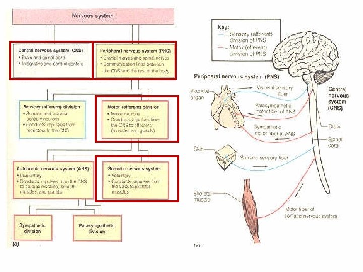

CNS Motor Sensory
 Descending Tracts Pyramidal tract")
Organization of Motor function Brain (Planning & initiation of movement) Descending Tracts Pyramidal tract extra-pyramidal tract (processing in the B. G. ) Alpha & Gamma motor n. motor fiber NMJ Initiating of movement & muscle spindle Regulating body posture
 1. Pyramidal tract. 2. Extra-pyramidal tracts. 3. Cerebellar tracts 4. Inter-connection")
Descending (Motor tracts) 1. Pyramidal tract. 2. Extra-pyramidal tracts. 3. Cerebellar tracts 4. Inter-connection between: Cerebellum. Basal ganglia. Thalamus These will produce movement, and coordinate movement at the level of the spinal cord.

Pyramidal system Started at the C. C. Discharge by them self. Or by input from sensory area Decussating at medulla oblongata Cortico-spinal tract The left hemisphere control the right side of the body

Pyramidal tract 80% of the fibers are crossed and descend As Lateral Cortico - spinal tract. 20% of the fibers are uncrossed and descend Anterior Corteco - spinal tract These will also cross to other side but the crossing occur just before termination.

Cortical representation 1. Left to Right 2. Upside down 3. Unilateral EXCEPT the upper part of the face (bilaterally represented). 4. Area of cerebral cortex that control part of the body is proportional to the fine movement.

Function of pyramidal system Skilled fine movement Cutting of this tract will cause: No paralysis, but there is: - No associated movement. - Clumsy grasp: move the muscle without precision, because there is no sensory feed back Hypotonia Dorsi-flexion of Babiniski's reflex

Extra-pyramidal system It concerned with gross movement and postural control Its defect will cause: - Positive signs: Like tremor - Negative signs: Like slow initiation of movement (Hypokinesia)

Origin of extra-pyramidal system Brain areas other than anterior motor area Like: 1. B. G. 2. Red nucleus 3. RAS 4. Vestibular nuclei 5. Superior colliculus

Programming & initiation of movement Basal ganglia: 1. Caudate nucleus. 2. Globus palidus. 3. Putamen. 4. Subthalamic nucleus. - Substantia nigra. - Red nucleus.

Tracts of the extra-pyramidal system 1. Tecto-spinal: from tectum and tegmentum to the spinal cord. 2. Rubro-spinal: from the red nuclei to the spinal cord. 3. Reticulo-spinal: from the reticular formation to the spinal cord. 4. Vestibulo-spinal: from the vestibular nuclei to the spinal cord

Function of the Extra-pyramidal 1. Programming & initiation of movement. 2. Control of Gross movement. 3. Regulation of body posture. 4. Regulation of tone. Regulation of movement can not be separated from posture.
")
Regulation of posture and tone Muscle spindle: Receptors at muscle (Parallel with the muscle) Regulate muscle contraction involuntary. Formed of: 1. Intrafusal muscle fibers 2. Sensory nerve: 3. Motor nerve:

Regulation of posture & tone Postural control occur through the activity of the muscle spindle by two types of reflexes. 1. Static reflex : Control posture at rest. 2. Phasic Reflex: Control posture on movement. Or sudden change in the movement.

Spinal cord Gray & White Matters 1. Nerve cells: Sensory cells. Motor cells. Interconnecting cells. Autonomic cells. Connective tissue cells. 2. Nerve fibers: Ascending n. f. Descending n. f. Interconnecting n. f.

Functions of the spinal cord 1. Carry motor impulses (from CNS to the peripheral N. S. ) 2. Carry sensory impulses (from peripheral N. S. to the CNS. ) 3. Presences of Centers. 4. Coordination of movement. 5. Reflexes.
 to peripheral N. S. Higher centers:")
1. Carry motor impulses From CNS (Higher Centers) to peripheral N. S. Higher centers: - Motor cortex - Basal ganglia Descend down in the spinal cord to: - Alpha (α ) motor neuron - Gamma (γ )motor neuron At the ventral horn of the spinal cord Then goes to the peripheral nerve fiber goes to the muscle.

1. Carry motor impulses Cont. motor impulse from the CNS to peripheral N. S. Cerebral motor cortex Basal ganglia Descending fibers Ca ++ Spinal cord Inter connecting neuron

Alpha motor neuron & peripheral motor nerve Motor nerve fiber Muscle Ventral horn Alpha motor neuron
 Sensory impulses from different receptors")
2. Carry sensory impulses (from peripheral to CNS. ) Sensory impulses from different receptors are send to CNS (Thalamus, Sensory cortex, Cerebellum) through ascending fibers The two main ascending tracts are: 1. Lateral spinothalamic tract. Carry pain, temperature & light touch sensation. 2. Dorsal column (F. gracilis & Cuneatus). Carry properioceptive, vibration & crud touch)
 Dorsal column Lateral spinothalamic tract")
2. Carry sensory impulses (from peripheral to CNS. ) Dorsal column Lateral spinothalamic tract

3. Presences of Centers Center: Is a collection of nerve cells within the CNS that do certain function like: Urination center. Defecation center.

Spinal cord centers Defecation center Urination center

4. Coordination of movement. 1. Descending Motor input from the higher centers + 2. Sensory nerve from the receptors 3. Interconnecting nerves Terminated at the α & γ cells Alphha neuron can be stimulated by: - Afferents of muscle spindles - Corticospinal - Cerebellum & Vestibular Neucli. where coordination of all these input to get the final output from motor cells

Coordination of movement. Impulses from Cerebral cortex Impulses from Cerebellum Impulses from Vestibular Nuclei Impulses from sensory input Impulses from interconnecting neurons

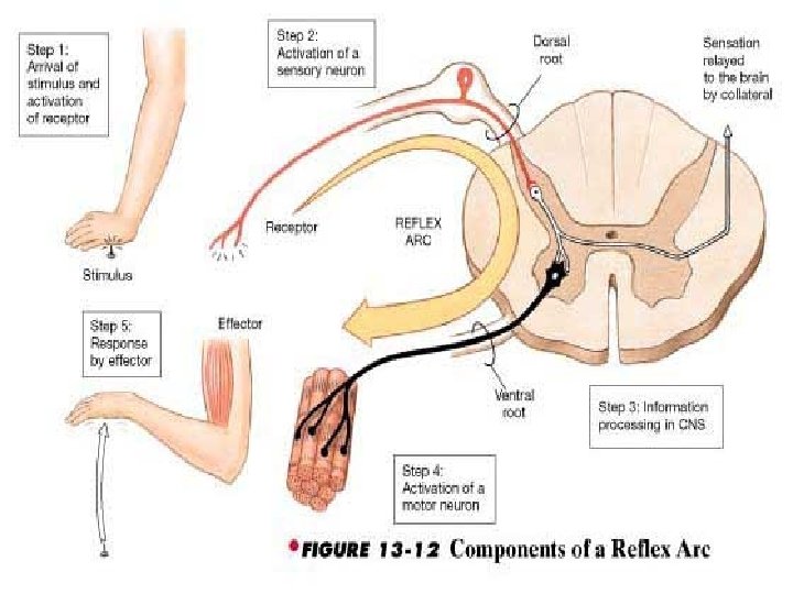
5. Reflexes. It is an involuntary response to a sensory input. The response could be: 1. Motor movement as in somatic reflex. 2. Glandular secretion as in autonomic reflex. To get a reflex we should have a reflex arc: 1. Receptor. 2. Sensory nerve. 3. Normal spinal cord sigment. 4. Motor nerve. 5. Effecter organ.

Reflex arc Somatic reflex arc for Tendon jerk Sensory nerve Receptor Motor nerve Effecter organ Muscle

Types of somatic reflex 1. Monosynaptic reflex: Single synapse between sensory and motor nerve like: Biceps jerk C 5 - C 6 Supinator jerk C 5 - C 6 Triceps jerk C 6 – C 7 Knee jerk L 2 – L 3 Ankle jerk L 5 – S 1 2. Polysynaptic reflex: Many synapse between sensory and motor nerve like: Painful reflex. Blinking reflex.

Monosynaptic reflex

Mono- & Poly-synaptic reflex
 L 2 – L 3 Supinator Jerk (Reflex) Biceps")
Monosynaptic reflex Knee Jerk (Reflex) L 2 – L 3 Supinator Jerk (Reflex) Biceps Jerk (Reflex) Ankle Jerk (Reflex) L 5–S 1 C 5 - C 6 Triceps Jerk (Reflex) C 5 - C 6

Polysynaptic reflex

Properties of the reflexes 1. Adequate stimulus 2. Specific & Stereotyping response 3. Central excitation & inhibition state 4. Habituation and sensitization of the response 5. Localization depending on position of the org. 6. Fractionation and occlusion. 7. Rebound phenomenon. 8. Irradiation of the stimulus.

Muscle Tone It is a partial state of involuntary muscle contraction It can be identified by muscle resistance against passive stretching Need intact Reflex arc

Decreased Tone Decreased tone occur when there is: 1. Cutting of the sensory nerve. 2. Cutting of the motor nerve. 3. Damage of aspinal segment at arc level. 4. Damage of the muscle. 5. Damage of the receptor. Decrease tone = Hypotonia = Flaccidity. With decrease in the reflex

Increased Tone Normally stretch reflex is suppressed by higher control So Cutting of the higher influence will cause Increase in tone (Hypertonia) With increase in the reflex as in UMN disease
- Slides: 39