Physiology of Excitable tissue L 11 Sensation II

Physiology of Excitable tissue L 11 Sensation II Prof. Fakhir Al-Ani fakeralani 2000@yahoo. com

Physiological role of Different diameter & conduction velocity Properioceptive sensation Fast conducting nf. To get fast information about leg position during running Acute sharp pain Fast conducting fibers To get protective response by the body Dull chronic pain Slow conduction fibers.

Grading of sensory signals Grading can be done by: 1. Temporal summation. frequency of firing impulses in the nerve. or 2. Spatial summation. number of nerve fibers excited.

Temporal Summation Signal strength is transmitted by using higher frequency of nerve impulses. The code in the CNS for increasing the strength of stimulus Is The frequency of impulse reaching the sensory area.

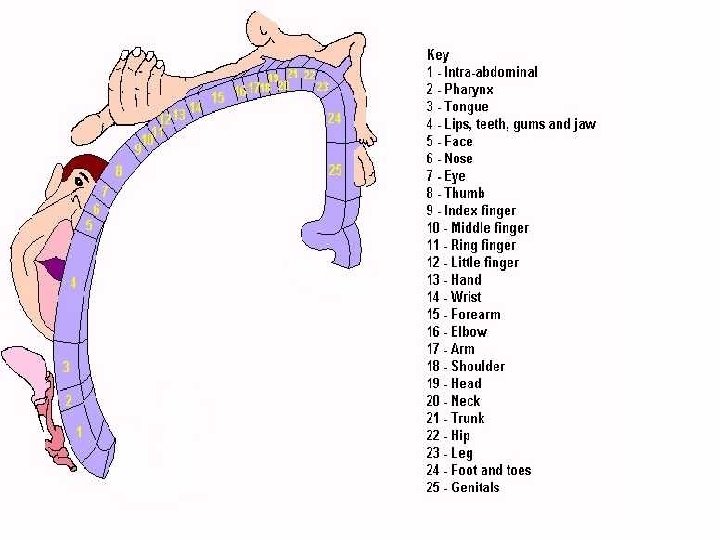
Spatial Summation Signal strength is by using progressively greater number of fibers are excited. Receptor field: Area of skin (5 cm diameter) supplied by number of branches related to one nerve. Nerve fibers are more at the center of the field So it is the most sensitive area in comparison to the peripheral areas.

Facilitation by successive stimuli One excitatory single pre-synaptic terminal never causes an A. P. in the post synaptic nerve But Large number of input terminals must discharge in the same neuron either simultaneously or in rapid successive discharge causes excitation

Facilitation by successive stimuli Facilitated zone Zone receives sub-threshold impulses So can not produce A. P. in the postsynaptic membrane unless it is supported by next. Discharge zone Zone receives Threshold or supra-threshold impulses can produce A. P. in the postsynaptic membrane.

Inhibitory neuronal pool Opposite to the facilitatory pool of neurons Some of the nerve terminals causes inhibitory effect on the post-synaptic membrane that will influence the impulse reaching another nerve and the post-synaptic membrane will not respond to this second stimulus

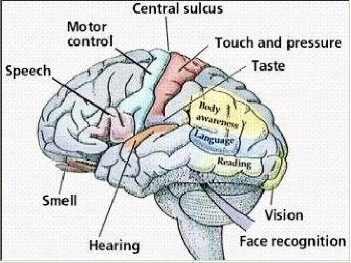
Dermatome Area of skin supplied by single spinal root Or Area of skin related to one single spinal segments Number of the dermatomes are the same as the number of the spinal root Or the same number of the spinal segments.

Dermatome

Spinal roots

Clinical significance of dermatome To assess the normal function of the spinal cord: 1. Sensory level: The level at which there is lose of sensation in spinal cord injury. 2. Reflexes: The defect in the tendon reflex is related to the site of injury in the spinal cord. Supinator tendon jerk: C 5 - C 6 Biceps tendon jerk: C 5 – C 6 Triceps tendon jerk: C 6 – C 7 Knee tendon jerk: L 2 L 4 Ankle jerk: L 5 – S 1

Sensory pathway There are 3 major somatic sensory pathways. 1. Posterior column pathway 2. Spino-thalamic pathway 3. Spino-cerebellar pathway Each pathway is formed of three neurons: 1. First order neuron. 2. Eecond order neuron. 3. Third order neuron.

Sensory pathways in the spinal cord 1. First order 2. Second order 3. Third order

1. Posterior column pathway

1. Posterior column pathway Carries Touch, Pressure, Vibration & Proprioception Tracts of the posterior column. 1. Fasciculus gracilis: carries sensory information from the inferior half of the body 2. Fasciculus cuneatus: carries sensory information from the superior half of the body

In this pathway The first order neuron extends all the way from Receptors Spinal cord medulla oblongata to the nucleus gracilis, & nucleus cuneatus It ascends ipsilateraly in the spinal cord First order neurons synapse with second-order neurons in the medulla oblongata.

The second order neuron The axons of the 2 nd order neurons start at the nuclei & cross to the opposite side in the medulla. Crossing over of an axons from one side to the opposite side is called decussation. These axons that have crossed over enter the medial lemniscal tract which carries them to the thalamus where there is the 3 rd order neuron

The Third order neuron The 2 nd order neurons synapse with the 3 rd order neurons in the thalamus. The thalamus sorts out the Type of the stimulus and the region of the body involved. Then The 3 rd order neurons extend their axons to the primary sensory cortex.

2. Spinothalamic tract Carry Pain, Thermal & Fine touch sensation This pathway begins at the peripheral receptor and ends at the primary sensory cortex.

Lateral spino-thalamic tract

The first order neuron The axon of the 1 st order neuron extends from the receptors to the spinal cord where it synapse with the 2 nd order neuron at the dorsal horn. At lamina II & Lamina III Gate control theory of pain transmission Impulses from touch fiber (Type A δ fibers) inhibit transmission of pain impulses

The second & third order neuron The second order neuron is started at the dorsal horn the its fiber cross the mid line Then it ascend in the contra lateral side up to the thalamus where we have third order neuron From the thalamus the third order neuron started to end at the cerebral cortex sensory area.

Spinothalamic tract

Lamellate pathway in the spinal cord In the posterior column: The fibers from the lower limb are present medial to the fibers that come from the upper limb. In the spino-thalamic tract: The fibers from the lower limb are present lateral to the fibers that come from the upper limb. Clinical significance?

Lamination of the sensory pathways
- Slides: 28