physiology of digestive System Function 1 Obtain resources

physiology of digestive System Function: 1 - Obtain resources from the external environment like Water, Minerals, Nutrients (Lipids, Carbohydrates, Proteins) and Vitamins 2 - Break down large particles into smaller ones transfer materials from external environment blood cells Anatomy of digestive tract: oral cavity, pharynx, esophagus, stomach, small intestine large intestine and anus

Digestive tract in hours

Digestive tract in dog

Digestive tract in frog

Digestive tract in bird

Digestive tract in sheep & goat

Gastrointestinal physiology: - The major processes of the GI system 1. Motility 2. Secretion 3. Digestion 4. Absorption

1 - Motility The gastrointestinal tract generates motility using smooth muscle , there are two type of contractions patterns A- The Tonic motility : Its contractions are occur in the sphincters of the tract, as well as in the anterior stomach. B- phasic contractions motility : It’s a periods of both relaxation and contraction, occurring in the posterior stomach and the small intestine. 1. Peristalsis : The contractions occur directly behind the bolus of food that is in the system, forcing it toward the anus into the next relaxed section of smooth muscle. 2. Segmentation : This process is carried out by the longitudinal muscles relaxing while circular muscles contract at alternating sections thereby mixing the food. This mixing allows food and digestive enzymes to maintain a uniform composition.

2 - Secretion • Every day, seven liters of fluid are secreted by the digestive system. This fluid is composed of four primary components: ions, digestive enzymes, mucus, and bile. About half of these fluids are secreted by the salivary glands, pancreas, and liver, which compose the accessory organs and glands of the digestive system. The rest of the fluid is secreted by the GI epithelial cells. • Release of substances to enhance breakdown of food called digestive juices which contended from enzymes, bile salts, mucus, etc. released by exocrine glands into GI tract. • Must break bonds with enzymes (various organs) Enzyme function aided by HCl (stomach) Bile (liver) Na. HCO 3 (pancreas)

3 - Digestion Physical and chemical break down nutrients into absorbable unit: 1 - Physical digestion (chewing, mixing) 2 - Chemical digestion (enzyme catalyzed) Carbohydrate polysaccharides monosaccharides proteins amino acids fats glycerol + fatty acids
 metabolism: - • The carbohydrates (starch) breakdown in")
3 - Digestion A. Carbohydrate (polysaccharides) metabolism: - • The carbohydrates (starch) breakdown in to oligosaccharides by pancreatic amylase, but the other carbohydrates pass undigested into the large intestine and further handling by intestinal bacteria. • The most poly-saccharides, such a scellulose, are not digested at all, despite being made of multiple glucose units.

3 - Digestion B. Protein metabolism: The biochemical processes responsible for the synthesis of proteins and amino acids, and the breakdown of proteins (and other large molecules, too) by catabolism. Which is the breakdown of proteins into amino acids and simple derivative compounds, for transport into the cell through the plasma membrane and ultimately for new proteins via the use of ribonucleic acids (RNA) and ribosomes, or undergo amino acid catabolism to be converted to other compounds via the Krebs cycle
 are degraded into fatty")
3 - Digestion C- Lipid metabolism • The lipids (fats) are degraded into fatty acids and glycerol. • The pancreatic lipase breaks down triglycerides into free fatty acids and monoglycerides.

4 - Absorption • Digested food is now able to pass into the blood vessels in the wall of the intestine through either diffusion or active transport. • The small intestine is the site where most of the nutrients from ingested food are absorbed. The inner wall, or mucosa, of the small intestine is has finger-like pieces of tissue called villi. • The epithelial cells of the villi transport nutrients from the lumen of the intestine into these capillaries (amino acids and carbohydrates) and lacteals (lipids). • The absorbed substances are transported via the blood vessels to different organs of the body where they are used to build complex substances such as the proteins required by our body. • The material that remains undigested and unabsorbed passes into the large intestine. • Absorption of the majority of nutrients takes place in the jejunum.

Accessory organs 1. 2. 3. 4. salivary glands pancreas liver gall bladder

Salivary glands 1. 2. 3. 4. Parotid glands Submandibular glands Sublingual glands Minor salivary glands • The salivary glands in mammals are exocrine glands, glands with ducts, that produce saliva , amylase and digestive enzyme that breaks down starch into maltose and glucose.

salivary glands

The Saliva is a watery substance located in the mouths of humans and animals, secreted by the salivary glands. The saliva is 99. 5% water, while the other 0. 5% consists of electrolytes, mucus, glycoproteins, enzymes, antibacterial, Functions 1. Maintenance of oral hygiene. 2. Lubricant of the bullets. 3. Digestion : . by enzyme amylase, which is breaking down starch into simpler sugars such as maltose and dextrin. 4. Ion reservoir, buffer function : typically p. H 6. 2– 7. 4. This prevents minerals in the dental hard tissues from dissolving. 5. Role in taste: 6. Wound licking : The saliva can be help to heal wounds in some species.

Pancreas Exocrine cells secrete pancreatic juice into duodenum are: - 1. Amylase - breaks down starch 2. Trypsinogen 3. lipase - digests triglycerides 4. Proteases and Nucleases 5. Na. HCO 3 (alkaline) - neutralizes stomach acidity

Liver 1. secretes bile • Bile salts used for lipid absorption 2. metabolic processing of absorbed materials 3. degradation of waste, hormones and drugs. 4. synthesizes plasma proteins 5. stores glycogen, fats, minerals and vitamins
 - Epithelial tissue. • Submucosa - Elastic")
Gastrointestinal Tract Structure • Mucosa (lumen side) - Epithelial tissue. • Submucosa - Elastic connective tissue contains lymph and blood vessels. • Muscularis - Smooth muscle. • Serosa - Outer layer of connective tissue - Secretes serous fluid

1 - Mouth Ingestion • Bringing food into the body • Tongue taste buds detect chemical composition of food Mastication • Chewing (physical digestion) by teeth and tongue Chemical digestion • Saliva which are moistens food, amylase - breaks down starch into maltose and lysozyme - antibacterial agent

2 - Pharynx and Esophagus • Transport food and water to stomach, secretes mucus – deglutition (swallowing) reflex moves food to stomach • Movement of food bolus in esophagus (and rest of GI tract) via peristalsis

3 - Stomach • The stomach is a muscular, hollow, dilated part of the digestive system which functions as an important organ of the digestive tract in vertebrates. It is involved in the second phase of digestion, following mastication (chewing). • It secretes protein-digesting enzymes called proteases and gastric acid to aid in food digestion. • Through the smooth muscular contractions before sending partially digested food (chyme) to the small intestines.
: – Sec. of")
Stomach Mucosal Cells 1 - Goblet cells (mucus neck cells ): – Sec. of the Mucus The Gastric Mucosal Barrier Mucus that protects stomach epithelium 2 - Parietal cells: – Sec. of the gastric acid HCl Kills bacteria and denatures proteins _ Sec. of Intrinsic factor (Ca++ absorption) 3 - Chief cells : Sec. of the Pepsinogen & Gastric lipase Pepsinogen activated by HCl pepsin which breaks down proteins

Stomach Mucosal Cells

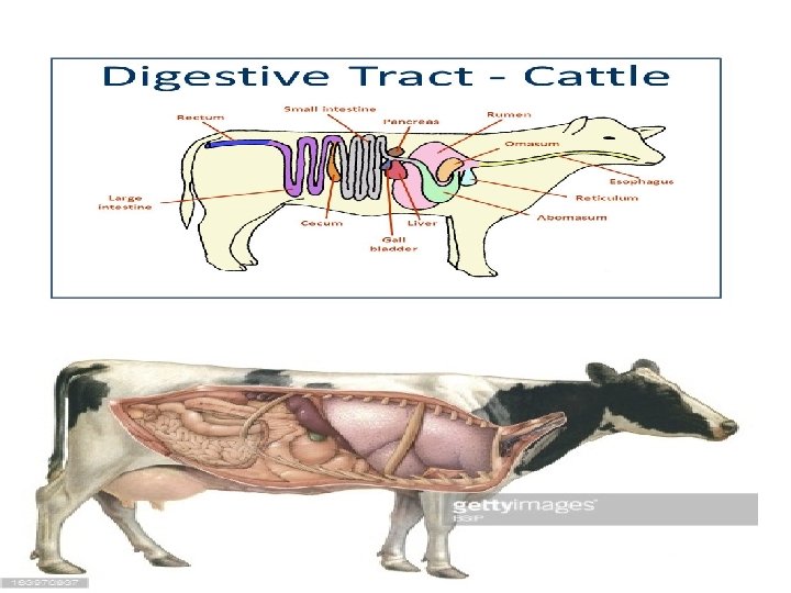
Ruminant • Ruminants are mammals that are able to acquire nutrients from plant-based food by fermenting it in a specialized stomach prior to digestion, principally through microbial actions. • The process typically requires the fermented ingesta (cud) to be regurgitated and chewed again. • The process of rechewing the cud to further break down plant matter and stimulate digestion is called rumination. • Ruminating mammals include cattle, goats, sheep, giraffes, yaks, deer.

Ruminant 1 -The Rumen: It’s a larger part of the reticulorumen, which is the first chamber in the alimentary canal of ruminant animals. It serves as the primary site for microbial fermentation of ingested feed. 2 - Reticulum: It’s smaller part, and a first chamber in the alimentary canal of a ruminant animal. When cleaned and used for food, it is called "tripe". 3 - Omasum : Its functions are absorption of water, magnesium, and the volatile fatty acids produced by rumen fermentation, that have not been absorbed into the bloodstream. 4 - Abomasum: It is a secretory stomach similar in anatomy and function as the monogastric stomach. It serves primarily in the acid hydrolysis of microbial and dietary protein, preparing these protein sources for further digestion and absorption in the small intestine.

Ruminant

Small Intestine • The small intestine or small bowel is the part of the gastrointestinal tract between the stomach and the large intestine, and is where most of the digestion and absorption of food takes place. The small intestine has three distinct regions – the duodenum, jejunum, and ileum. The duodenum receives bile and pancreatic juice through the pancreatic duct, controlled by the sphincter of Oddi. The primary function of the small intestine is the absorption of nutrients and minerals from food. • Most chemical digestion occurs here and most absorption occurs here • Large surface area – Plicae – folds in mucosa – Villi – fingerlike projections • Capillaries, central lacteal (absorption) – Microvilli ("brush border") on epithelium – hydrolyze disaccharides, polypeptides, etc. ( enterokinase - activates trypsin (pancreatic enz. )

Small Intestine

Large Intestine • The large intestine, also called the large bowel, is the last part of the digestive system in vertebrates. Water is absorbed here and the remaining waste material is stored as feces before being removed by defecation. • combination of the cecum, colon, rectum, and anal canal, it begins in the right iliac region of the pelvis, just at or below the waist, where it is joined to the end of the small intestine. It then continues up the abdomen, across the width of the abdominal cavity, and then down to its endpoint at the anus. • main function is to store undigested material (feces) • 30% dry weight of feces = bacteria (E. coli) live in large intestine (produce vitamin K) •
- Slides: 33