Physiology of Cell Body Fluids Excitable tissue Muscle

Physiology of Cell, Body Fluids, Excitable tissue & Muscle Choesnan Effendi Physiology Dep. Airlangga University 2012

Episode Kedua

Cair Tubuh & Transport bahan melewati membran Body Fluids & Transport of substances through the cell membrane

Cair Tubuh Extracellular Interstitial Plasma darah Transcellular
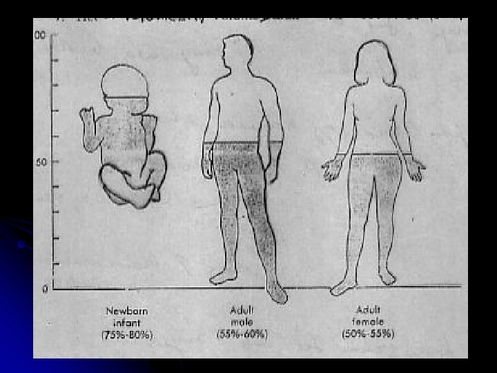
 Indikator Total body water (cair tubuh total)")
Volume % BB ( Berat Badan ) Indikator Total body water (cair tubuh total) 60 Deutrium ( D 2 O / 2 H 2 O ), Tritium ( 3 H 2 O ), Antipyrine Cair Ekstrasellular 20 Inulin *C 14 , Thiosulfate Cair Intrasellular 40 Total body water − Cair Ekstrasellular Plasma darah 5 Evans blue ( T- 1824 ) , 125 IAlbumin Darah 7– 8 red blood cells { Volume plasma darah : ( 100 % − Hct )} Cair Interstisial 15 Volume ekstrasellular − Volume plasma 51 Cr-labeled

Plasma darah Whole Hematocrit blood
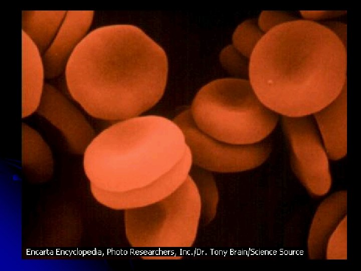

Volume Eritrosit Volume Darah X 100 % = 36 – 45 % = Hct
 Adalah volume kumpulan")
HCT = Hematocrit = PCV ( Packed Red Cell Volume ) Adalah volume kumpulan erithrocytes yang dinyatakan dengan % terhadap volume darah keseluruhan

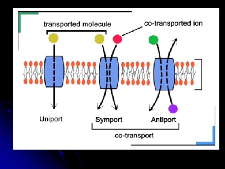
Beberapa cara masuk / keluarnya bahan melewati membran sel Interstitiel / Plasma darah 1. Osmosa 2. Diffusi sederhana 3. Diffusi fasilitasi 4. Transport aktif Cytoplasma 5. Exocytosis / endocytosis

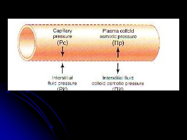
Pertukaran cairan didaerah kapiller

Ruang interstitiel Sitoplasma / sitosol Plasma darah

Fluid exchange : Arteriole capillary venule Arteriole Capillary Venule

Filtrasi / pertukaran cairan daerah kapiller Dipengaruhi oleh beberapa faktor : • Tekanan onkotik plasma • Tekanan onkotik interstisial • Tekanan hidrostatik plasma • Tekanan hidrostatik interstisial Tekanan hidrostatik plasma = tekanan darah

Tekanan kolloid osmotik = Tekanan onkotik plasma darah Oleh karena adanya Protein plasma ( p ) Protein plasma Gram % Albumin 4, 5 21, 8 Globulin 2, 5 6, 0 Fibrinogen 0, 3 0, 2 Total 7, 3 28, 0 P mm Hg

Dari ketiganya, jumlah terbanyak adalah ALBUMIN
 Interstisial Ponkotik -")
Sebagai contoh : Pint : 1 mm Hg ( hidrostatik ) Interstisial Ponkotik - int : 8 mm Hg Ponkotik - art ( ven ) : 28 mm Hg Arteriole Kapiller Pkap : 25 mm Hg Part : 37 mm Hg NFP ( Net Filtration Pressure ) Venule Pven : 17 mm Hg = Pkap – Pint - p kap + p int 25 – 1 – 28 + 8 = + 4 + ( positip ) : artinya cairan keluar dari kapiller, sisanya ini akan di absorbsi oleh limfe
 = Pkap – Pint - p kap +")
NFP ( Net Filtration Pressure ) = Pkap – Pint - p kap + p int 25 – 1 – 28 + 8 = + 4 + ( positip ) : artinya cairan keluar dari kapiller, sisanya ini akan di absorbsi oleh limfe

Mengapa hypoproteinemia udema Bagaimana mengenai tekanan oncotic protein plasma ? ? ?

Starving Children in Nigeria

Udem akan terjadi apabila 1. Bendungan vena : tumor, dekompensasi jantung kanan, bendungan aliran limfe 2. Cairan dari intersitial yang menuju plasma < dibanding yang masuk

Cairan dari intersitial yang menuju plasma << dibanding yang masuk O. K. Tekanan osmotik plasma yang rendah O. K. Kadar protein plasma yang rendah = HIPOPROTEINEMIA

HIPOPROTEINEMIA O. K. 1. Under nutrition : kurang gizi /rendah protein. 2. Sintesa protein ( terutama Albumin ) terganggu : a. l pada penyakit hati : cirrhosis hepatis 3. Sekresi protein : yang seharusnya tidak terjadi , yaitu terjadi proteinuria ( pada nephrotic syndrome )

Tekanan osmotik plasma Berperanan untuk reabsorbsi kembali cairan yang dari interstisial

Beberapa cara masuk / keluarnya bahan melewati membran sel

1. Osmosa 2. Diffusi sederhana 3. Diffusi fasilitasi 4. Transport aktif H 2 O yg bergerak dari larutan hipotonis kearah hipertonis Bahan yang terlarut bergerak dari tekanan tinggi ketekanan rendah Seperti No. 2, menggunakan mediator (carrier system) Bahan yang terlarut bergerak dari tekanan rendah ketekanan tinggi, menggunakan mediator, energi ( ATP )

1. Osmosa Contoh : H 2 O 2. Diffusi sederhana CO 2 , Ureum 3. Diffusi fasilitasi glukosa, asam amino 4. Transport aktif Na, K, Ca

Mediator = carrier system

Simple diffusion, facilitated diffusion & osmosis: are passive transport, without ATP Active transport, sodium potassium pump, calcium pump, exocytosis: are active, need ATP
 is a process of")
Facilitated diffusion (also known as facilitated transport or passive-mediated transport) is a process of passive transport, facilitated by integral proteins (mediator). Without energy (ATP)
 depends on the relative concentration of solute molecules")
Osmosis (movement of water across membranes) depends on the relative concentration of solute molecules on either side of the membrane Water move from low concentration to high concentration

How do about erythrocytes if in: - hypotonic solution - isotonic solution - hypertonic solution

Crenated / wrinkled ery in hypertonic medium Normal Ery structure in isotonic medium Swollen ery & rupture in hypotonic medium

Normal Ery structure in isotonic medium Crenated / wrinkled ery in hypertonic medium Swollen ery & rupture in hypotonic medium

Simple Diffusion; the flow substances or matter from a higher concentration to a lower concentration

Alveoli: O 2: Diffusion from alveoli into blood stream capillary CO 2: Diffusion from blood capillary into alveoli

PO alv : 104 mm. HG Pc. O alv : 40 mm. HG PO cap : 40 mm. Hg Pc. O cap : 46 mm. Hg 2 2

at alveoli or at respiratory membrane O 2 diffusion into blood capillary, then enter to the erythrocyte, bound by hemoglobin → Hb. O 2 at tissue; tissue membrane and endothelium capillary CO 2 diffusion into blood capillary, then enter to the erythrocyte, bound with H 2 O → H 2 CO 3 →dissociation Becomes: H+ + HCO 3 - (bicarbonate ion)
 flow out from erythrocyte into blood")
In blood stream: HCO 3 - (bicarbonate ion) flow out from erythrocyte into blood stream, to the capillary beds of respiratory membrane at respiratory membrane HCO 3 - (bicarbonate ion) flow in from blood stream into erythrocyte, then bind with H+ , become H 2 CO 3, H 2 CO 3 dissociation, Become H 2 O + CO 2
 and then")
at respiratory membrane CO 2 flow out to blood (exit from erythrocyte) and then diffusion into alveoli lumen

In blood stream: O 2 bound by hemoglobin → Hb. O 2 → to tissues and cells all the body

at tissue O 2 simple diffusion from Hb. O 2 into cytosol, and then into mitochondria. Glucose move into cytosol by glucose transporter (facilitated diffusion)

Facilitated Diffusion Like simple diffusion, but requires interaction of a carrier protein that bind the molecules or ions to aids passage through the membrane Carrier protein = mediator or transporter
 is a process of")
Facilitated diffusion (also known as facilitated transport or passive-mediated transport) is a process of passive transport, facilitated by integral proteins (mediator). Without energy (ATP)

Glut = Glucose transporter Skeletal Muscle requires GLUT – 4 , GLUT-4 stand-by in cytosol of muscle fiber, they ‘ll move into the membrane if insulin receptors are stimulated by insulin Glut = Glucose transporter is mediator/transporter of glucose enter into cytosol
 GLUT- 4 IRS-1 vesicle")
Insulin Glucose – facilitated diffusion Insulin Receptor ( IR ) GLUT- 4 IRS-1 vesicle contains GLUT- 4 PI 3 kinase translocation Cell membrane

Glucose enter into cytosol of skeletal muscle fiber by; Signal transduction by insulin Insulin activate insulin rec → form IRS 1 activates PI 3 -Kinase stimulate translocation vesicle, which contains GLUT-4 GLUT- 4 is mediator / transporter of glucose In skeletal muscle fiber

There are 2 processes: * Signal transduction by insulin ** Facilitated diffusion by GLUT- 4

Active Transport is the Pumping of Solutes Against their Gradients

Active Transport is the Pumping of Solutes Against their Gradients 1. Cell must expend ATP/ energy to pump a molecule across a membrane 2. Performed by embedded proteins 3. Na-K Pump (sodium-potassium)exchanges Na+ for K+ in animal cells when ATP changes protein conformation by transferring its terminal phosphate group to the transport protein
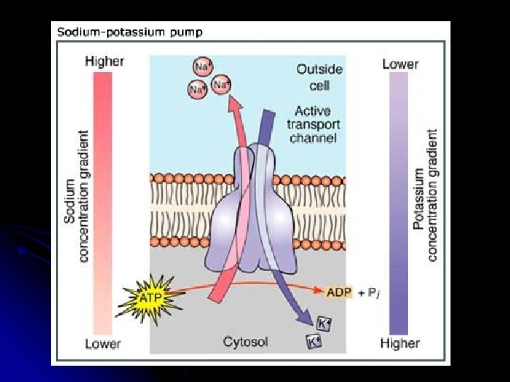

Active transport is the movement of a substance against its concentration gradient (from low to high concentration). active transport: energy-requiring, carrier-mediated transport system in which molecules can be moved across cell membrane against electrochemical gradient

Electrolyte inside & outside the cell membrane Resting Na+ 142 m. Eq/L Cl- 120 m. Eq/L + + + – – – K+ K+ 4 m. E/L + – 140 m. Eq/L Na+ 14 m. Eq/L CL- 5 m. Eq/L Axon

3 molecules Na+ carried out into extracellular, changed by 2 molecules K+ (carried into cytosol)

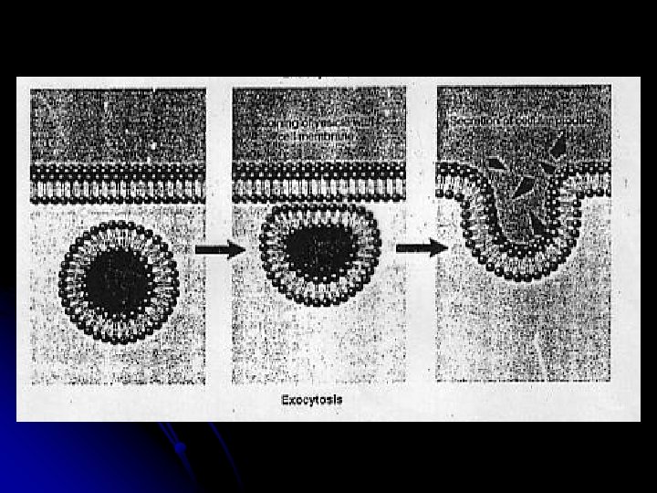
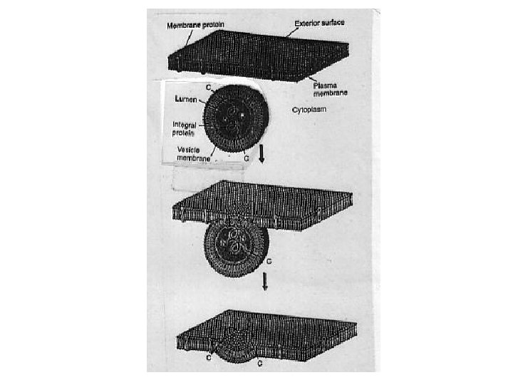
Exocytosis

Exocytosis is the cellular process in which intracellular vesicles in the cytoplasm fuse with the plasma membrane and release or "secrete" their contents into the extracellular space

Exocytosis is the process by which cells excrete waste products and other large molecules from the cytoplasm
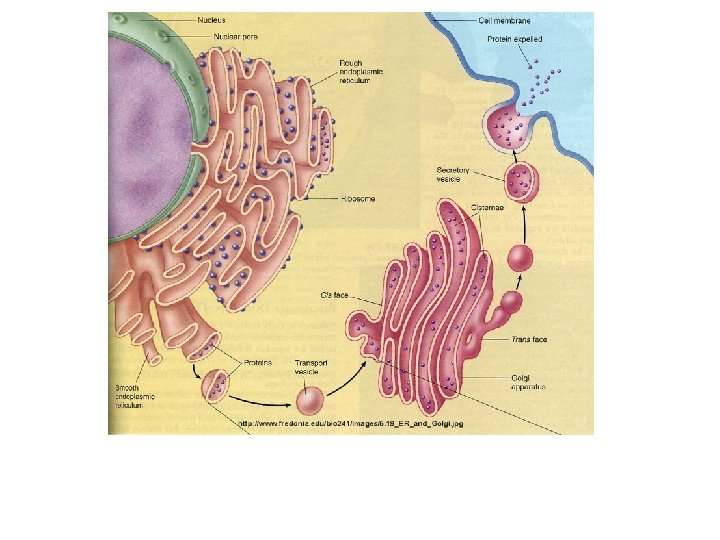

Exocytosis is the cellular process in which intracellular vesicles in the cytoplasm fuse with the plasma membrane and release or "secrete" their contents into the extracellular space Exocytosis is the process secretion substances into the extracellular space or into the blood stream.

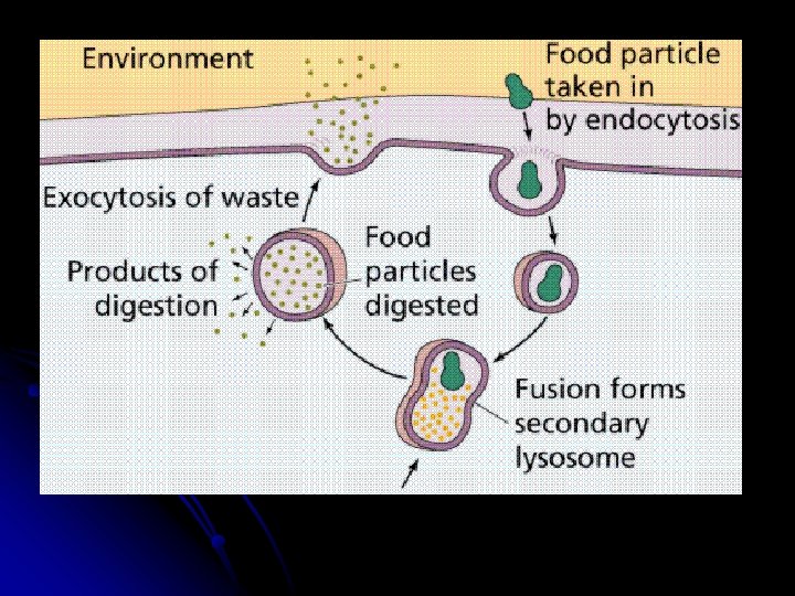
Exocytosis is the reverse of endocytosis. Endocytosis is like phagocytosis


Exocytosis
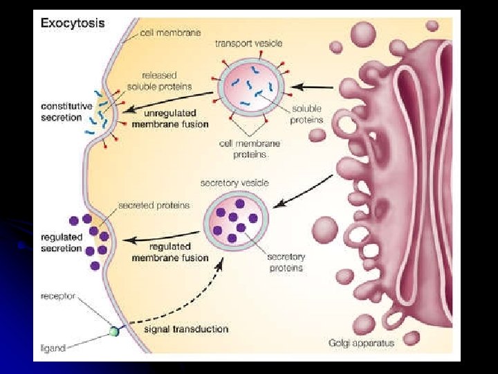

Exocytosis & Endocytosis Transport Large Molecules 1. Exocytosis- transport vesicles migrate to plasma membrane & fuse & release contents 2. Endocytosis- large molecules enter cells within vesicles pinched inward from the membrane --> Phagocytosis- cell engulfs particles “cell eating” --> Pinocytosis- cell engulfs droplets of extracellular fluid “cell drinking”

The other way of transport across membrane

Cotransport: also known as coupled transport or secondary active transport, refers to the simultaneous or sequential passive transfer of molecules or ions across biological membranes. - Symport - Antiport
")
Several types transport across membrane (facilitated diffusion)

Symport

Sodium – glucose symport / Na-Glucose co-transport
 is an integral membrane protein involved")
Antiport An antiporter (also called exchanger or countertransporter) is an integral membrane protein involved in secondary active transport of two or more different molecules or ions (i. e. , solutes) across a phospholipid membrane such as the plasma membrane in opposite directions. or called IONS EXCHANGE

Amino acid Na+ Glucose Na+ Ca++

For example, the Na+/Ca 2+ exchanger, used by many cells to remove cytoplasmic calcium, exchanges one calcium ion for three sodium ions
")
the Na+ - Ca 2+ exchanger (transporter)

The other example Na+ - H+ antiport

Contoh transduksi signal oleh insulin yang diikuti diffusi fasilitasi glukosa melalui GLUT - 4 GLUT – 4 : Glucose transporter – 4 PI-3 kinase : ( Phosphatidyl Inositol 3’ kinase ) Menyebabkan translokasi vesikel yang berisi GLUT – 4 menuju sel membran
 GLUT- 4 IRS-1 PI 3 kinase")
Diffusi fasilitasi glukosa Insulin Receptor ( IR ) GLUT- 4 IRS-1 PI 3 kinase vesikel yang berisi GLUT- 4 Translokasi Membran sel otot

Apa beda : Diffusi fasilitasi dengan Transport aktif ?

Acidosis ? Alkalosis ?

p. H darah 7, 35 – 7, 45 terlalu asam : disebut terlalu basa / alkali : disebut ASIDOSIS ALKALOSIS Diare yang berlebihan ( gastroenteritis ) pada anak dapat menimbulkan dehidrasi yang disertai asidosis o. k. Kehilangan cairan ( H 2 O ) + bikarbonat ( HCO 3 )

Sodium – glucose symporter / Na-Glucose cotranspor

Resume Cair Tubuh & Transport bahan melewati membran 1. Komposisi cair tubuh 2. Cara pengukuran 3. Pertukaran cairan didaerah kapiller 4. Mekanisme terjadinya udem 5. Pengertian osmosa, diffusi, diff fasilitasi, aktif transport

Modul / P. R. : Seorang ibu sedang membaca buku ilmiah populer, ada artikel yang menyebutkan bahwa sel pada manusia dapat membelah diri, juga artikel tersebut tertulis bahwa chromosome pria dan wanita berbeda, selanjutnya artikel itu menyebutkan bahwa tempat produksi energi terjadi didalam sel. Si ibu tersebut kesulitan memahami isi buku tersebut, kemudian bertanya pada anaknya, yang kebetulan kuliah di Universitas Airlangga.

Pertanyaannya : Bagaimana cara suatu sel dapat membelah diri ? Chromosome itu apa ? Dimana tempatnya, tersusun oleh apa, berapa jumlahnya, apakah berbeda antara pria dan wanita ? Apa yang dimaksud produksi energi didalam sel ?

Seorang anak wanita umur 12 tahun – siswa SD Kelas 6 akan menghadapi Ujian Nasional, belajar mengenai Biologi. Si anak bertanya pada ibunya, orang yang sedang berjalan dan berlari apakah membutuhkan sumber energi, darimana sumber energi tersebut. Kalau dari makanan bagaimana makanan tersebut bisa memberi energi tubuh orang yang sedang berjalan dan berlari tersebut. Ibunya kesulitan untuk menjawab dan menjelaskan, kemudian bertanya pada kakak anak tersebut yang sedang Kuliah di UNAIR

Pertanyaannya : Organ apa yang aktif sehingga seseorang dapat berjalan dan berlari. Bagaimana mekanismenya sehingga makanan dapat digunakan sebagai sumber energi sehingga dapat sampai ke sel-sel organ tersebut. Apakah glukosa dapat digunakan sebagai sumber energi ? Kalau bisa bagaimana caranya masuk kedalam sel ?

Sugar Crystals This electron microscope image of raw cane sugar reveals the shape of sugar crystals.

Sugar = sucrose Glucose – fructose

To Be Continued

NEXT EPISODE

Overshoot +30 m. V repolarization - 0 m. V depolarization - 55 m. V Firing level - 70 m. V Action potential

Local anesthesia Block konduksi potensial aksi / impuls Block impuls dengan cara : - menghambat pembukaan saluran ion Natrium ( Na channel penting untuk konduksi potensial aksi )
- Slides: 97