PHYSIOLOGY EXTERNAL AND INTERNAL RESPIRATION The Respiratory System


 cilia goblet cell – Tube connecting larynx to primary")


 l Asthma, emphysema, chronic bronchitis")
- Slides: 49
PHYSIOLOGY EXTERNAL AND INTERNAL RESPIRATION
The Respiratory System l The primary function of the respiratory system is to allow oxygen from the air to enter the blood and carbon dioxide from the blood to exit into the air. l Ventilation, or breathing, has two parts. – Inspiration, or inhaling, conducts air toward the lungs. – Expiration, or exhaling, conducts air away from the lungs.
The Respiratory System l The respiratory system and the cardiovascular system work together to accomplish 1. Exchange of gases (O 2 and CO 2) between air and the blood (external respiration). 2. Transport of gases to and from the lungs and the tissues. 3. Exchange of gases (O 2 and CO 2) between blood and tissue fluid (internal respiration).
4
5
6
7
Anatomy of the Respiratory System l Nasal Concha – Air eddies Air is cleaned l Warmed l Humidified l l Tonsils and Adenoids – Lymph nodes that filter the air – Located in the nose, back of the throat, below the tongue
The Respiratory Tract l As air moves in along the airways, it is: – Cleansed - by nostril hair and cilia and mucus along nasal cavities and trachea; – Warmed – by the heat given off by the blood vessels lying close to the surface of airway lining; – Moistened – by the wet surfaces of the air passages. l As air moves out, it cools and loses moisture.
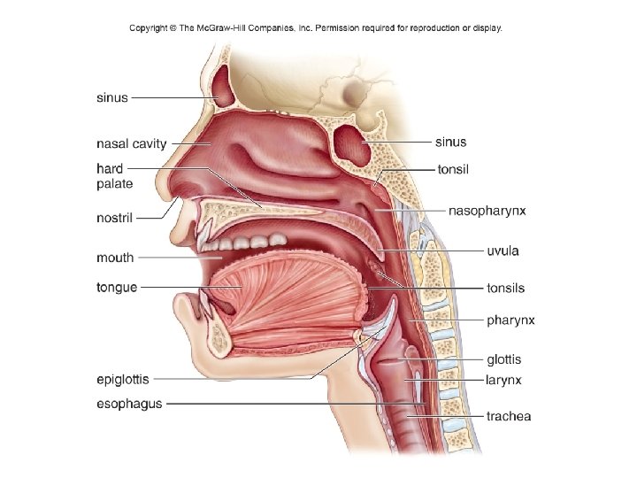
The Nose l The Nose – Part of upper respiratory tract l Includes nasal cavities, pharynx, and larynx – Air enters through nostrils (external openings) – Contains two nasal cavities Warms and moistens air during inhalation l Contains odor receptors l Tear glands drain into nasal cavity l Separated from mouth by hard and soft palate l
The Pharynx l Pharynx connects nasal and oral cavities to larynx – Three parts Nasopharynx – where the nasal cavities open posterior to soft palate l Oropharynx – where the mouth opens l Laryngopharynx – opens into the larynx l – Uvula - soft extension of soft plate projects into oropharynx – Tonsils - a protective ring l Lymphatic tissue that protects against inhaled microbes
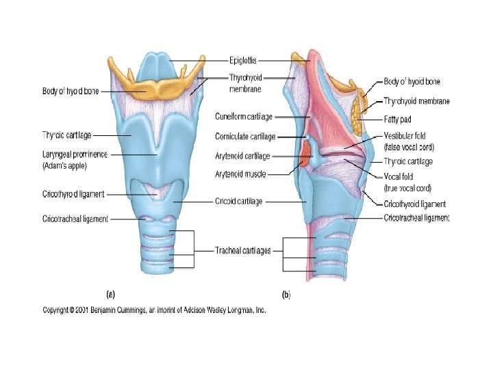
Larynx l Contains Vocal Cords – Connective tissue bands that tighten to create sound when air moves past them l Thyroid Cartilage – Sensitive to Testosterone levels
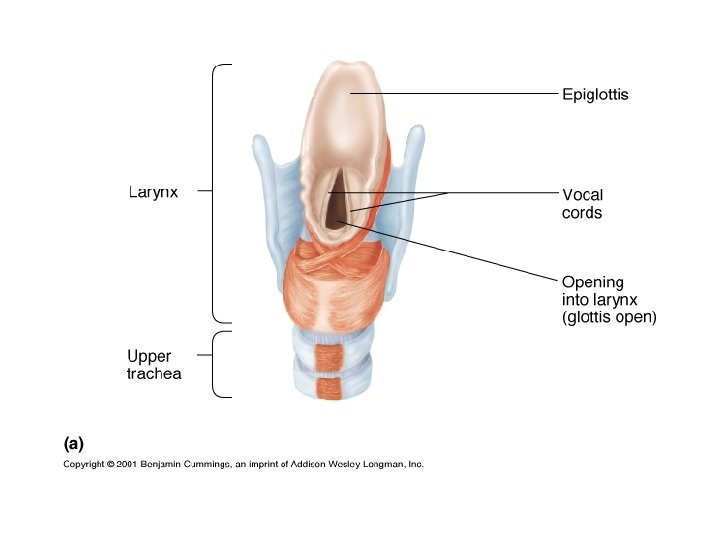
Copyright © The Mc. Graw-Hill Companies, Inc. Permission required for reproduction or display. base of tongue epiglottis vocal cords trachea © CNRI/Phototake l Larynx – cartilaginous structure – Passageway for air between pharynx and trachea – Vocal cords l Folds of mucosa that vibrate to make sounds – Glottis - opening between folds l Epiglottis – Flap preventing food from entering the respiratory tract Figure 15. 3
17
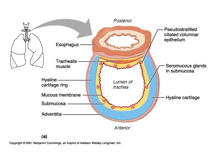
The Trachea l Trachea (windpipe) cilia goblet cell – Tube connecting larynx to primary bronchi – Held open by C-shaped cartilage rings – Cilia sweep mucus toward the pharynx l Smoking can destroy cilia 3. 5 m
20
Trachea l Conducts Air – Lined with pseudostratified ciliated columnar epithelium – Cilia can be paralyzed by cigarette smoke l Surrounded by C-shaped Cartilagenous rings and the trachealis muscle – Esophagus is dorsal to the trachea l Approximately 4 inches long
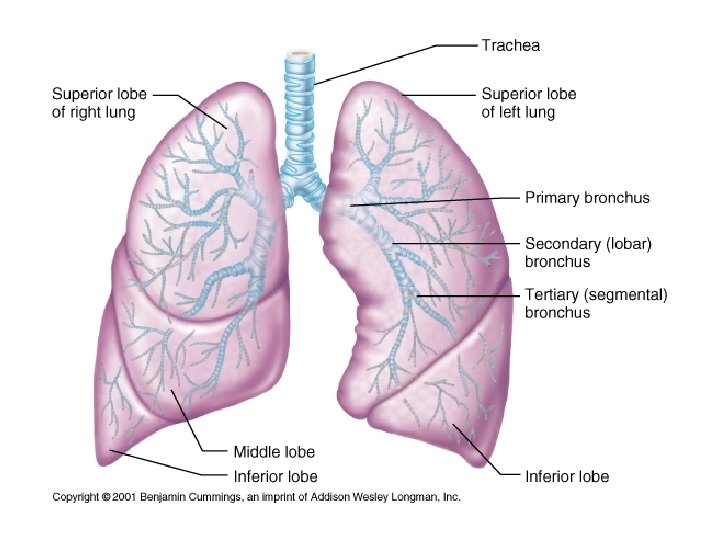
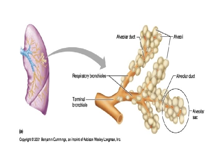
The Bronchial Tree l Trachea divides into right and left primary bronchi l Lead into right and left lungs – Branch to secondary bronchi l Eventually lead to bronchioles – As airways divide and subdivide, the walls become thinner l The small rings of cartilage are no longer present – Each bronchiole leads into sac called alveoli
Copyright © The Mc. Graw-Hill Companies, Inc. Permission required for reproduction or display. nasal cavity nostril pharynx epiglottis larynx trachea bronchus bronchiole lung diaphragm a.
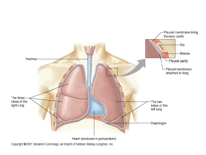
The Lungs l The lungs are paired, cone-shaped organs. – Occupy thoracic cavity l Diaphragm separates it from abdominal cavity – Right lung has 3 lobes – Left lung has 2 lobes l Allows room for heart – Each lobe subdivided into lobules l Each lobule has a bronchiole serving many alveoli
The Lungs – Each lung is covered by very thin serous membranes called pleura. – Another pleura covers the internal chest wall and diaphragm. – Both pleura produce lubricating serous fluid that helps the pleurae slide freely against each other during inspiration and expiration. – Surface tension holds the two pleura layers together when the lungs recoil in expiration.
Pleural Membranes l Visceral Pleura – Attached directly to the lungs l Parietal Pleura – Attaches to the visceral pleura – Also attaches to the thoracic cavity l Serous Fluid – Separates the two pleura and lubricates in order to decrease friction – Consistency of egg whites – Pleurisy occurs when the fluid decreases l The Function of the Pleural Membranes is to hold the lungs open
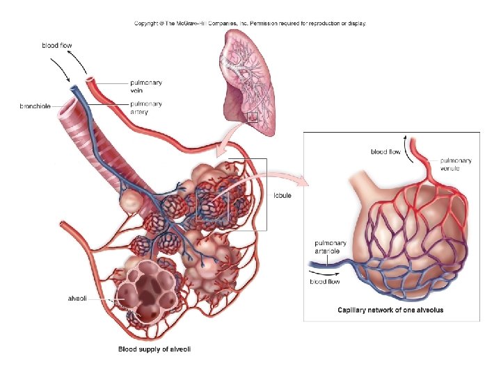
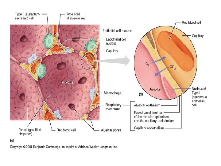
The Lungs l The alveoli are made up of simple squamous epithelium surrounded by blood capillaries. – Gas exchange occurs between the air in the alveolus and the blood in the capillaries. Oxygen diffuses across the walls into blood. l Carbon dioxide diffuses into alveoli. l – Alveoli must stay open to receive air. l Pulmonary surfactant helps prevent them from closing. – Infant respiratory distress syndrome – premature infants lack surfactant
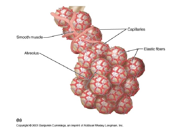
Alveoli l Clustered at the ends of the terminal bronchioles l Makes up the bulk of lung tissue l Primary function is the exchange of gases between themselves and the blood l Surrounded by elastic fibers – Creates Elastic Recoil
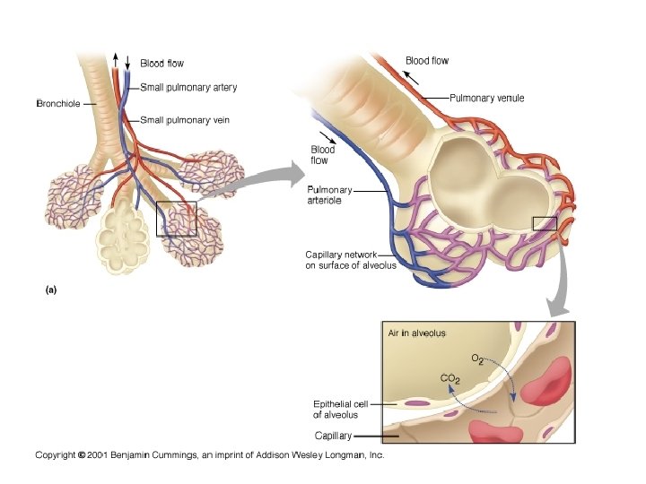
Copyright © The Mc. Graw-Hill Companies, Inc. Permission required for reproduction or display. blood flow pulmonary venule pulmonary arteriole blood flow Capillary network of one alveolus
35
Capillaries l The alveoli are closely associated with an extensive network of capillaries – Blood vessels cover 80 -90% of the alveolar surface forming a continuous “sheet” of blood in close contact with the air-filled alveoli
Respiratory Membrane l Consists of – The Wall of the Alveoli – The Respiratory Space This is a fluid filled space l Pneumonia may cause the space to fill with more fluid than normal l – This decreases the ability to exchange gases – The Wall of the Capillary
Alveoli Composed of a single layer of epithelium called Type I cells l Type II alveolar cells l – Secretes surfactant – Surfactant decreases the surface tension of the water within the alveoli – Coats the inside of the alveoli – Cortisol causes the maturation of the type II cells in the fetal stage of development l Dust Cells – Phagocytes
Copyright © The Mc. Graw-Hill Companies, Inc. Permission required for reproduction or display. Inspiration l The active phase – Diaphragm contracts l trachea intercostal muscles Rib cage moves up and out. Becomes flattened – Internal intercostals contract l lungs Raises rib cage up and out Diaphragm contracts and moves down. – Volume of thoracic cavity increases – Air pressure inside alveoli lowers – Air rushes in due to negative pressure Figure 15. 7 a air in lung rib cage pleurae Inspiration When lung volume increases, pressure decreases, and air comes rushing in.
Copyright © The Mc. Graw-Hill Companies, Inc. Permission required for reproduction or display. Expiration l The passive phase – Diaphragm and internal – – Rib cage moves down and in. intercostals relax Recoil returns them to original shape Volume of thoracic cavity decreases Air pressure inside alveoli increases Air rushes out Figure 15. 7 b Diaphragm relaxes and moves up. air out When lung volume decreases, pressure increases, and air is pushed out. b. Expiration
43
44
Gas Exchanges in the Body l Respiration includes the exchange of gases in the lungs (external respiration) and the exchange of gases in the tissues (internal respiration). l Most of the O 2 carried in the blood is attached to the iron-containing heme portion of the protein hemoglobin.
46
47
Emphysema l Loss of elastic fibers for elastic recoil during expiration – Elastin is destroyed by elastase l An enzyme released by immune cells l Have more difficulty exhaling than inhaling
Chronic Obstructive Pulmonary Disease (COPD) l Asthma, emphysema, chronic bronchitis