PHYSIOLOGY DEPARTMENT CENTRAL NERVOUS SYSTEM PHYSIOLOGY LECTURE 1


 2. Withdrawal reflex (tonic) 3. Reflex of")
 scheme")


. 2. Heart and vessel activity")
. 2. Postural reflexes. Labyrinthal tonic Neck tonic Static")


- Slides: 38
PHYSIOLOGY DEPARTMENT
CENTRAL NERVOUS SYSTEM PHYSIOLOGY. LECTURE #1. SPINAL CORD PHYSIOLOGY OF MEDULLA OBLONGATA AND MIDBRAIN Ass. Prof. VASTYANOV Rooslan
ATTENTION !!!
SPINAL CORD PHYSIOLOGY
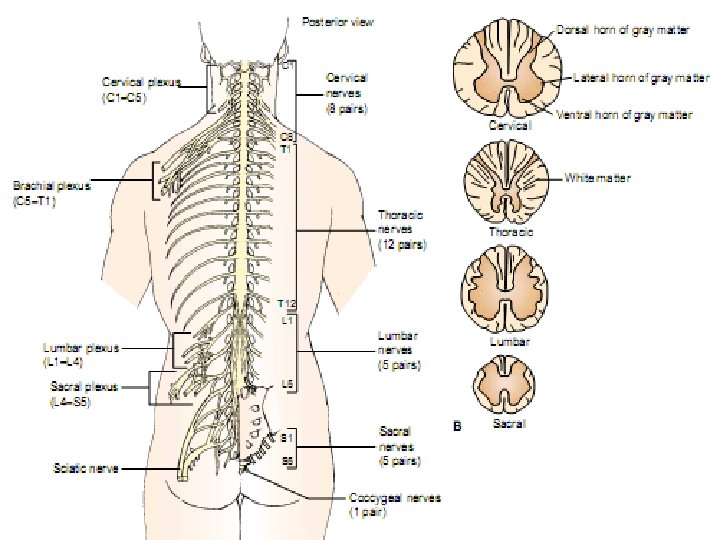
SPINAL CORD COMPOSITION The law of Bell - Mazhandi The ventral roots have motor (efferent) fibers whereas the spinal cord dorsal roots have sensitive (afferent) fibers.
Spinal Cord Cross-section
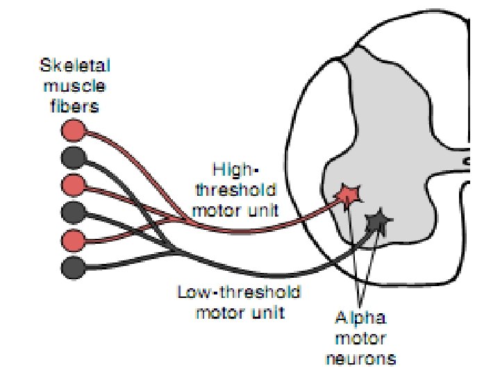
Spinal Cord Neurones Motor neurons Alpha-motor neurons, HT Alpha-motor neurons, LT Gamma-motor neurons Sensitive neurons are located in the back horns
Spinal Cord Functions REFLECTORY CONDUCTIVE
Spinal Cord Reflexes 1. Stretch reflex (phasic) 2. Withdrawal reflex (tonic) 3. Reflex of muscle-antagonists 4. Viscero-motor reflexes 5. Vegetative reflexes.
Stretch reflex (knee reflex) scheme
Withdrawal reflex scheme
Muscle – antagonists reflex scheme
Spinal Cord Functions REFLECTORY CONDUCTIVE
Spinal Cord Cross-Section. The view of the conductive pathways.
The uscendent pathways of the spinal cord 1 Tractus spino-thalamicus lateralis Tractus spino-thalamicus ventralis 2 Tractus spino-cerebellaris dorsalis Tractus spino-thalamicus ventralis 3
The descendent pathways of the spinal cord Tractus cortico-spinalis lateralis Tractus cortico-spinalis frontalis Tractus rubro-spinalis after Monakov Tractus vestibulo-spinalis Tractus reticulo-spinalis Tractus tecto-spinalis
Tractus corticospinalis
Descending motor tracts lesions SPINAL SHOCK - temporary pathologic condition characterizes by spinal cord connections rupture with upper brain after trauma or surgical intersection that follows by the lower spinal reflexes dissappearing.
BRAINSTEM PONS Cerebellum Medulla oblongata
FOSSA RHOMBOIDALIS
RETICULAR FORMATION OF THE MEDULLA OBLONGATA
Brain sagittal cross-section on the level of medulla oblongata
Functions of reticular formation 1. Respiration regulation (1885, Mislavsky). 2. Heart and vessel activity regulation (1871, Ovsyannikov). 3. Brain cortex activation via thalamus. Reaction of desynchronization. 4. Spinal reflexes activation. N. Reticularis lateralis. 5. Spinal reflexes inhibition. 1862, Sechenov. Reticular giant nucleus.
BRAINSTEM reflexes 1. Chain reflexes (digestive). 2. Postural reflexes. Labyrinthal tonic Neck tonic Static 3. Orientative reflexes. Stato – kinetic
BRAINSTEM reflexes 2. Postural reflexes. Neck tonic Labyrinthal tonic Stato – kinetic
MIDBRAIN PHYSIOLOGY
MIDBRAIN
ONE OF THE VERY IMPORTANT MIDBRAIN FUNCTION ! ! ! eyeballs movement regulation
N. Ruber of the midbrain Starts the extrapyramidal motor pathway - Tr. rubrospinalis Receives motor inputs from motor cortex, cerebellar nuclei, substantia nigra. FUNCTIONS: 1. Skeletal muscles tone regulation. 2. Unconsciousness instinctive and precise consciousness movements.
Lamina quadrigemina of the midbrain Colliculus superior Visual subcortical centers Colliculus inferior Audial subcortical centers
BLACK SUBSTANCE of the midbrain Pars reticulata Neurons use GABA as neurotransmitter Pars compacta Neurons use dopamine as neurotransmitter
DECEREBRATIVE RIGIDITY - pathological conditions induced in experiment after brain tissue section with red nucleus connections to vestibular nuclei complete dissection (Ch. Sherringtone experiment). This condition characterizes by extreme extensor tone prevailing on the flexor tone of the muscles.
DECEREBRATIVE RIGIDITY
DECEREBRATIVE RIGIDITY • chaws are clenched; • neck muscles are extended ; • the arms are adducted; • and stiffly extended at the elbows with the forearms pronated; • wrists and fingers are flexed.