Physiology BIO 240 Lecture No 3 Tonicity Genetics

Physiology BIO 240 Lecture No. 3 Tonicity Genetics Spring 2014 Dr. Ana M Jimenez

Membrane Transport Passive and Active Transport, osmosis, tonicity

Thinking About Cell Membranes – Most of the cell membrane is made of phospholipids, – Cell membranes are hydrophobic, – How do hydrophilic molecules such as water cross the membrane? – What forces water to move across the cell membrane?

Figure 3. 6 a

Cells have a selectively permeable membrane – Allow some but NOT other molecules to go through, – Factors: size, charge, polarity, hydrophilic/hydrophobic Passive Movement: does not require cell to spend energy (ATP), – Simple Diffusion – Facilitated Diffusion – Filtration • Active Movement: requires cell to spend cellular energy (ATP) – Active Transport Membrane Transport

Simple Diffusion • Movement of particles from an area of HIGH concentration to an area of LOW concentration ([]=concentration), [High] [Low] Concentration gradient difference between area of [high] and area of [low]

Factors that Affect Diffusion Rate • Temperature: Higher temperature, faster rate. • Molecular weight: Higher molecular weight, slower rate. • ‘Steepness of concentration gradient: Larger difference between concentrations, faster rate. • Membrane surface area: Larger surface area, faster rate. • Membrane permeability: Increased permeability, faster rate.
 pressure – Forces water")
Filtration vs. Osmosis of Fluid • Filtration - hydrostatic (water) pressure – Forces water to go through a filter, – Kidney filtration is important in removing waste from the blood, – Filtration of fluid at capillaries is governed by Starling Law of capillary diffusion. • Osmosis – tonicity (solute) pressure – Forces water to go through a membrane, – Water does not cross plasma membrane easily, but crosses through water pores called aquaporins.

Facilitated Diffusion • Carrier or pore - mediated transport – DOES NOT require energy, • Cells transport hydrophilic molecules that would not go through the membrane – such as glucose or water, • Specific for each molecule, transport is dependent on solute concentration: – Large concentration gradient= faster rate – Small concentration gradient= slower rate • Some carriers glucose – saturate (glucose)
![High [molecule] Low [molecule]](http://slidetodoc.com/presentation_image_h2/eb08890f0574feb221328189eadc2762/image-10.jpg "High [molecule] Low [molecule]")
High [molecule] Low [molecule]

Osmosis • Only molecules that cannot cross plasma membrane determine ‘tonicity’, • Tonicity – force of solutes that moves water across a membrane, – Large particles (albumin) cannot move through the membrane, • No transporters for proteins only for amino acids. – Small particles and water, glucose can move through membrane • due to transporters found on membrane (Ex. Aquaporins, glucose transporter - GLUT)

Side A = 5% albumin, Side B = 10% albumin • Side A has a LOW [albumin] as compared to side B, • Albumin can NOT move through the membrane, • In order for albumin to reach equilibrium, water is forced to move from side A into side B, • At equilibrium, albumin concentration is the same on both sides, – water has moved from A to B (not albumin), A B

Osmolarity vs. Tonicity • Osmolarity- Concentration of particles in solution – defined as number of osmoles in 1 Liter of solution – 1 Mol of glucose =1 Osm glucose, – BUT 1 Mol Na. Cl=1 Osm Na+ and 1 Osm Cl-, therefore 2 Osm (salts ionize in solution not sugars), – Blood ~300 m. Osm - Range is 280 -295 m. Osm/L • Tonicity - Difference in osmolarity of solutions across a membrane yields a force to ‘push’ or ‘pull’ water across a membrane in the process of osmosis. – When referring to tonicity – you are comparing two solutions (Ex. Extracellular fluid vs. Intracellular fluid)
: – due to lower concentration of non-permeating")
Tonicity of Solutions • Hypotonic Solution (hypo=low): – due to lower concentration of non-permeating solutes. – Cells in distilled water will gain water, swell and burst (because water moves toward hypertonic solution). • Hypertonic Solution (hyper=high): – due to high concentration of non-permeating solutes. – Cells in 2% Na. Cl will loose water, shrink, cellular membrane may rupture and cell dies (water moves toward hypertonic solution). • Isotonic Solution (iso=same): – equal concentration on non-permeating molecules. – Cells in 0. 9% saline solution retain their volume.

Hypotonic Solution Cells Swell with Water from Solution Isotonic Solution Hypertonic Solution Cells Remain the same Volume Cells Shrink due to Water loss

Active Transport - Requires ATP • Carrier mediated transport that spend ATP, • Molecules move against their concentration gradient from: [Low] [High]. – Carrier binds to ligand, – Carrier is phosphorylated by ATP, – Carrier changes conformation (shape), – Carrier releases ligand to other side of membrane.

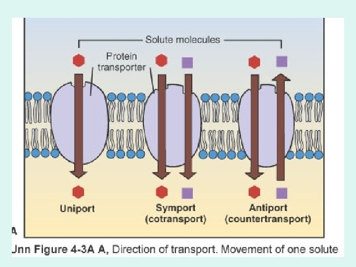
Co-transport • Carrier that moves 2 different types of molecules at the same time. – Symport: • Molecules move in the same direction, • Ex, sodium-glucose transport proteins (SGLTs), – Move Na+ and glucose into the cell. – Found in cells of digestive system and kidney tubules – Antiport: • One molecule moves in one direction, the other molecule in the opposite direction, • Ex. Na+/K+ pump (3 Na+ out, and 2 K+ in).

Sodium-potassium ATPase Pump • Found in all cells, • Uses ATP • Pumps 3 Na+ ions out of the cell and 2 K+ ions into cell, • Maintains –High Na+ in ECF (low in ICF) –High K+ in ICF (low in ECF) • Is Electrogenic – produces a charge

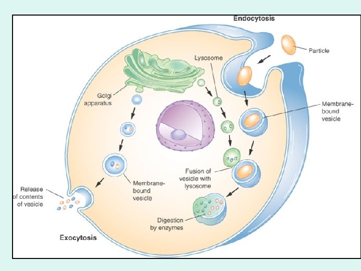
Bulk Transport • • Large number of molecules are moved in one direction in ‘vesicles’ across cell, There are two types, – Endocytosis: • • (endo=inside, cyto=cell, sis=process) Molecules are internalized into the cell. Phagocytosis – molecules are moved in bulk Pinoctytosis – fluid is moved in bulk – Exocytosis: • • (exo=out) Molecules are externalized out of the cell.

Is it possible to drink too much water? – What is water toxicity? – Loss of electrolytes during exercise and water retention cause hyposmolarity of ECF. – What would happen to cells? Billy wants to know!
 – most cells remain")
Life Cycle of a Cell Interphase • Growth (G 1) – most cells remain in this stage most of the time, • DNA Replication, • Growth (G 2) Cellular Division Mitosis • Prophase • Metaphase • Anaphase • Telophase Cytokinesis

How and When do Cells Divide? Cytokinesis separates one cell into two cells How are organelles separated? How do cells get the same set of chromosomes?

Karyotype – picture of chromosomes during cell division Diploid Human Cells have 46 chromosomes Autosomal chromosomes are pairs No. 1 -22, Sex determining chromosome pair is No. 23 Female - XX Male – XY ‘Y’ carries ‘male’ genes, Genes found on the ‘X’ No. 23 do not have a counterpart on the ‘Y’ (hemizygous).

Replicated Chromosomes Sister Chromatid
 Cells have? 46 46 46 Synthesis")
How Many Chromosomes do Human Diploid (2 n) Cells have? 46 46 46 Synthesis Phase – Duplication of Chromosomes

Prophase completely,
")
Metaphase plate (in animal cell)

Chromosomes look like ‘X’s when they are replicated and linked by centromeres. This is actually two identical chromosomes which will divide in anaphase.

Anaphase Telophase
 vs. Germ-line Cells (meiosis) Purves, et al.")
Human Sexual Life Cycle Somatic Cells (mitosis) vs. Germ-line Cells (meiosis) Purves, et al. Life 7 th ed. 2004

Mitosis vs. Meiosis • Mitosis – is cellular division that conserves the number of chromosomes. – Almost all cells are diploid and undergo mitosis, – Results in two daughter cells each with 46 chromosomes, – Each daughter cell is genetically identical to each other and to the mother cell. • Meiosis – is cellular division to divide chromosome number in half. – Germ line cells undergo meiosis to form gametes - ovum (egg) and sperm, – Results in four daughter cells with half the number of chromosomes (23 chromosomes each), – Each daughter cell has genetic combinations that are different from each other and from mother cell.

Independent Assortment Why are you not identical to your siblings of the same gender? Gametes are either ovum or sperm. These potential gametes are each different from each other due to the genetic recombination that takes place in meiosis.

Unique Features of Meiosis

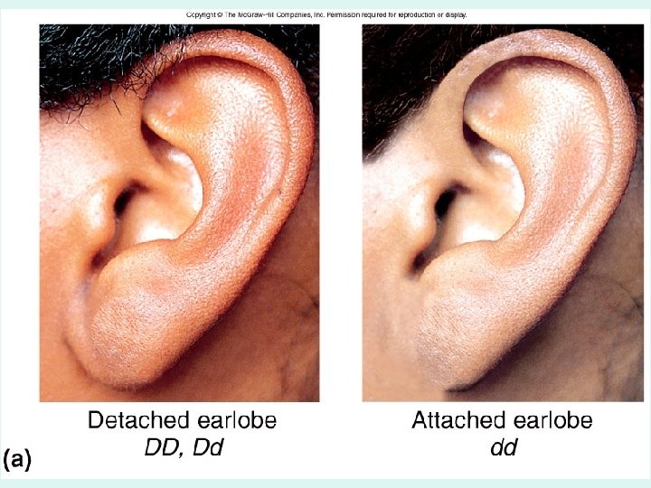
Do you have attached or detached ear lobe? – This trait is determined by one developmental gene, – Since you have two chromosomes in that pair – which genes do you have? – Which genes do your parents have? Billy wants to know!

MOM A punnet square – is a statistical tool to determine genetic probability DAD

Genetics Principles Overview of Human Genetics

Thinking About Genetics – Expression of a gene product, such as hair color or genetic predisposition to cancer is called - phenotype, – The set of nucleotides in DNA that ‘code’s for the phenotype is called genotype, – Genotype is half from mom and half from dad.

What is Phenotype and Genotype? Fig. 4. 18 • Phenotype for this trait is cleft or uncleft chin • Genotype for this trait is the letter representation of the allele that encodes the trait such as CC, Cc, cc (there are two alleles for each gene)

How to build a Punnet Square to determine probabilities of inheritance Shown are the possible phenotypes and genotypes of the possible children from a mother with a genetic genotype of Cc and a father with a genetic genotype of of Cc Fig. 4. 18

Genotype and Phenotype • Gene - portion of DNA that can be expressed such as eye-color, • Alleles - possible genes in a population such as brown, blue, green, hazel, etc • Genotype - is the actual alleles on DNA. – If the same = homozygous – If different = heterozygous – If only one allele (‘Y’)= hemizygous • Phenotype - is the ‘expression’ of a gene in the form of a protein.
 Phenotype")
Some Human Genetic Traits Governed by One Gene • Cleft/uncleft chin (compete dominance) Phenotype Genotype Cleft Chin Homozygous Dominant (CC) Cleft Chin Heterozygous (Cc) Uncleft Chin Homozygous Recessive (cc)

Dominance • Complete dominance – One allele is dominant over the other – Ex. Widow’s peak • Incomplete dominance – Both alleles ‘blend’ into each other – Ex. Skin color • Co-dominance – Both alleles are dominant at the same time – Ex. Blood types
: – Huntington’s Disease (Dominant) – Sickle Cell")
Human Genetic Diseases • Complete Dominance (Autosomal): – Huntington’s Disease (Dominant) – Sickle Cell Anemia (Recessive) • Complete Dominance (Sex Linked): – Hemophilia

Huntington's Disease • Inherited as an autosomal dominant disease, • Males and females have equal chances of getting it, – Homozygous dominant has the disease – Heterozygous has the disease – Homozygous does not have the disease (normal)
")
Huntington's Disease Example of Autosomal Dominant Disease Abbreviations: (Huntington’s = H Normal = h) Woman with Huntington’s disease (Heterozygous) and normal man. Haploid Mother (Hh) (Possible gametes) Gametes H h • h Hh hh • Father (hh) h Hh hh • (Possible gametes) Possible zygotes are diploid • Couple has 2 in 4 or 50% chances of having a child with Huntington’s disease (Hh) and 50% chances of a normal child.

Sickle Cell Anemia • Inherited as an autosomal recessive disease, • Men and women have equal chances of getting it, • Homozygous dominant (normal) does not have disease, • Heterozygous (carrier) has one copy of mutated gene but not the disease (sickle trait), • Homozygous recessive has the disease: – If two carriers for the sickle cell trait marry what chances do they have of having a child with the disease? – What chances do they have of having a child who is a carrier?

Normal RBCs ~200 -300 hemoglobin molecules Anemia non-functional erythrocytes Sickle Cell Anemia Non-functional hemoglobin (inherited condition homozygous recessive = both parents must carry one gene)

Sickle Cell and Malaria – is a disease transmitted by a mosquito that causes liver disease and eventually death. Sickle cell anemia carriers are ‘protected’ against malaria and tend to survive infection, while malaria is lethal to individuals who are normal for sickle cell anemia.

Punnet Square: Probabilities Example of heterozygous parents S=Normal and s=Sickle cell anemia Mom is heterozygous (Carrier) Ss Dad is Heterozygous (Carrier) Ss S s S___s SS Ss Ss ss Possibilities for each child: ¼ SS (Normal), ½ Ss (Carrier), ¼ ss (sickle cell anemia).

Human Genetics: Hemophilia • Hemophilia is a sex-linked recessive disease, • Men have a higher chance of getting the disease than women (Why? ): – Female homozygous dominant (normal) – Female heterozygous = carrier (normal) – Female homozygous recessive (hemophiliac) – Male homozygous (normal) – Male hemophiliac

Figure 4. 20

Punnet Square: Probabilities For Gender, Female = XX and Male = XY Mom is female XX Dad is male XY X XX XY Possibilities for each child: ½ XX (Female), ½ XY (Male).
 XNXn Dad is Normal")
Punnet Square: Probabilities N=Normal and n=hemophiliac Mom is heterozygous (Carrier) XNXn Dad is Normal X NY XN X NX N X NY Xn X NX n X n. Y Possibilities for each child: ¼ XNXN (Female normal), ¼ XNXn (Female, Carrier), ¼ XNY (male normal), ¼ (male hemophiliac).

Is it possible for a human baby to have blood type ‘O’ if both his parents are blood type ‘AB’? – What are the human blood types? – What is responsible for ‘inserting’ the blood antigen unto the erythrocyte membrane? Billy wants to know!

Genetics of Blood Types • Co-dominant alleles • Antigen: Glycoprotein expressed on the surface of cells, • Antibody: Protein that binds to antigen, • Possible Human Blood Antigen Alleles – ‘A’ represented as A allele – ‘B’ represented as B allele – No A or B called ‘O’ represented as ‘i’ allele

Blood antigens are glycoproteins on the membrane of red blood cells

Blood Types Genotype • • Blood type A = Blood type B = Blood type AB = Blood type O = AA or Ai BB or Bi AB ii

ABO Blood Groups: Co-dominance

Blood Typing for Paternity • Mom is blood type AB and Dad is blood type AB what are possible blood types for their child? Mom is AB A B Dad is A AA AB AB BB • Possible blood types A or AB or B
 Blood Type")
Genotype for Blood Types • • • Blood Type O (ii genotype) Blood Type AB (AB genotype) Blood Type A (AA genotype) Blood Type A (Ai genotype) Blood Type B (BB genotype) Blood Type B (Bi genotype) = = = __0___% __50__% __25__% __0___% Is it possible for this couple to have a child with blood type O?

Beyond Mendelian Inheritance • Polygenic inheritance: many genes are involved in controlling one trait – The phenotype depends on the cumulative contribution of many alleles – The traits show continuous variation (are called quantitative traits) § Human height § Blood pressure § Eye color
 are the result of more than one gene Fig. 4. 19")
Some traits (phenotypes) are the result of more than one gene Fig. 4. 19

Eye color in humans depends on the presence of several genes that code for specific enzymes. Melanin is a brown pigment also found in skin, retina and inner ear. Fig. 4. 22
. Do you know your blood")
Dogs have 11 different blood types (5 are antigenic). Do you know your blood type? – In humans - there are 4 ABO blood types, which ones are they? – If we add the Rh-factor, how many possible blood types in humans? – What does antigenic mean? Billy wants to know!
- Slides: 67