
Physiologic Basis for Hemodynamic Monitoring Circulation to Perfusion

Physiologic Basis for Hemodynamic Monitoring 臺大醫院麻醉部 鄭雅蓉
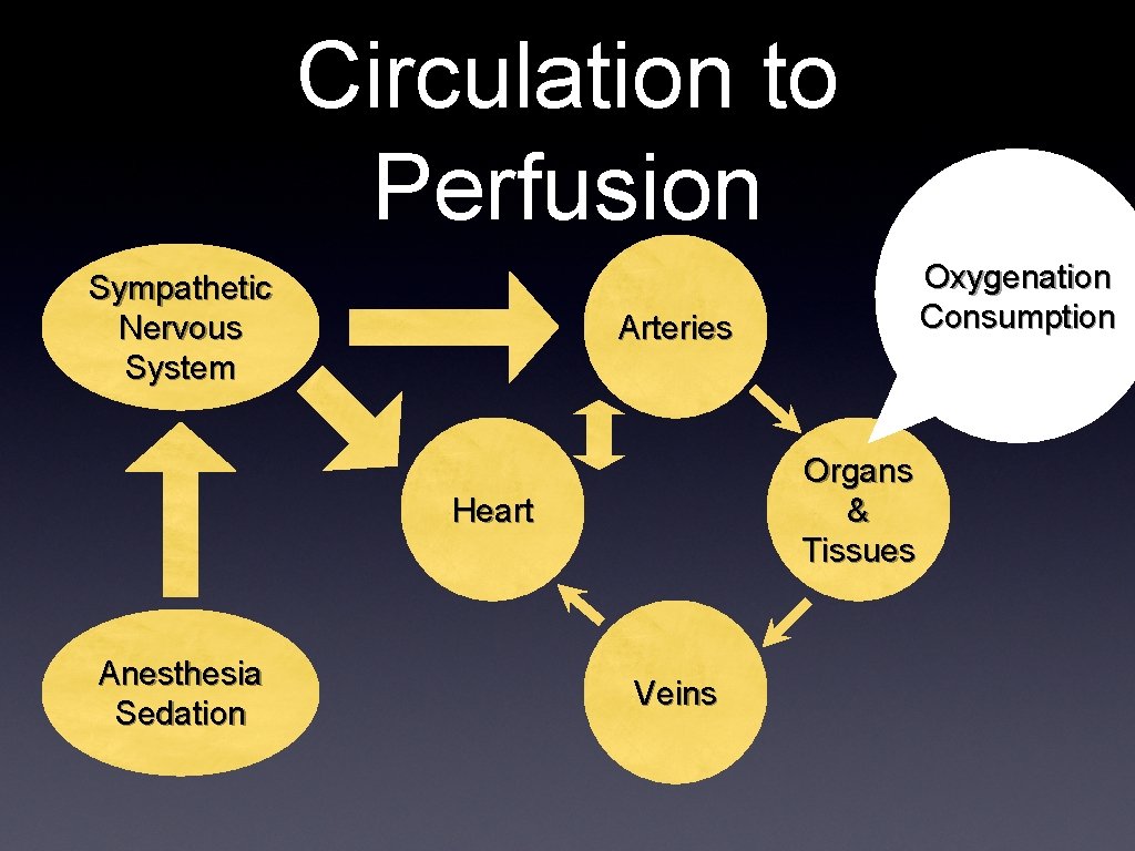
Circulation to Perfusion Sympathetic Nervous System Arteries Organs & Tissues Heart Anesthesia Sedation Oxygenation Consumption Veins
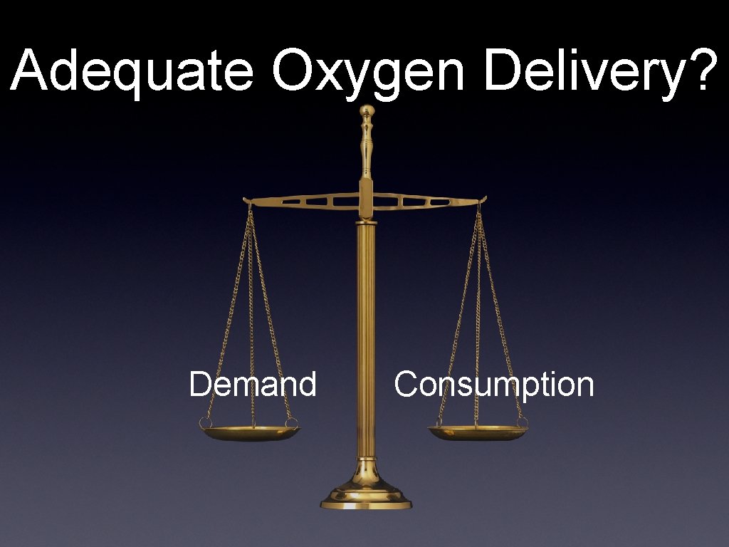
Adequate Oxygen Delivery? Demand Consumption
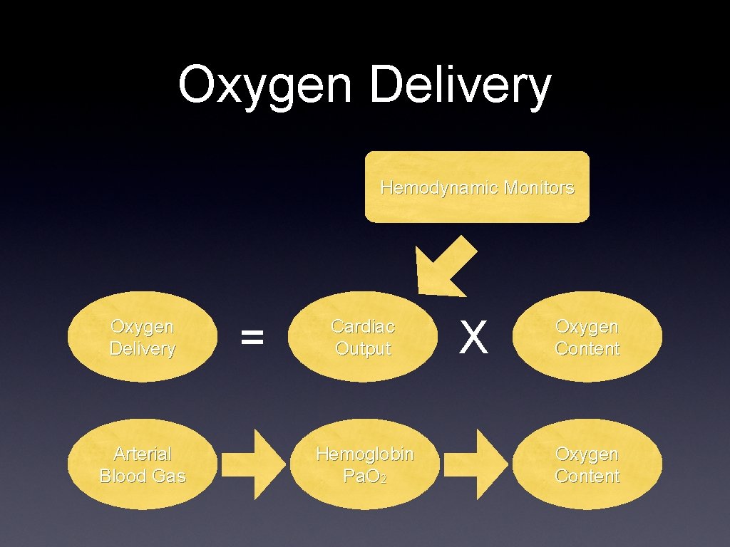
Oxygen Delivery Hemodynamic Monitors Oxygen Delivery Arterial Blood Gas = Cardiac Output Hemoglobin Pa. O 2 X Oxygen Content
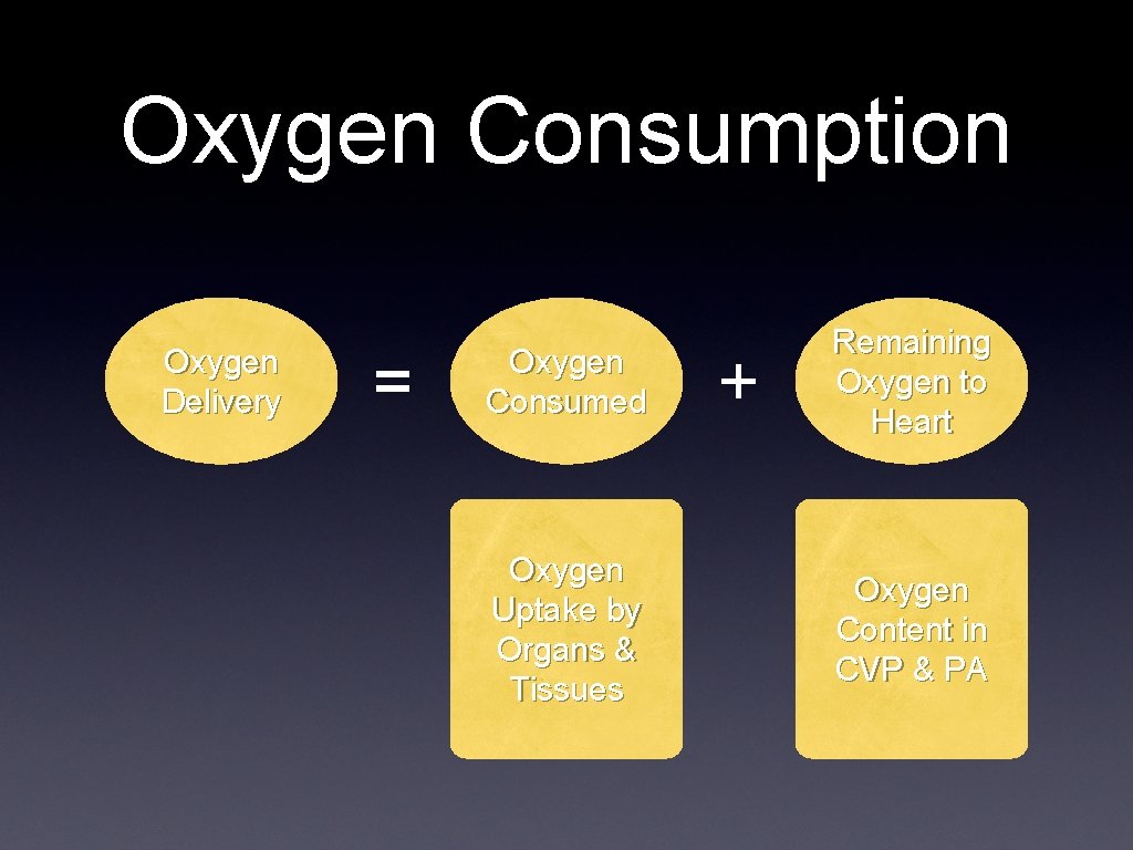
Oxygen Consumption Oxygen Delivery = Oxygen Consumed Oxygen Uptake by Organs & Tissues + Remaining Oxygen to Heart Oxygen Content in CVP & PA

Physiological Truth
Physiological Truth • There is no such thing as a “Normal Cardiac Output” • Cardiac output is either - Adequate to meet the metabolic demands - Inadequate to meet the metabolic demands Absolute values can only be used as minimal levels below which some tissue beds are probably under perfused •

History of Monitoring • 1960 s: golden age of vasopressors • • • Pressure, arterial line & CVP 1970 s: golden age of inotropes Cardiac output, PA catheter 1980 s: Sv. O 2 , relative balance between oxygen supply and demand 1990 s till now: Better understanding of tissue oxygenation, right ventricular function Functional monitoring, Pi. CCO, continuous CO Less invasive, TEE
Hemodynamic Monitoring Truth • No monitoring device, no matter how simple or complex, invasive or noninvasive, inaccurate or precise will improve outcome • Unless coupled to a treatment, which itself improves outcome Pinsky & Payen. Functional Hemodynamic Monitoring, Springer, 2004
Goals of Monitors To assure the adequacy of perfusion Early detection of inadequacy of perfusion To titrate therapy to specific hemodynamic end point To differentiate among various organ system dysfunctions Hemodynamic monitoring for individual patient should be physiologically based and goal oriented.
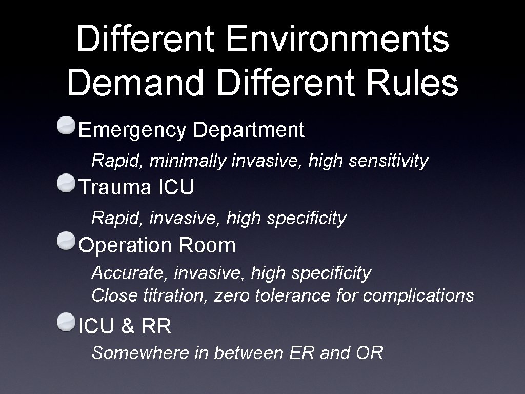
Different Environments Demand Different Rules Emergency Department Rapid, minimally invasive, high sensitivity Trauma ICU Rapid, invasive, high specificity Operation Room Accurate, invasive, high specificity Close titration, zero tolerance for complications ICU & RR Somewhere in between ER and OR

Hemodynamic monitors (1) • Traditional invasive monitors Arterial line CVP & Scv. O 2 • PA catheter, CCO, Sv. O 2 Functional pressure variation Pulse pressure variation Stroke volume variation
Hemodynamic monitors (2) • • Alternative to right-side heart catheterization • Pi. CCO Echocardiography • • Transesophageal echocardiography (TEE) Esophageal doppler monitor

Is Cardiac Output Adequate? Is blood flow adequate to meet metabolic demands? Pump function ? Adequate intravascular volume? Driving pressure for venous return?
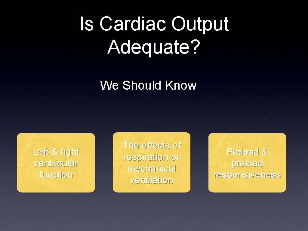
Is Cardiac Output Adequate? We Should Know Left & right ventricular function The effects of respiration or mechanical ventilation Preload & preload responsiveness
Ventricular Function • Left ventricular function • Right ventricular function • Depressed right ventricular function was further linked to more severely compromised left ventricular function. Nielsen et al. Intensive care med 32: 585 -94, 2006
Respiration and RV function • Spontaneous ventilation • Mechanical positive pressure ventilation
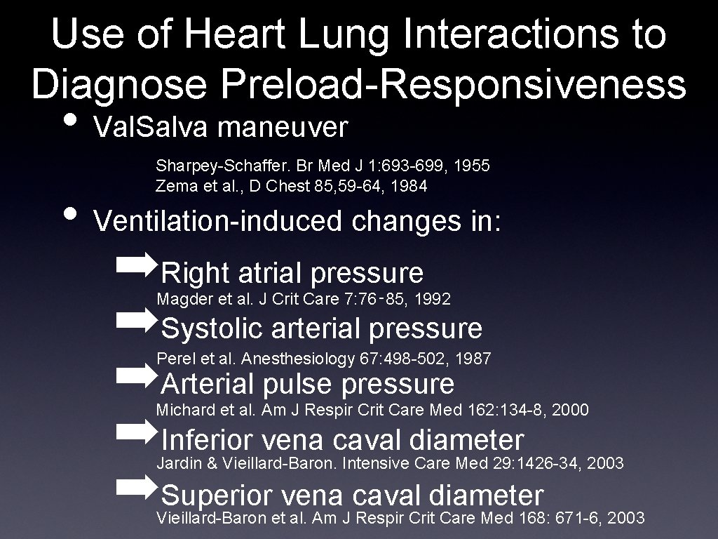
Use of Heart Lung Interactions to Diagnose Preload-Responsiveness • Val. Salva maneuver Sharpey-Schaffer. Br Med J 1: 693 -699, 1955 Zema et al. , D Chest 85, 59 -64, 1984 • Ventilation-induced changes in: ➡Right atrial pressure ➡Systolic arterial pressure ➡Arterial pulse pressure ➡Inferior vena caval diameter ➡Superior vena caval diameter Magder et al. J Crit Care 7: 76‑ 85, 1992 Perel et al. Anesthesiology 67: 498 -502, 1987 Michard et al. Am J Respir Crit Care Med 162: 134 -8, 2000 Jardin & Vieillard-Baron. Intensive Care Med 29: 1426 -34, 2003 Vieillard-Baron et al. Am J Respir Crit Care Med 168: 671 -6, 2003
Mechanical positive pressure ventilation ØIncrease RV outflow impedance, reduce ejection, increase RVEDV, tricuspid regurgitation ØTEE: SVC diameter: the effect of venous return? ØCVP may be misleading
Preload & Preload Responsiveness Starling’s law is still operated. If end diastolic volume ( EDV ) increased in response to volume loading, then stroke volume increased as well. CVP, PAOP and their changes: Did not respond with EDV, but Provide a stable route for drug titration and fluid infusion
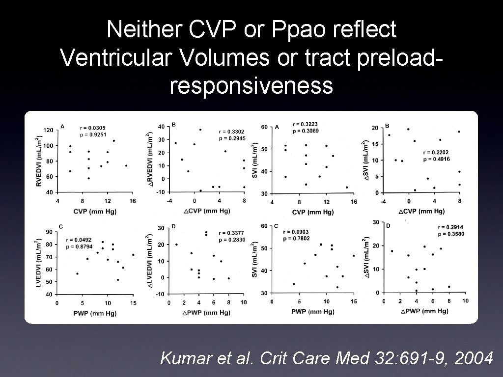
Neither CVP or Ppao reflect Ventricular Volumes or tract preloadresponsiveness Kumar et al. Crit Care Med 32: 691 -9, 2004
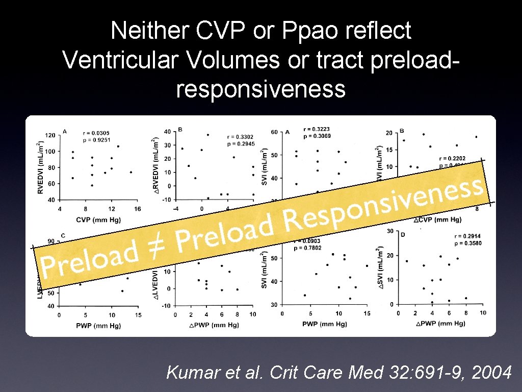
Neither CVP or Ppao reflect Ventricular Volumes or tract preloadresponsiveness Kumar et al. Crit Care Med 32: 691 -9, 2004
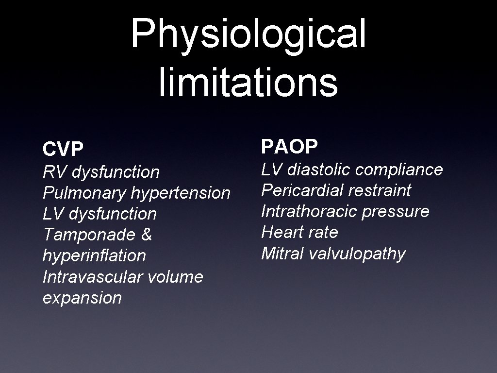
Physiological limitations CVP RV dysfunction Pulmonary hypertension LV dysfunction Tamponade & hyperinflation Intravascular volume expansion PAOP LV diastolic compliance Pericardial restraint Intrathoracic pressure Heart rate Mitral valvulopathy
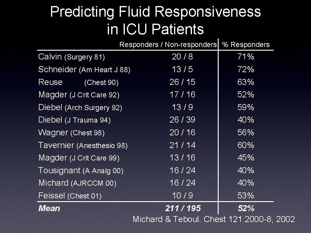
Predicting Fluid Responsiveness in ICU Patients Responders / Non-responders % Responders Calvin (Surgery 81) Schneider (Am Heart J 88) Reuse (Chest 90) Magder (J Crit Care 92) Diebel (Arch Surgery 92) Diebel (J Trauma 94) Wagner (Chest 98) Tavernier (Anesthesio 98) Magder (J Crit Care 99) Tousignant (A Analg 00) Michard (AJRCCM 00) Feissel (Chest 01) Mean 20 / 8 71% 13 / 5 72% 26 / 15 63% 17 / 16 52% 13 / 9 59% 26 / 39 40% 20 / 16 56% 21 / 14 60% 13 / 16 45% 16 / 24 40% 10 / 9 53% 211 / 195 52% Michard & Teboul. Chest 121: 2000 -8, 2002
• Can CVP Be Use for Fluid Management? Relatively Yes on most counts • Absolutely Yes for hypovolemia (10 mm. Hg cut-off) • Does apneic CVP predict preload responsiveness? No, but then neither does Ppao or direct measures of LV end-diastolic volume Michard et al. Am J Respir Crit Care Med 162: 134 -8, 2000
Thermodilution Cardiac Output The meaning of cardiac output ➡Mean (steady state) blood flow ➡Functional significance of a specific cardiac output value ➡Cardiac output varies to match the metabolic demands of the body Pinsky, The meaning of cardiac output. Intensive Care Med 16: 415 -417, 1990
Mixed Venous Oximetry • Sv. O is the averaged end-capillary 2 oxygen content (essential for VO 2 Fick) • Sv. O is a useful parameter of hemodynamic status is specific conditions 2 ➡If Sv. O 2 < 60% some capillary beds ischemic ➡In sedated, paralyzed patient Sv. O parallels CO 2
Adequate Oxygen delivery? • Sv. O : mixed venous oxygen saturation 2 • Consumption & delivery C(a-v)O 2: arterial-venous oxygen content difference Consumption & cardiac output • Lactate: the demand need of the use of oxygen Consumption & demand
Central Venous and Mixed Venous O 2 Saturation • Scv. O on CVP monitor • Sv. O on PA catheter • Sv. O is a sensitive but non-specific 2 2 2 measure of cardiovascular instability • Although Scv. O tracked Sv. O 2, it is tended to 7 ± 4 % higher. 2
Arterial Catheterization • Directly measured arterial blood pressure • Baroreceptor mechanisms defend arterial pressure over a wide range of flows • Hypotension is always pathological • Beat-to-beat variations in pulse pressure reflect changes in stroke volume rather than cardiac output
Pulmonary Arterial Catheterization • • Pressures reflect intrathoracic pressure Ventilation alters both pulmonary blood flow and vascular resistance ➡Resistance increases with increasing lung volume above resting lung volume (FRC) ➡changes Right ventricular output varies in phase with respiration-induced in venous return ➡Spontaneous inspiration increases pulmonary blood flow ➡flow Positive-pressure inspiration decreases pulmonary blood
Functional Hemodynamic Monitors • • • Arterial pulse contour analysis A better monitors for preload responsiveness: ➡a significant correlation between the increase of cardiac index by fluid loading by pulse pressure variation and stroke volume variation Peripheral continuous cardiac output system (Pi. CCO): arterial pulse contour and transpulmonary thermal injection: ➡intrathoracic volume and extravascular lung water
Conclusions Regarding Different Monitors • Hemodynamic monitoring becomes more effective at predicting cardiovascular function when measured using performance parameters ➡CVP and arterial pulse pressure (ΔPP) variations predict preload responsiveness ➡CVP, Scv. O and PAOP, Sv. O 2 predict the adequacy of oxygen transport 2
The Truths in Hemodynamics • • • Tachycardia is never a good thing. Hypotension is always pathological. There is no normal cardiac output. CVP is only elevated in disease. A higher mortality was shown in patients with right ventricular dysfunction and an increase of pulmonary vascular resistance.

The Truths in Hemodynamic Monitoring • Monitors associate with inaccuracies, misconceptions and poorly documented benefits. • A good understanding of the pathophysiological underpinnings for its effective application across patient groups is required. • Functional hemodynamic monitors are superior to conventional filling pressure. • The goal of treatments based on monitoring is to restore the physiological homeostasis.
- Slides: 36