PHYSICAL EXAMINATION OF THE SPINE Prof Dr afak


 - Rotations - Translations -")


")


: The patient lies on his")

,")


- Slides: 70
PHYSICAL EXAMINATION OF THE SPINE Prof. Dr. Şafak Sahir KARAMEHMETOĞLU, MD. İU/CMF/PMRD
CERVICAL SPINE
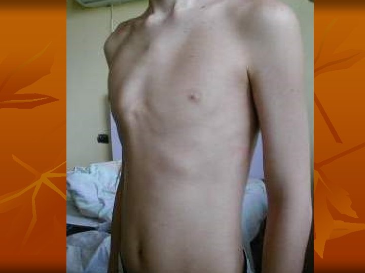
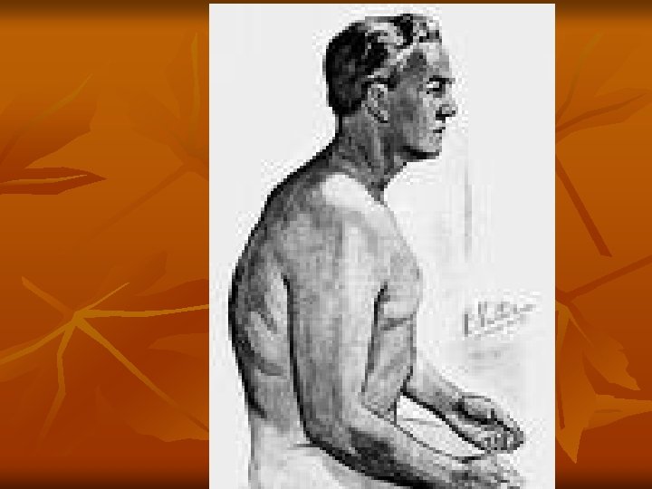
INSPECTION-1 - Lordosis - Scoliosis - Swelling - Torticollis - Muscle atrophy
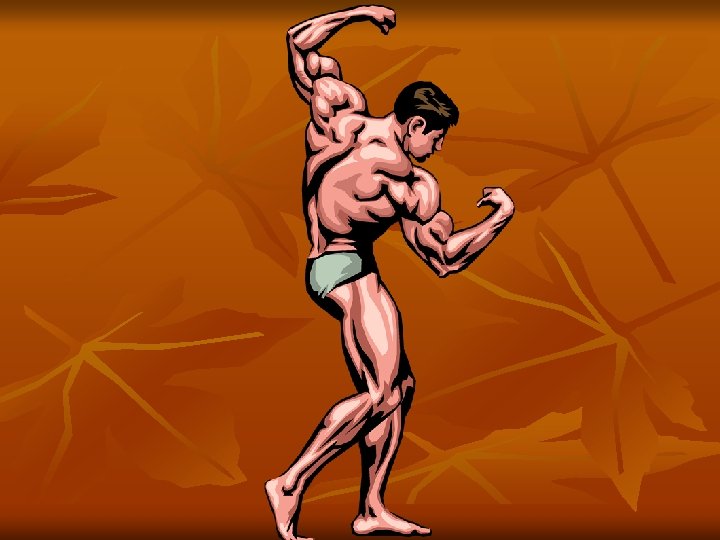
INSPECTION-2 - Muscle hypertrophy - Color changes - Arterial pulse - Postural changes
PALPATION-1 * Bone-Joint - Spinous process - Interspinous space - Foramen
PALPATION-2 * SOFT TISSUES - Swelling - PVM - SCMM - Spinal nerves - Ligaments
PALPATION-3 * Neighbouring structures - Arterial pulse - Lymph nodules - Thyroid - Trachea - Others
ROM - Flexion - Extension - Lateral Flexion (45°) - Rotations - Translations - Circumflexion
NEUROLOGIC EXAMINATION - Cervical Plexus C 1 -C 4 - Brachial Plexus C 5 -T 1
Cervical Plexus Has no key muscle, flexors, extensors, lateral flexors and rotators are tested in groups. C 1: Mainly motor fibres. C 2: Key point: protuberantia occipitalis externa. C 3: Key point: middle of the fossa supraclavicularis. C 4: Key point: acromio-clavicular joint.
Brachial Plexus C 5: KM: biceps, brachialis, KP: lateral of the antecubital fossa, DTR: biceps. C 6: KM: extensor carpi radialis longus and brevis, KP: middle of the dorsum of the first phalanx of the thumb, DTR: brachioradialis. C 7: KM: triceps, KP: middle of the dorsum of the first phalanx of the middle finger, DTR: triceps C 8: KM: flexor digitorum profundus, KP: middle of the dorsum of the first phalanx of the little finger. T 1: KM: abductor digiti minimi, KP: medial of the antecubital fossa.
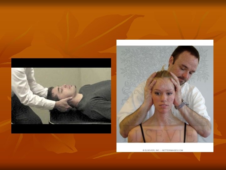
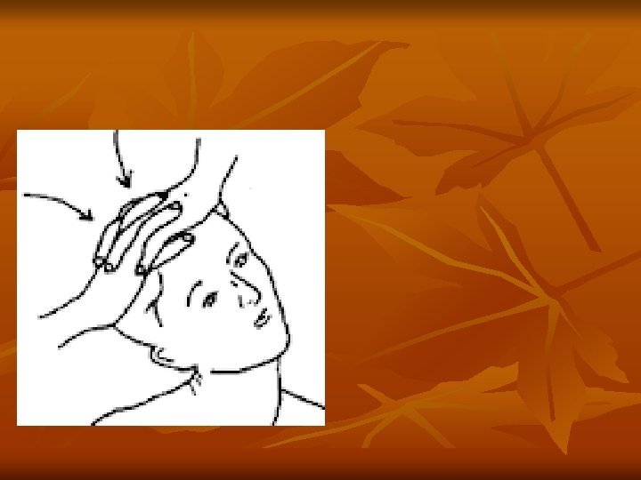
Special Tests -1 Distraction: To perform this test, place the open palm of one hand under the patient’s chin, and the other hand under the occiput. Then, gradually lift (distract) to remove its weight from the neck, if the neck and/or arm pain decreases or disappaers, the test is positive. It demonstrates the effect that neck traction might have in relieving pain by widening the foramen, decreasing pressure on the joints capsules around the facet joints. In addition it may help to alleviate muscle spasm by relaxing the contracted muscles.
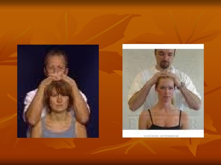
Special Tests -2 Compression: To perform this test, press down upon the top of the patient’s head while he is either sitting or lying down, if there is an increase in pain in the neck and/or arm(s), then the test is considered to be positive. A narrowing of the neural foramen, pressure on the facet joints or muscle spasm can cause increased pain. In addition, this test may reproduce pain referred to the upper extremity from the cervical helping to locate the neurological level of any existing pathology.
Special Tests -3 Valsalva: To perform this test, have the patient hold his breath and bear down as he/she were moving his/her bowels. Then, ask the patient whether he/she feels any increase in pain, and if so, whether he/she can describes the location. If the response is accurate, the test is positive. This test increases intrathecal pressure. If a space occupying lesion such as a herniated disc or a tumor, is present in the cervical canal, the patient may develop pain in the cervical spine. The pain may also radiate according to the neurological level.
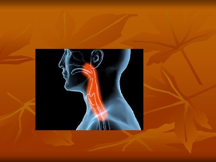
Special Tests -4 Swallowing: Difficulty or pain upon swallowing can sometimes be caused by cervical spine pathology such as bony protuberances, osteophytes, or by soft tissue swelling due to hematomas, infection, or tumor in the anterior portion of the cervical spine.
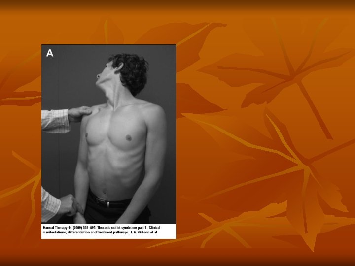
Special Tests -5 Adson: To perform this test, take the patient’s radial pulse at the wrist. As you continue to feel the pulse, abduct, extend and externally rotate his arm. Then istruct him/her to take a deep breath and to turn his/her head toward the arm being tested. If there is compression of the subclavian artrey, you will feel a marked diminution or absence of the radial pulse, then the test is positive. This test is used to determine the state of the subclavian artery, which may be compressed by an extra cervical rib or by tightened scalenus anticus and scalenus medius muscles, which can compress the artery where it passes between them on its way to the upper extremity.
Special Tests -6 Spurling: To perform this test, instruct the paient to extend, lateral flex and rotate his/her head. Then, press down upon the top of the patient’s head while he is either sitting or lying down, if there is an increase in pain in the neck and/or arm(s), then the test is considered to be positive. A narrowing of the neural foramen, pressure on the facet joints or muscle spasm can cause increased pain. In addition, this test may reproduce pain referred to the upper extremity from the cervical helping to locate the neurological level of any existing pathology.
Special Tests -7 Slump: This test is a progressive series of maneuvers designed to place the sciatic nerve roots under increasing tension. The patient sitting on the examining table, flexes the cervical, thoracic and lumbar spine, extends one of the knees and dorsifelexes the foot on the same side. If the patient experiences pain in low-back and/or leg(s), the test is positive.
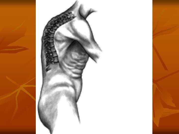
THORACIC SPINE
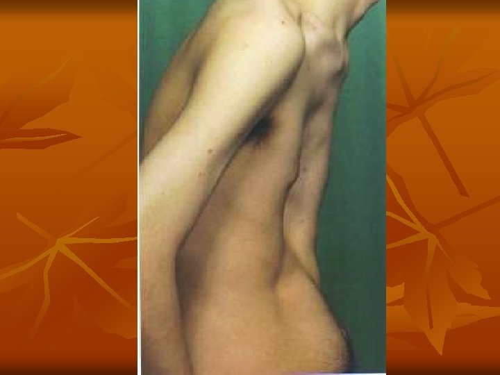
INSPECTION-1 * The patient must be undressed, * Posture * Supine, prone and side-lying
INSPECTION-2 * Spina scapula T 3 * End of scapula T 7 -9 * Medial border of the scapula and spinous processes 5 sm. * End of the ears, acromions and, iliac crests must be horizontal
PALPATION and PRESSION * PVM * Facet joints * Spinous processes * Interspinous spaces
ROM-1 * Flexion 20 -45° * Extension 20 -45° * Lateral flexion 20 -40° * Rotation 35 -50°
ROM-2 * Sitting position, * C 7 -T 12: 3 sm. , C 7 -S 1 15 sm. * Structural scoliosis does not change in flexion
Neurologic Examination The level of the lesion by key points There is no key muscle Beevor’s sign
Key Points T 2: KP: Apex of the axilla T 3: KP: Third intercostal space (mid-clavicular line) T 4: KP: Fourth intercostal space (nipple level, mid-clavicular line) T 5: KP: Fifth intercostal space (mid-clavicular line) T 6: KP: Sixth intercostal space (xiphoid level, mid-clavicular line) T 7: KP: Between T 6 and T 8 (mid-clavicular line) T 8: KP: Between T 7 and T 9 (mid-clavicular line) T 9: KP: Between T 8 and T 10 (mid-clavicular line) T 10: KP: Umblicus (mid-clavicular line) T 11: KP: Between T 10 and T 12 (mid-clavicular line) T 12: KP: Superior of the middle of the inguinal ligament (mid-clavicular line)
Special tests * Slump * Passive scapular approximation * First thoracal nerve stretching
Special tests - 2 Passive scapular approximation test: The patient lies prone. The shoulders are stretched backwards with the approximation of scapulae. If there is pain or pain worsening the test is pozitive. This test stretches the first thoracal spinal nerve.
Special tests - 3 First thoracal nerve stretching test: The patient abducts the shoulder to 90°, flexes the elbow and holds the occipital region of the head and the shoulder is forced to extention. If there is pain or pain worsening the test is pozitive. This test stretches the first thoracal spinal nerve.
Special tests - 4 Beevor's sign is the movement of the belly button towards the head on flexing the neck. It is caused by weakness of the lower abdominal muscles. Beevor’s sign is characteristic of spinal cord injury at the T 10 level. It has also been described in amyotrophic lateral sclerosis and facioscapulohumeral muscular dystrophy.
LUMBAR SPINE
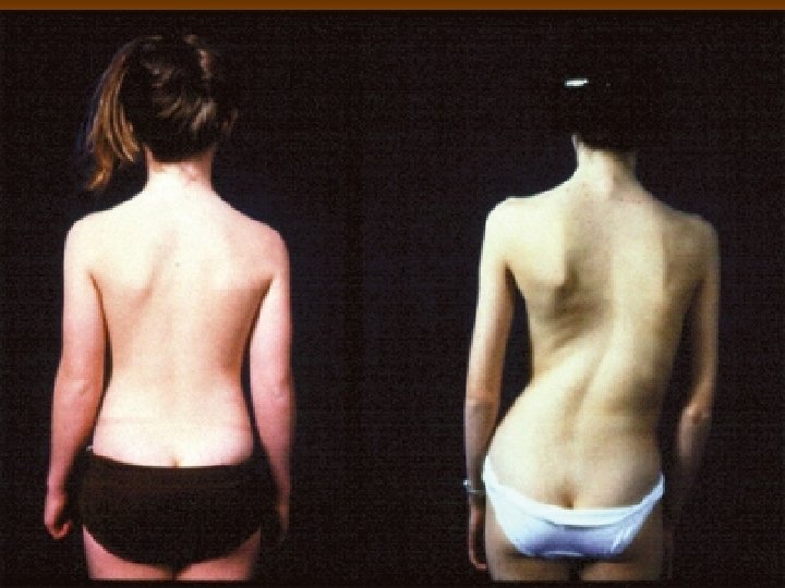
INSPECTION - 1 - Lordosis - Scoliosis - Swelling - Deviation - Muscle atrophy
INSPECTION - 2 - Muscle spasm - Color changes - Lipomas - Abnormal hair - Café au lait spots - Postural changes
PALPATION-1 Bone – Joint: - Spinous processes - Interspinous spaces - Facet joints - İliac crests - Coccyx
PALPATION-2 SOFT TISSUES - Swelling - PVM - Ligaments - Umblicus (L 3 -4) - Sacral promontorium
PALPATION-3 VALLEIX POINTS: 1. Middle of the trochanter major ve ischial tuberosity 2. Middle of the posteroir thigh 3. Middle of the popliteal fossa 4. Middle of the gastrocnemius muscle 5. Middle of the Achilles tendon
ROM - Flexion - Extension - Lateral Flexion - Rotations - Circumduction
NEUROLOGIC EXAMINATION - DTR - Muscle testing - Sensation
Neurologic examination L 1: KM: None, KP: inferior of the middle of the inguinal ligament. L 2: KM: iliopsoas, KP: midway of the KPs L 1 and L 3: KM: quad. femoris, KP: medial femoral condyl, DTR: patella. L 4: KM: tibialis anterior, KP: medial malleolus L 5: KM: extensor hallucis longus, KP: third metatarsophalangeal joint at the dorsum of the foot. S 1: KM: gastrocnemius-soleus, KP: lateral of the heel, DTR: Achilles.
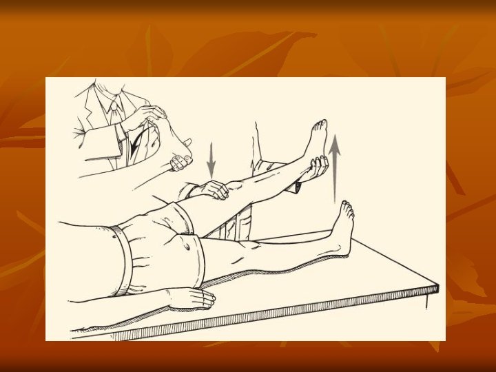
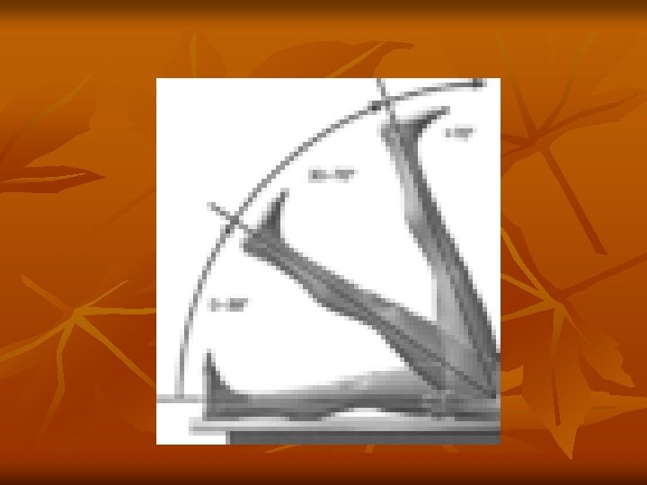
Special tests – 1 Straight Leg Raising Test (SLRT): The patient lies on his back (supine). The examiner raises the leg with the knee extended straight. Normally, the angle between the leg and the bench can reach 70° – 80° without any discomfort. If the patient experiences pain before, the test is positive. To differentiate between sciatic nerve stretching and hamstring muscles tightness, lower the leg a few angle and dorsiflex the foot in order to relax the hamstrings and to stretch sciatic nerve. If the patient reexperiences pain along the sciatic nerve, this is due to sciatic nerve stretching.
Special tests - 2 Contralateral SLRT: If there is low-back and/or contralateral leg pain when the uninvolved leg is raised the test is positive. This test may be associated with a considerable disc hernation or a space occupying lesion.
Special tests - 3 Kernig’ sign: The patient lies supine (flat on the back), flexes his head with hands on the occipital region. If the patient experiences neck pain and/or pain along the vertebral column the test is positive. This may be a sign of meningeal or spinal nerve roots irritation.
Special tests - 4 Naffziger’s test: Pressure is applied on the jugular vein of the patient lying on his/her back for 10 seconds. When the face of the paient flushes he/she is requested to cough. If he/she experiences low-back and/or leg pain the test is positive. Pressure on the jugular vein results in increased cerebrospinal fluid pressure and may cause pain in the case of a herniated disc.
Special tests - 5 Valsalva test: The patient takes a deep breath, holds and attempts exhalation against a closed glottis and closed mouth and nose. If there is low-back and/or leg pain the test is positive. Valsalva test increases the intrathecal pressure.
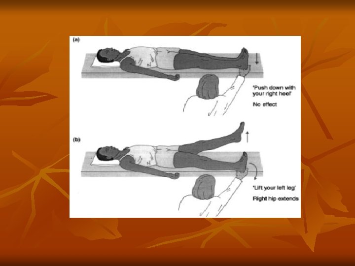
Special tests - 6 Hoover test: This test determines whether the patient is malingering when he states that he/she cannot raise his leg, and should be performed in conjuction with straight leg raising test. When a patient is genuinely trying to raise his/her leg, he/she puts pressure on the calcaneus of his/her opposite leg to gain leverage; you can feel downward pressure on your hand. If there is no pressure the patient is malingering.
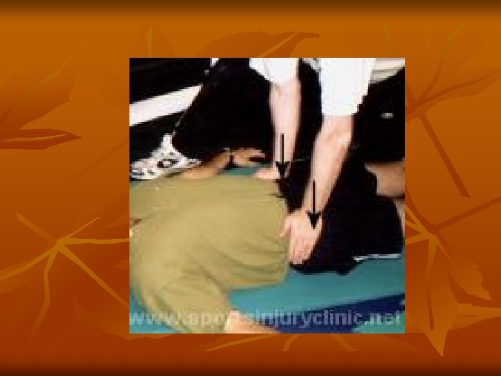
Special Tests - 7 Pelvic Rock: The patient lying supine, the examiner places hands on the iliac crests of the patient with thumbs on the anterior superior iliac spines, and palms on the iliac tubercles. Then, forcibly compresses the pelvis toward the midline of the body. If the patient complains of pain around the sacroiliac joint, there may be pathology in the joint itself, such an infection or problem secondary to trauma.
Special Tests - 8 Gaenslen’s sign: Have the patient lie supine on the table and ask him to draw both legs onto the chest. Then, shift the patient to the side of the table so that one buttock extends over the edge of the table while the other remains on it. Allow the unsupported leg to drop over the edge, while the opposite leg remains flexed. Complaints of subsequent pain in the area of the sacroiliac joint give another indication of pathology in that area.
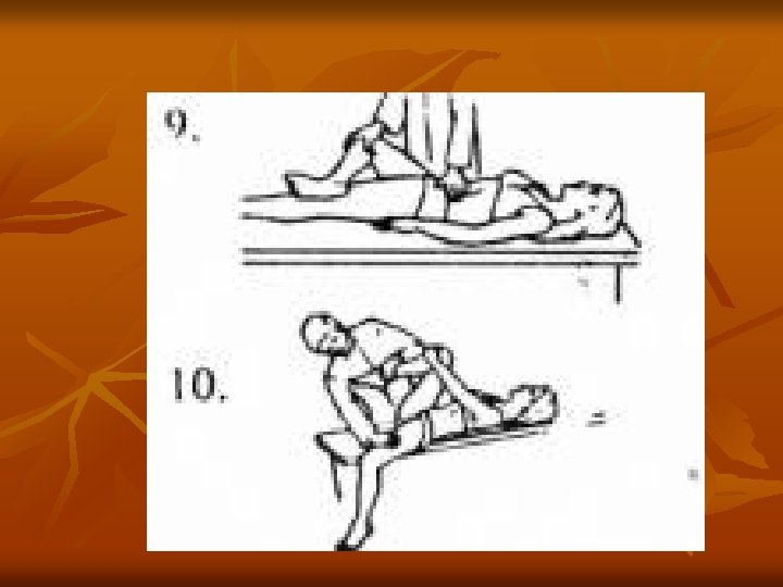
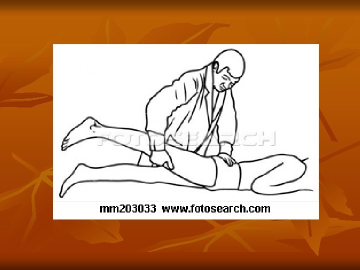
Special Tests - 9 Femoral nerve stretch: The patient lying prone, the examiner fully flexes the knee and extends the hip. If the patient experiences pain in the anterior area of the thigh, the test is positive.
Special Tests - 10 Slump: This test is a progressive series of maneuvers designed to place the sciatic nerve roots under increasing tension. The patient sitting on the examining table, flexes the cervical, thoracic and lumbar spine, extends one of the knees and dorsiflexes the foot on the same side. If the patient experiences pain in low -back and/or leg(s), the test is positive.