Physical Examination of the Skin Hair and Nails


- Slides: 31
Physical Examination of the Skin, Hair, and Nails
Skin Function Protective barrier Mechanical barrier Temperature regulator Sensor Vitamin D producer Repairer Excreter Expresser
Skin Anatomy
History Past Medical History previous problems systemic disease Family History skin CA, psoriasis, allergy, infestations and infections Psychosocial personal habits exposures
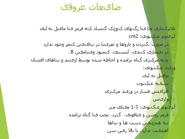
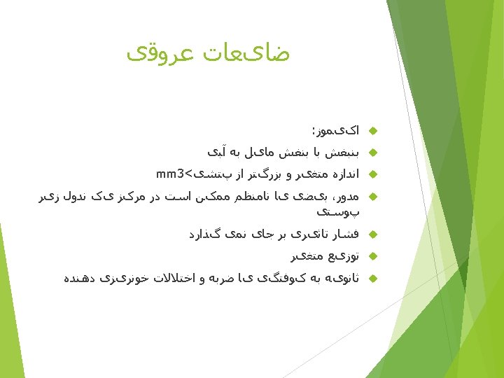
I. Primary lesions = lesions which have not been altered by external forces or time A. Flat, non-palpable lesions (demarcated only by color change from the surrounding skin; cannot be distinguished by touch) 1. Macule – smaller than 2 cm. In diameter 2. Patch – larger than 2 cm. B. Solid, palpable lesions 1. Papule – smaller than 1 cm. , elevated above the skin surface. 2. Plaque – larger than 1 cm. , flat-topped and elevated above the skin surface. Plaques may be very large, covering extensive areas. Smaller plaques may be composed of grouped, confluent papules. 3. Nodule – Usually spherical, a nodule may be palpated deeper than a papule or plaque and may be below the skin surface. 4. Wheal – A papule or plaque which is formed by edema in the skin, and which typically disappears after a short time.
II. Fluid-filled lesions 1. Vesicle – smaller than 1 cm and raised above the skin surface; a vesicle usually contains a clear serous fluid, but may also contain blood. Vesicles are commonly called blisters. 2. Bulla – Larger than 1 cm with the same contents as a vesicle. 3. Cyst – A firm-walled lesion usually containing a semisolid material. A cyst may be distinguished from a nodule by its softer, more rubbery feel. 4. Pustule – Small lesion raised above the skin which contains purulent (opaque) material (pus). Lesions which contain pus and that are larger and extend deeper are called in order of the increasing size, furuncles, carbuncles (made up of multiple furuncles) and abscesses.
III. Depressed lesions 1. Erosion – An area of skin loss, usually with a moist, erythematous base. An erosion is fairly superficial and does not extend below the epidermis. 2. Ulcer – An area of skin loss extending into the dermis or deeper. 3. Fissure – A linear erosion or ulcer.
IV. Secondary lesions – These occur as the result of change in primary lesions over time, or from exogenous manipulation of the skin. 1. Scale – White flakes from the top of the epidermal layer (stratum corneum) which are retained on the skin surface. 2. Crust (scab or eschar) – A solid, brownish covering over a lesion which is composed of old dried serum, blood or exudate. 3. Erosion – As described above. An erosion may be secondary if it results from the rupture of a vesicle or bulla. An excoriation is erosion created by scratching and is usually linear or angular. 4. Lichenification – Thickening of the skin with accentuation of skin lines. Lichenification results from repeated rubbing and scratching.
Morphology
MACULE PATCH
Morphology
PLAQUE WHEAL
NODULES
Morphology
Morphology
VESICLE BULLA
PUSTULE CYST
Morphology
SCALING CRUSTING
Morphology
EXCORIATION EROSION
FISSURE ULCER
ATROPHY LICHENIFICATION
HYPERTROPHIC SCAR KELOID
Distribution